Comprehensive Oncology: Neoplasia, Carcinogenesis, Molecular Pathogenesis, and Clinical Tumor Markers
1. Foundational Concepts of Neoplastic Transformation
Key Definitions in Oncology
- Neoplasm (Tumor): An abnormal mass of tissue that develops when cells proliferate in an autonomous, uncoordinated fashion, persisting in the same excessive manner even after cessation of the initiating stimuli. The growth pattern is pathologically irreversible, uncontrolled, unlimited, progressive, and purposeless.
- Cancer: A generalized medical designation describing malignant states of aberrant, deregulated, and aggressive cellular multiplication, with the intrinsic capacity to breach tissue boundaries and infiltrate surrounding or distant anatomical structures.
- Carcinogen: Any exogenous biological, chemical, or physical factor capable of inflicting genomic injury, inducing somatic mutations, and precipitating neoplastic change. If these agents trigger malignancy in utero, they function as teratogenic carcinogens.
- Co-carcinogen (Promoter): Non-mutagenic assisting factors that do not independently transform cells, but significantly accelerate and facilitate the transition of initiated, latent cells into frank malignancy.
The progression toward clinical malignancy follows a sequential chain of cellular events: aberrant and unchecked cell division triggers the aggregation of a non-physiological cellular mass (a tumor), which subsequently undergoes invasive local expansion and malignant transformation.
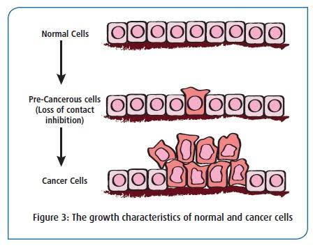
Progression from Regulated Normal Cellular Division to Uncontrolled Tumor Growth
1.1 Divergence Between Benign and Malignant Neoplasms
Tumors diverge significantly across their growth dynamics, border demarcation, and capacity for systemic dissemination.
| Pathological Attribute | Benign Tumors | Malignant Tumors (Cancer) |
|---|---|---|
| Growth Mechanism | Expansive, localized push; compresses adjacent parenchyma | Infiltrative, destructive, and erosive radial penetration |
| Capsular Integrity | Typically enclosed within a discrete fibrous connective tissue capsule | Non-encapsulated with poorly defined, irregular invasive margins |
| Metastatic Potential | Strictly absent; stays localized to tissue of origin | Present; disseminates systemically via vascular and lymphatic channels |
| Prognostic Threat | Generally non-life-threatening (unless strategically located in closed spaces like the cranium) | Life-threatening through tissue destruction, cachexia, and systemic compromise |
| Clinical Examples | Cutaneous melanocytic nevi, verrucae (warts), uterine leiomyomas | Bronchogenic carcinoma, acute myeloid leukemia, osteosarcoma |
1.2 Histogenetic Classification of Malignancies
Malignancies are categorized based on their underlying embryonic germ layer and histological lineage of origin:
- Carcinoma: Malignant tumors derived from epithelial cell layers (e.g., adenocarcinomas, squamous cell carcinomas).
- Sarcoma: Malignancies originating from mesenchymal or connective tissues (e.g., osteosarcoma, liposarcoma, chondrosarcoma).
- Lymphoma: Neoplastic proliferation confined to lymphoid nodes and associated extranodal lymphoid tissues.
- Leukemia: Diffuse malignancies arising from hematopoietic stem cells within the bone marrow, overflowing into the circulating blood pool.
2. Phenotypic, Morphological, and Biochemical Hallmarks of Cancer Cells
2.1 Morphological and Ultrastructural Derangements
Malignant transformation fundamentally remodels cellular architecture:
- Pleomorphism and Anaplasia: The cell loses its specialized differentiation and normal geometric boundaries. Cells exhibit significant variation in size and shape, associated with a loss of baseline structural receptors.
- Nuclear Atypia: Marked nuclear enlargement with hyperchromasia, irregular nuclear envelopes, coarse chromatin aggregation, distinct prominent nucleoli, and an increased nuclear-to-cytoplasmic (N:C) ratio with scarce, disorganized cytoplasm.
- Loss of Contact Inhibition: While physiological cells arrest division upon physical contact with neighbors, transformed cells continuously divide in a disordered fashion, creating multilayered clusters.
- Loss of Anchorage Dependence: Cancer cells bypass the physiological requirement of attachment to basement membranes or extracellular matrix proteins to survive and expand.
- Accelerated Cell Cycle Kinetics: Transformed cells demonstrate an approximate 2- to 5-fold expansion in the active mitotic phase relative to baseline tissues.
- Enzymatic Invasiveness: Downregulation of cell-to-cell adhesion molecules (e.g., E-cadherin) accompanied by hypersecretion of proteolytic enzymes (collagenases, matrix metalloproteinases) that actively digest extracellular matrix components to facilitate local invasion and systemic metastasis.
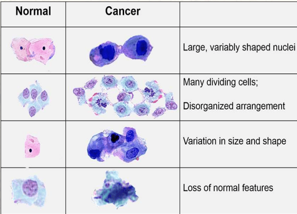
Cytomorphological Differences: Normal Versus Malignant Cells
2.2 Detailed Physiological Comparison: Malignant vs. Physiological Cells
| Biological Feature | Malignant Cells | Normal Physiological Cells |
|---|---|---|
| Cellular Morphology | Enlarged, irregular, and markedly pleomorphic | Predictable, uniform, and regular architecture |
| Nuclear-Cytoplasmic Architecture | Irregular hyperchromatic nucleus, prominent nucleoli, diminished cytoplasm | Proportionate, uniform nucleus and balanced cytoplasm |
| Proliferation Control | Uncontrolled, autonomous, and self-sufficient | Strictly homeostatic, regulated, and coordinated |
| Intercellular Communication | Impaired or absent (disrupted gap junctions) | Active coordination via tight, adhering, and gap junctions |
| Immunological Visibility | Evades immune surveillance (downregulates MHC-I, upregulates PD-L1) | Recognizable and visible to host immune checkpoints |
| Angiogenic Drive | Sustained tumor neoangiogenesis via autonomous VEGF secretion | Transient angiogenesis confined to wound repair or cyclic states |
| Oxygen Dependence | Can thrive in severe hypoxia; shifts away from oxidative phosphorylation | Obligate oxygen reliance for efficient energy generation |
| Glucose Consumption | Massive glucose uptake (glucose craving) | Moderate, feedback-inhibited glucose intake |
| Bioenergetic Efficiency | Extremely low (yields only 2 ATP per glucose molecule) | High efficiency via oxidative phosphorylation (up to 36–38 ATP) |
| Extracellular Microenvironment | Acidic (lactate-rich, promoting matrix breakdown) | Physiologically buffered, slightly alkaline pH (7.35–7.45) |
| Primary Nutrient Substrate | Preferentially consumes glucose (and glutamine) | Versatile utilization of fatty acids, ketone bodies, and glucose |
2.3 Metabolic Reprogramming (The Warburg Effect) and Biochemical Shifts
- Elevated Nucleic Acid Turnover: Marked upregulation in both DNA and RNA synthesis rates to accommodate uncontrolled cell divisions.
- Membrane Surface Reconfiguration: Altered density, branching, and distribution of cell-surface glycoproteins and glycolipids, disrupting contact cues and driving invasion.
- Antigenic Remodeling: Loss of tissue-specific differentiation antigens alongside the appearance of novel tumor-specific or re-expressed embryonic antigens (oncofetal antigens).
- Ectopic Endocrine Production: Paraneoplastic, inappropriate autonomous synthesis of hormones, cytokines, and growth factors by non-endocrine malignancies.
- Aerobic Glycolysis and Lactic Acidosis: Despite the presence of ambient oxygen, malignant cells shift metabolic fluxes toward glycolysis (Warburg effect), converting pyruvate to lactic acid. This provides metabolic intermediates for rapid biosynthesis and acidifies the tumor microenvironment to favor invasion.
2.4 The Universal Hallmarks of Malignancy
As formalized by Hanahan and Weinberg, cancer progression relies on acquiring ten distinct capabilities (eight primary hallmarks and two enabling characteristics):
| Hallmark / Enabling Feature | Underlying Biological Mechanism |
|---|---|
| 1. Sustaining Proliferative Signaling | Autonomous growth factor synthesis, receptor overexpression, or constitutive intracellular pathway firing (e.g., mutant RAS, EGFR). |
| 2. Evading Growth Suppressors | Inactivation of checkpoint gatekeepers such as retinoblastoma protein (RB) and TP53. |
| 3. Resisting Programmed Cell Death | Downregulation of pro-apoptotic factors (BAX, BAK), loss of TP53, or overexpression of anti-apoptotic protectors (BCL-2). |
| 4. Enabling Replicative Immortality | Reactivation of telomerase, preventing progressive telomere erosion and bypassing cellular senescence. |
| 5. Inducing Angiogenesis | Tipping the angiogenic switch via sustained release of VEGF and bFGF to build chaotic, permeable neovessels. |
| 6. Activating Invasion & Metastasis | Epithelial-to-mesenchymal transition (EMT), loss of E-cadherin, secretion of MMPs, and intravasation into blood/lymph. |
| 7. Deregulating Cellular Energetics | Reprogramming energy metabolism toward aerobic glycolysis (Warburg effect) to fuel rapid biomass assembly. |
| 8. Avoiding Immune Destruction | Disabling cytotoxic T-lymphocytes and NK cells by shedding antigens, losing MHC-I expression, or engaging PD-1 checkpoints. |
| Enabling Factor A: Genome Instability | Defects in DNA repair machinery that accelerate the rate of random mutations (mutator phenotype). |
| Enabling Factor B: Tumor-Promoting Inflammation | Infiltrating inflammatory cells that paradoxically supply ROS, growth factors, pro-angiogenic signals, and matrix-degrading enzymes. |
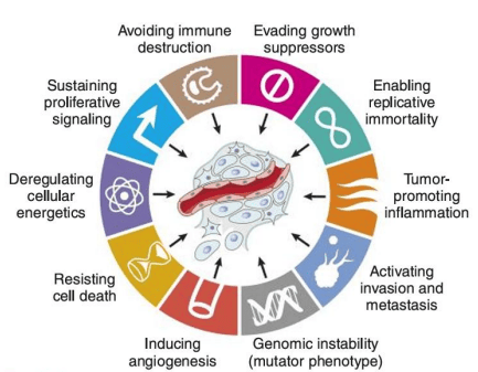
The Hallmarks of Cancer (Adapted from Hanahan & Weinberg)
3. Etiological Landscape and Predisposing Factors
Carcinogenesis is a complex, multifactorial process driven by endogenous predispositions and environmental exposures:
| Predisposing Domain | Clinical & Pathological Association | Underlying Mechanism |
|---|---|---|
| Age | Incidence peaks between 55 to 75 years; nearly universal histologic prostate cancer by age 100. | Progressive lifetime accumulation of somatic mutations and spontaneous chemical events (e.g., deamination of cytosine to uracil). |
| Sex Predilection | Overall cancer incidence is higher in males; specific carcinomas predominate in females (breast, thyroid). | Differential hormonal exposures, occupational risk profiles, and variable X-linked tumor suppressor gene expression. |
| Inherited Genetics | Familial retinoblastoma, Li-Fraumeni syndrome, hereditary breast/ovarian cancer (BRCA1/2). | Germline inheritance of a mutated tumor suppressor allele, predisposing every somatic cell to transformation upon a second hit. |
| Dietary Patterns | High-fat, low-fiber diet linked to colorectal cancer; smoked meats/fish linked to gastric adenocarcinoma. | Increased secondary bile acid formation; dietary nitrates and polycyclic aromatic hydrocarbons forming reactive carcinogens. |
| Blood Group & HLA | Gastric carcinoma occurs with higher frequency in individuals with blood group A. | Genetically linked alterations in gastric mucin secretion and mucosal susceptibility to chronic H. pylori colonisation. |
| Endocrine Factors | Elevated estrogens promote endometrial and breast carcinoma; elevated androgens promote prostatic neoplasia. | Steroid hormones act as potent mitogens, stimulating sustained replication of labile epithelial cells without directly damaging DNA. |
3.1 Chronic Irritation, Inflammation, and Precancerous States
Persistent tissue injury forces sustained compensatory cellular regeneration. In labile or stable cell populations, this increased replication significantly increases the risk of copying errors during DNA synthesis.
| Pathologic Clinical Condition | Associated Neoplasm | Etiologic / Inflammatory Trigger |
|---|---|---|
| Asbestosis & Silicosis | Malignant mesothelioma; bronchogenic lung carcinoma | Inhaled asbestos fibers and silica activating macrophage inflammasomes |
| Inflammatory Bowel Disease | Colorectal adenocarcinoma | Chronic ulcerative colitis and Crohn disease-induced mucosal turnover |
| Lichen Sclerosus | Vulvar squamous cell carcinoma | Persistent chronic anogenital epithelial inflammation |
| Chronic Pancreatitis | Pancreatic ductal adenocarcinoma | Prolonged alcohol abuse; inherited germline PRSS1/SPINK1 mutations |
| Chronic Cholecystitis | Gallbladder carcinoma | Mechanical trauma and chronic irritation from cholelithiasis (gallstones) |
| Barrett Esophagus | Esophageal adenocarcinoma | Gastric acid reflux inducing glandular intestinal metaplasia |
| Autoimmune Thyroiditis & Sjogren | Extranodal marginal zone B-cell lymphoma (MALT lymphoma) | Persistent autoimmune B-cell hyperactivation and proliferation |
| Opisthorchiasis & Clonorchiasis | Cholangiocarcinoma (bile duct cancer) | Chronic infestation of biliary trees by parasitic liver flukes |
| Chronic Gastritis / Peptic Ulcer | Gastric adenocarcinoma; gastric MALT lymphoma | Persistent mucosal colonization by Helicobacter pylori |
| Viral Hepatitis B and C | Hepatocellular carcinoma (HCC) | Chronic necroinflammation, hepatocyte regeneration, and cirrhosis |
| Chronic Draining Osteomyelitis | Squamous cell carcinoma of draining sinus tract (Marjolin ulcer) | Recurrent refractory bacterial infection of bone and surrounding skin |
| Urinary Schistosomiasis | Squamous cell carcinoma of the urinary bladder | Deposition of calcified Schistosoma haematobium ova causing chronic cystitis |
Additionally, pre-existing premalignant lesions show a high rate of malignant transformation:
- Hyperplastic and Metaplastic Changes: Oral leukoplakia (progresses to squamous cell carcinoma), atypical endometrial hyperplasia (progresses to endometrial adenocarcinoma), and complex mammary fibrocystic hyperplasia with atypia.
- Benign Neoplastic Precursors: Villous adenomas of the urinary bladder, colonic adenomatous polyps (tubulovillous and villous architectures carry the highest risk), and dysplastic melanocytic nevi.
- Chronic Stasis, Metabolic & Malposition States: Hepatic cirrhosis, chronic venous stasis ulcers (Marjolin ulcer transformation), Paget disease of bone (predisposes to secondary osteosarcoma), and cryptorchidism (undescended testis) which dramatically raises the risk of testicular seminoma and non-seminomatous germ cell tumors.
4. Chemical, Physical, and Biological Carcinogens
4.1 Physical Carcinogenesis: Radiation Mechanisms
Radiation functions through two distinct, complementary mechanisms:
- Direct Radiation Injury: High-energy photons directly strike DNA, inducing single-strand nicks, severe double-strand breaks, purine/pyrimidine base deletions, and aberrant intra- or inter-strand cross-linkages.
Non-ionizing Ultraviolet (UV) Rays: Sunlight exposure induces covalent bond formation between adjacent pyrimidines, producing cyclobutane pyrimidine (thymine) dimers. Unrepaired dimers disrupt transcription and replication, predisposing to cutaneous basal cell carcinoma, squamous cell carcinoma, and malignant melanoma. - Indirect Radiation Injury: Ionizing radiations (γ-rays, X-rays) radiolytically split water molecules within cells, generating reactive oxygen species (ROS) such as hydroxyl free radicals (OH•). These free radicals attack DNA bases and trigger membrane lipid peroxidation and severe oxidative stress.
Clinical Associations with Ionizing Radiation Exposure
- Early Radiologists: High incidence of cutaneous squamous carcinomas on hands and acute/chronic myeloid leukemias due to unshielded fluoroscopy.
- Head and Neck Irradiation: Markedly elevated long-term incidence of papillary thyroid carcinoma in children irradiated for tonsillar enlargement or tinea capitis.
- Atomic Bomb Survivors: Significant spike in acute myeloid leukemia and chronic myeloid leukemia (interestingly, chronic lymphocytic leukemia showed no associated increase).
- Radium Watch-Dial Painters: Ingestion of radium paint via brush-tipping caused direct bone deposition, driving osteosarcoma.
4.2 Chemical Carcinogenesis: Classification and Molecular Activation
Chemical carcinogens are functionally divided into two broad classes:
- Direct-Acting Carcinogens: Electrophilic compounds that require no enzymatic, metabolic conversion to interact with and damage cellular nucleophiles (e.g., chemotherapeutic alkylating agents like cyclophosphamide, busulfan).
- Indirect-Acting Carcinogens (Procarcinogens): Parent compounds that are biologically inert until metabolically transformed by endogenous host enzymes (predominantly the Cytochrome P-450 monooxygenase system) into an active, highly reactive electrophilic intermediary termed the ultimate carcinogen.
Molecular Mechanism of Attack: Ultimate carcinogens are electron-deficient (electrophilic) compounds that target electron-rich (nucleophilic) sites on cellular macromolecules. In DNA, they form covalent DNA adducts primarily targeting the N2, N3, and N7 positions of guanine, altering base pairing and causing point mutations.
| Chemical Agent / Class | Primary Exposure Source | Associated Human Malignancy |
|---|---|---|
| Polycyclic Aromatic Hydrocarbons (e.g., Benzo[a]pyrene) | Cigarette tobacco smoke; recycled/burned cooking oils; charcoal-broiled meats | Bronchogenic squamous/small-cell lung carcinoma; cutaneous squamous carcinoma |
| Aromatic Amines & Azo Dyes (e.g., β-naphthylamine) | Industrial rubber manufacturing, textile dyes, and printing facilities | Transitional cell carcinoma of the urinary bladder (mucosal glucuronidases cleave detoxified hepatic glucuronides, releasing free β-naphthylamine directly into the urine). |
| Aflatoxin B1 | Fungal mycotoxin from Aspergillus flavus contaminating improperly stored peanuts and grains | Hepatocellular carcinoma (HCC) (characteristically produces an arginine-to-serine transversion mutation at codon 249 of the TP53 gene). |
| Benzene | Industrial solvents, petroleum processing, chemical synthesis | Acute myeloid leukemia (AML); aplastic anemia/marrow failure |
| Vinyl Chloride Monomer | Polyvinyl chloride (PVC) plastics production | Hepatic angiosarcoma (hemangiosarcoma) |
| Thorotrast (Thorium Dioxide) | Historical radiographic contrast agent deposited in reticuloendothelial cells | Hepatic angiosarcoma, cholangiocarcinoma |
| Diethylstilbestrol (DES) | Synthetic non-steroidal estrogen historically administered to prevent miscarriage | Clear cell adenocarcinoma of the cervix and vagina in daughters exposed to the drug transplacentally in utero. |
| Asbestos Fibers | Insulation, shipyards, pipefitting, construction demolition | Bronchogenic carcinoma (magnified by cigarette smoking); pleural and peritoneal mesothelioma |
| Nitrosamines & Nitrosamides | Nitrate-cured, pickled, and smoked food items; formed endogenously in the stomach | Gastric adenocarcinoma, esophageal carcinoma |
| Inorganic Arsenic | Contaminated well water; agricultural pesticides; alloy manufacturing | Cutaneous squamous cell carcinoma and basal cell carcinoma (frequently on palms/soles) |
| Heavy Metals (Nickel, Chromium, Uranium) | Mining, metal refining, industrial welding, glass manufacturing | Bronchogenic carcinoma of the lung; sinonasal carcinomas |
| Alkylating Agents | Chemotherapeutic regimens (e.g., cyclophosphamide, melphalan) | Secondary therapy-related acute myeloid leukemia (t-AML) |
4.3 Mutagens and Antimutagens
- Mutagens: Physical or chemical substances that accelerate the basal mutation frequency of cellular DNA, directly escalating cancer incidence. Consequently, virtually all mutagens possess carcinogenic properties (e.g., ionizing rays, free-radical generators, alkylating compounds, viral integrants).
- Antimutagens (Chemopreventive Agents): Dietary or synthetic compounds capable of neutralizing mutagens, trapping reactive electrophiles, or blocking tumor promotion. Examples include:
- Vitamin A and Carotenoids: Promote physiological differentiation and repress clonal expansion.
- Vitamins C and E: Free radical scavengers that terminate lipid peroxidation chains.
- Polyphenolic Phytochemicals: Curcumin, resveratrol, and flavonoids from cruciferous and leafy vegetables that downregulate inflammatory NF-κB pathways and induce Phase II detoxification enzymes.
4.4 Biological and Infectious Carcinogenesis
Infectious organisms are responsible for up to 20% to 25% of malignancies worldwide. Microbes trigger transformation primarily through two major pathways:
- Chronic Inflammation and Regenerative Repair: Continuous, pathogen-induced tissue turnover increases the risk of acquiring somatic driver mutations (e.g., HBV/HCV in hepatocytes; H. pylori in gastric mucosa).
- Direct Expression of Viral Oncoproteins: Viral genes integrate into or express within host cells to directly neutralize tumor suppressor networks or stimulate mitogenic pathways.
| Infectious Agent | Genome / Class | Associated Malignancies | Molecular Mechanism |
|---|---|---|---|
| Human Papillomavirus (HPV High-Risk: 16, 18, 31, 33) | Double-stranded DNA virus | Squamous cell carcinoma of the cervix, vulva, penis, anus, tonsils, and head & neck | Viral E6 oncoprotein binds and promotes ubiquitin-mediated degradation of p53; viral E7 oncoprotein binds and inactivates retinoblastoma protein (RB), displacing E2F transcription factors to drive cell cycle progression into S-phase. |
| Epstein-Barr Virus (EBV / HHV-4) | Double-stranded DNA virus | Endemic Burkitt lymphoma; nasopharyngeal carcinoma; Hodgkin lymphoma; B-cell lymphomas in immunosuppressed states | Infects B lymphocytes via CD21; expresses LMP-1 (Latent Membrane Protein 1), which mimics constitutively active CD40 signaling, stimulating NF-κB and JAK/STAT pathways to prevent apoptosis. |
| Hepatitis B Virus (HBV) | Partially double-stranded DNA virus | Hepatocellular carcinoma (HCC) | Integration of viral DNA; expression of HBx protein, which disrupts growth control and inactivates p53, combined with chronic immune-mediated necroinflammation. |
| Hepatitis C Virus (HCV) | Single-stranded positive-sense RNA virus | Hepatocellular carcinoma (HCC) | Does not integrate into host DNA; causes persistent chronic immune-mediated hepatitis, oxidative DNA injury, and cirrhosis. |
| Human Herpesvirus 8 (HHV-8 / KSHV) | Double-stranded DNA virus | Kaposi sarcoma; Primary effusion lymphoma; Multicentric Castleman disease | Encodes viral homologs of cellular proteins, including viral cyclin D and viral GPCRs, driving endothelial proliferation and vascular spindle cell growth. |
| Human T-Cell Lymphotropic Virus 1 (HTLV-1) | Retrovirus (single-stranded RNA) | Adult T-cell leukemia / lymphoma (ATLL) | Encodes the Tax protein, an essential viral transactivator that upregulates host interleukin-2 (IL-2) and IL-2 receptors, establishing an autocrine loop that drives polyclonal T-cell expansion. |
| Helicobacter pylori | Microaerophilic, spiral Gram-negative bacterium | Gastric adenocarcinoma; Extranodal marginal zone B-cell lymphoma of the stomach (gastric MALToma) | Injects CagA and VacA virulence toxins into epithelial cells, stimulating chronic inflammation, oxygen free-radical generation, and continuous polyclonal B-cell recruitment. |
| Schistosoma haematobium | Blood trematode (Parasite) | Squamous cell carcinoma of the urinary bladder | Granulomatous reaction to parasite ova deposited in the bladder wall, resulting in chronic cystitis, squamous metaplasia, and malignant transformation. |
| Opisthorchis viverrini & Clonorchis sinensis | Biliary flukes (Parasites) | Cholangiocarcinoma | Mechanical irritation, chronic parasitic obstruction, and excretion of mitogenic secretomes into the biliary tree. |
5. Molecular Pathogenesis: Oncogenes, Tumor Suppressors, and Genomic Networks
Key Genetic Definitions
- Proto-oncogenes: Highly conserved physiological genes that orchestrate normal cell division, embryogenesis, and tissue repair (e.g., growth factors, receptor tyrosine kinases, cytoplasmic signal transducers, nuclear transcription factors). More than 100 proto-oncogenes operate in humans.
- Oncogenes: Mutated, constitutively activated alleles of proto-oncogenes that induce autonomous, unregulated cellular proliferation. Oncogenes act dominantly; a mutation in a single allele (gain-of-function) is sufficient to promote transformation. They are designated with a three-letter prefix preceded by c- (cellular) or v- (viral).
- Oncoproteins: Protein products encoded by oncogenes that are structurally devoid of physiological regulatory domains, functioning independently of extracellular growth cues.
- Anti-oncogenes (Tumor Suppressor Genes): Genes encoding proteins that normally slow down cell proliferation, enforce DNA damage checkpoints, or trigger apoptosis. Loss-of-function requires inactivation of both alleles (two-hit model) to eliminate growth suppression.
5.1 Four Molecular Mechanisms of Proto-Oncogene Activation
- Promoter and Enhancer Insertion: Integration of a retroviral provirus harboring powerful Long Terminal Repeats (LTRs) adjacent to a cellular proto-oncogene. These LTRs override native transcriptional controls and drive massive overexpression of the downstream gene (e.g., avian leukosis virus integrating adjacent to the host c-MYC locus, driving uncontrolled transcription factor synthesis).
- Chromosomal Translocation: Physical breakage and reciprocal exchange of chromosomal segments. This mechanism activates proto-oncogenes through two distinct paths:
- Transcriptional Overexpression via Gene Relocation: A proto-oncogene is juxtaposed next to a highly active, tissue-specific promoter/enhancer. In Burkitt lymphoma, the balanced translocation t(8;14)(q24;q32) relocates the c-MYC oncogene from chromosome 8 adjacent to the highly active immunoglobulin heavy-chain (IgH) gene locus on chromosome 14, driving constitutive MYC transcription factor synthesis.
- Chimeric Fusion Oncoprotein Formation: In Chronic Myeloid Leukemia (CML), the Philadelphia chromosome translocation t(9;22)(q34;q11) fuses the ABL1 non-receptor tyrosine kinase gene on chromosome 9 with the BCR gene on chromosome 22. This creates a chimeric BCR-ABL fusion kinase that is constitutively active and resistant to physiological autoinhibition.
- Gene Amplification: Duplication and multiplication of a proto-oncogene into dozens or hundreds of extra copies per cell, seen cytogenetically as extrachromosomal double minutes (dmin) or intrachromosomal homogeneous staining regions (HSRs).
- Clinical Example: Administration of the antifolate chemotherapeutic methotrexate can lead to amplification of the dihydrofolate reductase (DHFR) gene, producing drug resistance.
- Oncogene Amplification: Amplification of HER2/neu (c-erb-B2) in approximately 20% of invasive breast carcinomas drives aggressive tumor behavior and poor baseline prognosis.
- Point Mutation: A single nucleotide base substitution that alters a critical regulatory amino acid residue within the oncoprotein.
- The Classical RAS Paradigm: Point mutations in the RAS oncogene family (HRAS, KRAS, NRAS) typically affect codon 12, 13, or 61. Normally, wild-type Ras-p21 functions as an intracellular molecular switch: it is activated by binding guanosine triphosphate (GTP) and rapidly inactivated when its intrinsic GTPase hydrolyzes GTP into inactive GDP, a process accelerated by GTPase-Activating Proteins (GAPs). Codon 12 mutations disable intrinsic GTPase catalytic activity and prevent GAP interaction, locking the mutant Ras protein in a permanently active, GTP-bound conformation that continuously fires downstream MAPK and PI3K/AKT proliferation pathways. RAS mutations occur in roughly 25% to 30% of all human malignancies.
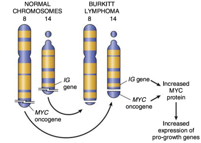
Reciprocal Chromosomal Translocation t(8;14) Juxtaposing MYC and Immunoglobulin Enhancers in Burkitt Lymphoma
5.2 Functional Classes of Oncogenes and Growth Factor Cascades
Oncogenes mimic physiological components of signaling cascades:
- Secreted Growth Factors: The v-sis oncogene encodes an aberrant homolog of the platelet-derived growth factor β-chain (PDGF-β), driving autocrine stimulation in glioblastomas and sarcomas.
- Receptor Tyrosine Kinases (RTKs):
- v-erb / c-erb-B1 (EGFR): Truncated or overexpressed epidermal growth factor receptor that phosphorylates cytoplasmic targets independent of ligand binding.
- c-neu / HER2 (c-erb-B2): Human EGFR-2, frequently amplified in breast carcinoma.
- v-fms: Mutated, constitutively firing homolog of the colony-stimulating factor-1 receptor (CSF-1R).
- Non-Receptor Tyrosine Kinases: The v-src oncoprotein of Rous sarcoma virus, functioning as an uninhibited cytoplasmic tyrosine kinase.
- GTP-Binding Signal Transducers: Membrane-associated Ras-p21 proteins.
- Nuclear Transcription Regulators: Oncoproteins localized strictly to the cell nucleus that directly drive expression of cell-cycle progression genes, including c-MYC, FOS, and MYB.
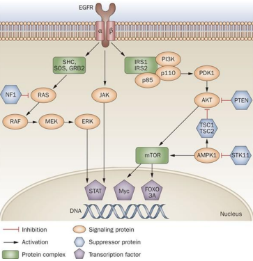
The EGFR Signal Transduction Network: Coupling Receptor Activation to Proliferation and Survival
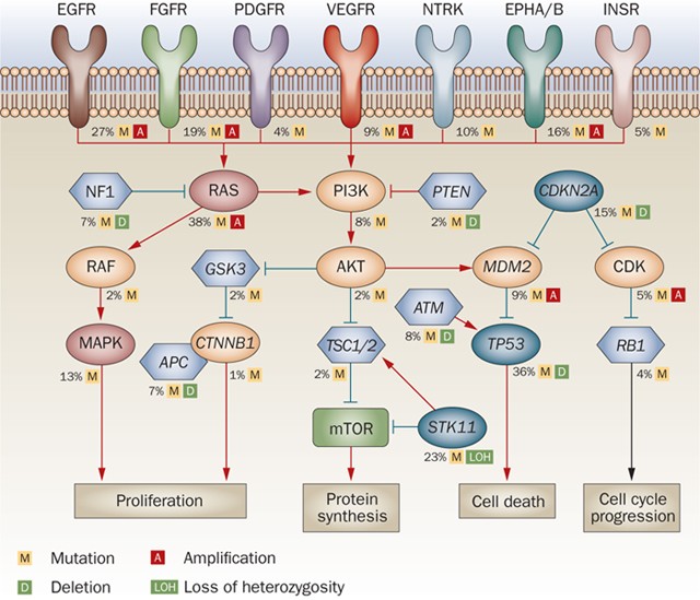
Recurrent Driver Alterations in Lung Adenocarcinoma Signaling Networks
5.3 Tumor Suppressor Genes and the Knudson Two-Hit Paradigm
Unlike oncogenes, tumor suppressor genes (TSGs) inhibit abnormal proliferation and ensure genomic stability. Knudson's "two-hit" hypothesis dictates that complete loss of a tumor suppressor requires inactivation of both functional alleles:
- Familial (Inherited) Cancer Syndromes: An individual inherits one defective mutant allele through the germline (Hit 1), present within every somatic cell. Although functionally heterozygous and phenotypically normal, the loss or somatic mutation of the single remaining wild-type allele (Hit 2 / Loss of Heterozygosity, LOH) completely eliminates tumor suppressor function, resulting in early-onset and often bilateral or multifocal tumors.
- Sporadic Malignancies: Both non-functional hits must occur somatically within the exact same cell lineage over an individual's lifetime, explaining the later age of onset and solitary presentation.
| Inherited Syndrome | Gene(s) Involved | Pattern | Functional Biological Defect |
|---|---|---|---|
| Retinoblastoma | RB1 | Autosomal Dominant | Loss of G1/S cell-cycle checkpoint control; free E2F drives transcription. |
| Li-Fraumeni Syndrome | TP53 | Autosomal Dominant | Severe genomic instability; failure to arrest the cell cycle or trigger apoptosis following DNA injury. Predisposes to sarcomas, breast cancers, leukemias, and adrenocortical carcinomas (SBLA). |
| Familial Melanoma | CDKN2A (encodes p16-INK4A, p14-ARF) | Autosomal Dominant | Inability to inhibit cyclin-dependent kinases (CDK4/6), causing uninhibited RB phosphorylation. |
| Familial Adenomatous Polyposis (FAP) | APC | Autosomal Dominant | Failure of the destruction complex to degrade β-catenin; leads to hyperactive Wnt signaling and development of thousands of colonic adenomas. |
| Neurofibromatosis Types 1 & 2 | NF1 (Neurofibromin), NF2 (Merlin) | Autosomal Dominant | NF1 functions as a Ras-GAP; its loss leads to constitutively active Ras signaling. NF2 coordinates contact inhibition. |
| Hereditary Breast & Ovarian Cancer | BRCA1, BRCA2 | Autosomal Dominant | Impaired homologous recombination repair of DNA double-strand breaks. |
| Hereditary Nonpolyposis Colorectal Cancer (HNPCC / Lynch) | MSH2, MLH1, MSH6, PMS2 | Autosomal Dominant | Defective DNA mismatch repair (MMR) machinery, leading to microsatellite instability (MSI). |
| Nevoid Basal Cell Carcinoma (Gorlin Syndrome) | PTCH1 | Autosomal Dominant | Loss of PTCH-mediated repression of Smoothened; leads to uncontrolled Hedgehog pathway signaling. |
| Xeroderma Pigmentosum | XP gene family (XPA through XPG) | Autosomal Recessive | Defective nucleotide excision repair (NER); inability to excise UV-induced thymine-thymine crosslinks. |
| Ataxia-Telangiectasia | ATM | Autosomal Recessive | Defective kinase sensor for double-strand DNA breaks, causing extreme sensitivity to ionizing radiation. |
| Bloom Syndrome | BLM | Autosomal Recessive | Defective RecQ-like DNA helicase, causing elevated sister chromatid exchanges and chromosomal breakage. |
| Fanconi Anemia | FANC core complex genes | Autosomal Recessive | Inability to repair interstrand DNA cross-links, predisposing to aplastic anemia and acute myeloid leukemia. |
5.4 Guardian of the Genome: The Central Role of TP53
The TP53 gene encodes a 53-kDa phosphoprotein that functions as the central genomic surveillance node. Under physiological conditions, p53 is maintained at low levels through continuous ubiquitination by its negative regulator, MDM2.
- DNA Damage Response: DNA double-strand breaks activate sensor kinases (ATM/ATR and Chk2), which phosphorylate p53. This prevents MDM2 binding, stabilizing and accumulating p53 in the nucleus.
- Cell Cycle Arrest (Quiescence): Stabilized p53 transcriptionally activates p21 (CDKN1A), an inhibitor of cyclin-dependent kinases (CDK4/6 and CDK2). This halts the cell cycle at the G1/S transition, allowing repair enzymes (e.g., GADD45) time to correct DNA lesions.
- Induction of Apoptosis (Cellular Suicide): If genomic damage is too extensive to repair, p53 transcriptionally upregulates pro-apoptotic factors (BAX, PUMA, NOXA) and represses anti-apoptotic BCL-2. This permeabilizes the mitochondrial membrane to trigger apoptosis.
- Prevalence in Human Cancer: More than 50% to 70% of human tumors harbor homozygous loss or missense mutations in TP53. These mutations eliminate its DNA-binding capacity, allowing cells to accumulate mutations unchecked.
5.5 Regulated DNA Rearrangements: Physiological Recombination Gone Awry
During normal lymphocyte maturation, developing B and T cells employ somatic DNA breakage and rejoining via V(D)J recombination to generate diverse antigen receptors, alongside somatic hypermutation (activation-induced cytidine deaminase, AID) to improve antibody affinity. Errors in these physiological cutting and rejoining events can aberrantly juxtapose oncogenes with immune enhancers, driving B- and T-cell lymphomas and leukemias.
6. Tumor Progression, Clonal Evolution, and Epigenetics
Carcinogenesis is a dynamic, multistep evolutionary process governed by Darwinian natural selection:
- Monoclonality and Stepwise Progression: Malignancies arise from a single transformed ancestral precursor cell (monoclonal origin). Over time, this founding cell accumulates complementary driver mutations, giving rise to distinct sublineages.
- Subclonal Heterogeneity: As tumor cells proliferate, random genetic and epigenetic errors generate subclones with variable traits. Subclones with superior fitness (e.g., accelerated replication, nutrient scavenging, resistance to hypoxia) outcompete their neighbors. Consequently, tumors are genetically and phenotypically heterogeneous at diagnosis.
- Driver Mutations vs. Passenger Mutations:
- Driver Mutations: Pathogenic alterations directly involved in initiating and maintaining malignancy by conferring an active growth or survival advantage (often 2 to 8 per tumor).
- Passenger Mutations: Neutral, non-pathogenic mutations scattered across the genome that do not directly drive growth. They outnumber drivers (especially in mutagen-driven tumors like melanoma and smoking-associated lung cancers). However, passengers can alter host-tumor interactions:
- Therapeutic Resistance: Rare dormant passenger mutations may confer resistance to a specific chemotherapeutic agent; exposure to therapy eliminates sensitive clones, allowing resistant subclones to expand.
- Tumor Neoantigens: Passenger mutations can alter protein coding sequences to create novel neoepitopes recognizable by cytotoxic T lymphocytes, making high-mutation-burden tumors prime candidates for immune checkpoint inhibitor therapy.
- Oncogene Addiction: Despite harboring complex mutational landscapes, some cancers remain dependent on the activity of a single activated oncogene for survival. Pharmacological inhibition of this target often leads to rapid tumor collapse (e.g., targeting BCR-ABL with imatinib, or mutant EGFR with osimertinib).
- Epigenetic Remodeling: Heritable shifts in gene expression that occur without altering primary DNA base sequences:
- Global DNA Hypomethylation: Causes genomic fragility and aberrant reactivation of silent oncogenes and transposable elements.
- Focal Promoter Hypermethylation: Dense methylation of CpG islands within tumor suppressor gene promoters, causing transcriptional silencing (e.g., silencing of MLH1 in sporadic MSI colorectal cancers, or BRCA1 in triple-negative breast cancers).
- Post-Translational Histone Alterations: Dysregulated histone acetylation, methylation, and chromatin remodeling that lock genomic loci into inactive states.
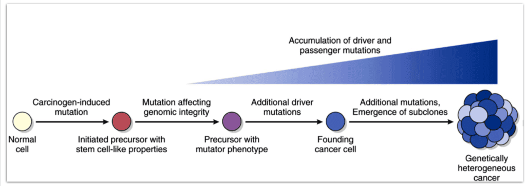
Stepwise Clonal Evolution and Driver Mutation Accumulation in Carcinogenesis
7. Histopathologic Grading, Clinical Staging, and Diagnostic Modalities
Tissue Sampling Requirement
Regardless of how strongly malignancy is suspected on physical exam or advanced radiographic imaging, definitive cancer diagnosis requires obtaining viable tissue specimens for histopathologic and cytological confirmation by a pathologist.
7.1 Histopathologic Grading Versus Clinical Staging
- Grading (G1 to G4): A microscopic histopathological evaluation based on the degree of cellular differentiation and structural resemblance to parent tissue, alongside the frequency of mitotic figures (Grade 1 = well-differentiated; Grade 4 = undifferentiated/anaplastic).
- Staging (Stage 0 to Stage IV): A clinical and surgical determination of the anatomical extent and physical spread of the tumor throughout the body. Staging relies on primary tumor size, invasiveness, regional lymph node status, and distant metastasis.
- Prognostic Value: While both metrics are clinically valuable, clinical and pathological staging correlates significantly better with overall patient survival and prognosis than histopathologic grading.
7.2 The International TNM Staging Framework (AJCC / UICC)
The American Joint Committee on Cancer (AJCC) staging manual classifies solid tumors based on three parameters:
- T (Primary Tumor):
- T0: No evidence of a primary tumor, or carcinoma strictly limited in situ without basement membrane invasion.
- T1, T2, T3, T4: Progressive increase in tumor diameter, depth of wall infiltration, and local radial invasion.
- N (Regional Lymph Node Involvement):
- N0: Complete absence of regional lymph node metastasis.
- N1, N2, N3: Progressive involvement of regional lymph nodes based on the absolute number, anatomic chains, and distance of involved nodes.
- NX: Regional lymph node status cannot be assessed.
- M (Distant Metastasis):
- M0: No evidence of distant hematogenous or non-regional lymphatic metastasis.
- M1 (or M2): Presence of confirmed metastatic seeding in distant organs (e.g., liver, lung, bone, brain).
- MX: Presence of distant metastasis cannot be evaluated.
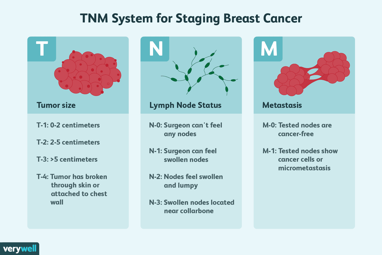
The AJCC TNM Classification Schema
7.3 Organ-Specific Staging Systems
The Historical Dukes Classification for Colorectal Carcinoma (1932)
Established by Dr. Cuthbert E. Dukes, this system staged colorectal tumors based on the depth of bowel wall invasion and lymphatic spread. While largely superseded by TNM staging, its principles remain relevant to prognostic outcomes:
| Dukes Stage | Pathological Extent of Colorectal Invasion | Proportion at Diagnosis | 5-Year Survival Rate |
|---|---|---|---|
| Dukes A | Carcinoma confined to the mucosa and submucosa; does not breach the muscularis propria | ~10% | 80% |
| Dukes B | Infiltrates into or through the muscularis propria into perirectal/pericolic soft tissues; no lymph node spread | ~35% | 60% – 70% |
| Dukes C | Presence of regional lymph node metastasis, irrespective of depth of bowel wall invasion | ~25% | 30% – 60% |
| Dukes D | Widespread distant hematogenous metastasis (e.g., to the liver, lungs, or peritoneum) | ~30% | 15% (2-year survival) |
The Ann Arbor Staging System for Lymphomas (Hodgkin and Non-Hodgkin)
- Stage I: Involvement of a single lymph node region (I) or localized involvement of a single extralymphatic organ/site (IE).
- Stage II: Involvement of two or more lymph node regions on the same side of the diaphragm, or localized contiguous extralymphatic extension (IIE).
- Stage III: Involvement of lymph node regions on both sides of the diaphragm, with or without localized extralymphatic extension (IIIE) or splenic involvement (IIIS).
- Stage IV: Diffuse or disseminated involvement of one or more distant extralymphatic organs (e.g., bone marrow, liver, lungs), with or without associated nodal disease.
- Systemic Constitutional Modifiers:
- A: Complete absence of systemic B-symptoms.
- B (B-Symptoms): Presence of unexplained fever (>38°C), drenching night sweats, or unexplained weight loss exceeding >10% of baseline body mass within the preceding 6 months.
- X (Bulky Disease): Single mass >10 cm in maximal diameter, or mediastinal widening exceeding one-third of the maximum transverse transthoracic diameter on chest radiography.
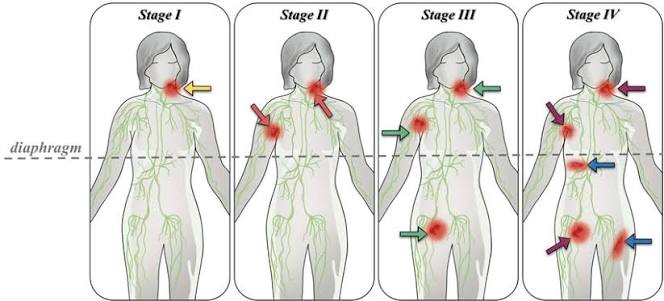
The Ann Arbor Anatomical Staging Schema for Malignant Lymphomas
Atypical Presentation of Advanced Colonic Adenocarcinoma
Patient History: A 27-year-old male presented with high-grade daily fevers (>38°C), drenching night sweats, significant unintentional weight loss exceeding 10% of total body mass, and rapid, massive abdominal distension. Initial differential diagnoses focused on systemic infections and hematologic malignancies.
Diagnostic Workup: Serial screening for Tuberculosis, HIV, Hepatitis viruses, and Lymphoma was entirely negative. Empirical anti-tuberculosis therapy was initiated without clinical improvement. Paracentesis was performed, and ascitic fluid was processed for cell block histopathology.
Pathological Findings: Ascitic cell block hematoxylin and eosin (H&E) staining revealed a mucinous (colloid) adenocarcinoma characterized by clusters of malignant epithelial cells floating in pools of extracellular mucin. Immunohistochemical staining was strongly positive for CDX2, confirming an intestinal primary. Postmortem autopsy identified extensive mucinous, gelatinous polypoid masses arising from the sigmoid colon with diffuse serosal and peritoneal carcinomatosis.
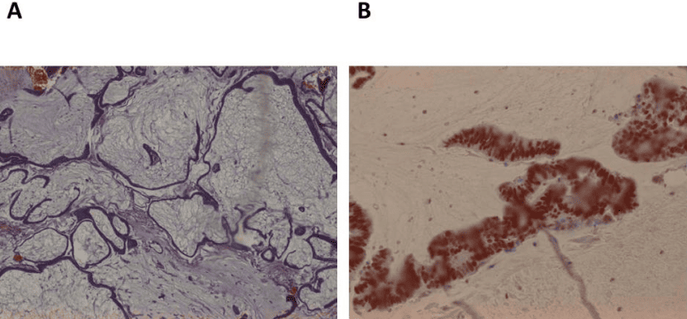
Histopathology and CDX2 Immunohistochemistry of Metastatic Colonic Adenocarcinoma
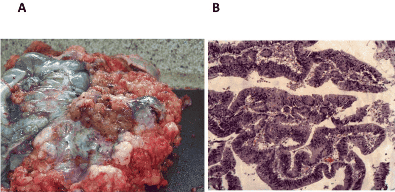
Gross Autopsy and Histopathology of Advanced Sigmoid Adenocarcinoma
8. Biochemical and Molecular Tumor Markers
Tumor Marker Definition & Scope
Tumor Marker: A measurable biochemical substance (enzyme, hormone, oncofetal antigen, protein, or receptor) synthesized directly by neoplastic cells or produced by non-neoplastic host tissues in response to the presence of a tumor. These markers can be identified and quantified in circulating blood, serous fluids, or tissue sections.
- Clinical Roles:
- Population Screening: Detection of occult disease in asymptomatic cohorts (e.g., PSA in select male populations).
- Diagnostic Aid: Helping differentiate benign from malignant masses alongside imaging and biopsy.
- Monitoring Therapy: Serial measurements to evaluate therapeutic response (levels drop following successful resection or cytotoxic therapy).
- Prognostication & Staging: Pretreatment marker concentrations often reflect total tumor burden.
- Surveillance for Recurrence: A secondary rise in marker levels after remission frequently signals early subclinical recurrence months before radiographic detection.
8.1 Characteristics of an Ideal Tumor Marker
- Absolute Specificity: Levels should rise exclusively in the presence of malignancy, remaining undetectable or at baseline in healthy individuals and benign disorders.
- High Sensitivity in Early Stages: Elevated levels should be detectable in early-stage disease to allow for curative interventions.
- Direct Correlation with Tumor Burden: Marker titers should correlate with tumor mass, falling with remission and rising with recurrence.
- Practical Clinical Utility: Tests should be straightforward, cost-effective, reproducible, and easily measured using standardized serum assays.
- Crucial Limitation: No single universal tumor marker exists that can reliably detect all malignancies or all stages of a specific cancer type.
8.2 Master Reference Guide of Clinically Utilized Tumor Markers
| Category | Biochemical Marker | Associated Human Malignancy | Clinical & Diagnostic Utility |
|---|---|---|---|
| Oncofetal Antigens | Alpha-Fetoprotein (AFP) | Hepatocellular carcinoma (HCC); non-seminomatous testicular germ cell tumors (yolk sac tumors) | Screening cirrhotic patients for HCC; monitoring response in testicular germ cell tumors. |
| Carcinoembryonic Antigen (CEA) | Colorectal adenocarcinoma; pancreatic, gastric, and bronchogenic lung carcinoma | Not used for screening; primary marker for surveillance of colorectal cancer recurrence post-resection. | |
| Cancer Antigen 125 (CA-125) | Epithelial ovarian carcinoma (serous cystadenocarcinoma) | Monitoring treatment response and recurrence in ovarian cancer; can also be elevated in benign states like endometriosis. | |
| Prostate-Specific Antigen (PSA) | Adenocarcinoma of the prostate | Used for screening, diagnostic workup, and monitoring post-prostatectomy recurrence. | |
| Cancer Antigen 19-9 (CA 19-9) | Pancreatic ductal adenocarcinoma; cholangiocarcinoma | Distinguishing pancreatic cancer from benign pancreatitis and monitoring treatment response. | |
| Enzymatic Markers | Alkaline Phosphatase (ALP) | Osteosarcoma; osteoblastic bone metastases (e.g., from prostate cancer); liver metastases | Marker of osteoblast activation or biliary duct obstruction due to hepatic metastases. |
| Prostatic Acid Phosphatase (PAP) | Prostate cancer (locally advanced or metastatic) | Historical marker for prostate cancer, largely replaced by the more sensitive PSA. | |
| Hormones & Metabolites | Human Chorionic Gonadotropin (β-hCG) | Gestational choriocarcinoma; hydatidiform moles; testicular choriocarcinoma/seminoma | Highly sensitive diagnostic and surveillance marker for gestational trophoblastic disease and germ cell tumors. |
| Calcitonin | Medullary thyroid carcinoma (derived from parafollicular C-cells) | Diagnostic marker and surveillance tool; used to screen asymptomatic family members in MEN 2A/2B syndromes. | |
| 5-Hydroxyindoleacetic Acid (5-HIAA) | Metastatic neuroendocrine (Carcinoid) tumors | 24-hour urine metabolite reflecting excessive serotonin turnover in carcinoid syndrome. | |
| Paraproteins | Monoclonal Immunoglobulins & Bence-Jones Proteins | Multiple myeloma; Waldenström macroglobulinemia | Serum protein electrophoresis (M-protein spike) and urine free light chains (Bence-Jones proteinuria). |
| Receptor Markers | Estrogen Receptor (ER) & Progesterone Receptor (PR) | Invasive breast carcinoma; endometrial adenocarcinoma | Predictive markers: positive expression indicates likely response to endocrine therapies (e.g., tamoxifen, aromatase inhibitors). |
| HER2/neu (c-erb-B2) | Invasive breast carcinoma; gastric adenocarcinoma | Predicts clinical benefit from targeted anti-HER2 monoclonal antibody therapy (trastuzumab). |
9. Principles of Cancer Therapeutics and Preventive Oncology
9.1 Primary Therapeutic Modalities
- Surgical Resection: The primary curative modality for localized solid malignancies, aimed at debulking or removing the primary tumor with negative microscopic margins.
- Radiation Therapy: Delivers localized high-energy radiation to targeted tissue volumes, inducing lethal double-strand DNA breaks that trigger mitotic catastrophe and p53-dependent apoptosis.
- Cytotoxic Chemotherapy: Systemic administration of pharmacological agents aimed at rapidly dividing cells. Primary mechanistic categories include:
- Antimetabolites: Structural analogs of normal cellular metabolites that competitively inhibit essential biosynthetic enzymes, blocking DNA and RNA synthesis.
- DNA Alkylating & Platinum Agents: Form covalent intrastrand and interstrand cross-links on DNA, preventing strand separation during replication.
| Therapeutic Agent | Chemical / Biological Class | Primary Mechanism of Action |
|---|---|---|
| Methotrexate | Folic acid analog (Antimetabolite) | Competitively inhibits dihydrofolate reductase (DHFR), depleting intracellular tetrahydrofolate reserves and arresting purine and thymidylate synthesis during S-phase. |
| 6-Mercaptopurine (6-MP) | Purine analog (Antimetabolite) | Metabolized by HGPRT to TIMP, which blocks de novo purine synthesis by inhibiting conversion of IMP to AMP and GMP. |
| 5-Fluorouracil (5-FU) | Pyrimidylate / Uracil analog (Antimetabolite) | Metabolically converted to 5-FdUMP, which irreversibly complexes with and inhibits thymidylate synthase, halting dUMP conversion to dTMP. |
| Cisplatin | Platinum coordination complex | Forms covalent intrastrand cross-links, primarily at the N7 positions of adjacent guanine bases, bending DNA to block replication and trigger apoptosis. |
| Trastuzumab | Recombinant humanized monoclonal antibody | Selectively binds the extracellular domain IV of the HER2/neu receptor tyrosine kinase, blocking ligand-independent downstream survival signaling and tagging cells for antibody-dependent cellular cytotoxicity (ADCC). |
| Imatinib | Small-molecule tyrosine kinase inhibitor (TKI) | Competitively occupies the ATP-binding pocket of the constitutively active BCR-ABL fusion kinase in CML, blocking downstream phosphorylation cascades. Also inhibits c-KIT and PDGFR. |
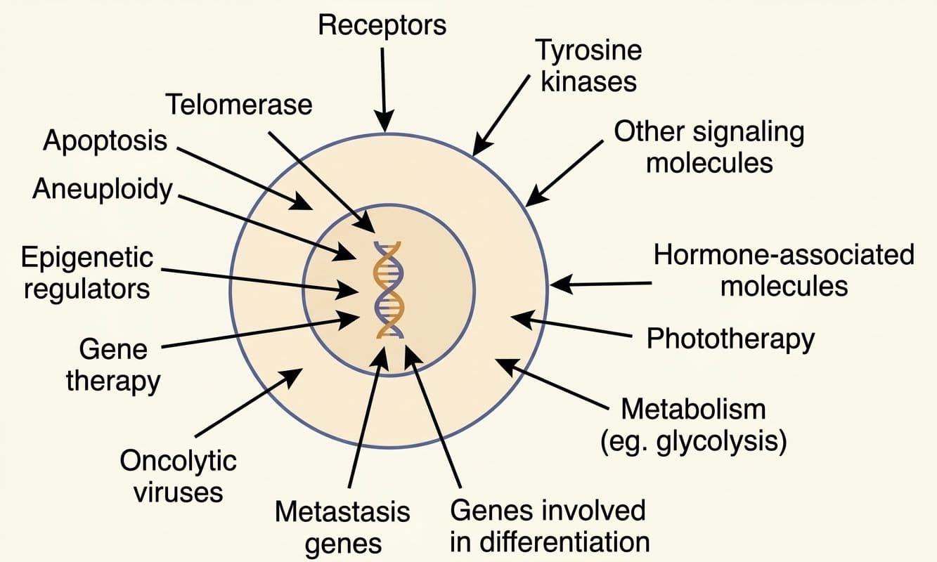
Emerging Molecular Targets and Frontiers in Precision Oncology
9.2 Preventive Oncology and Risk Mitigation Strategies
- Lifestyle Optimization: Engaging in regular physical exercise and maintaining a healthy body mass index (BMI) to reduce insulin resistance and mitogenic signaling.
- Elimination of Tobacco Products: Complete cessation of cigarette smoking and tobacco chewing to prevent exposure to polycyclic hydrocarbons and nitrosamines.
- Occupational and Environmental Controls: Enforcing strict safety limits for industrial pollutants, volatile organic solvents (benzene), asbestos insulation, and ionizing radiation.
- Dietary Modifications: Reducing consumption of high-fat foods, processed meats, and charcoal-broiled products, while adopting a diet rich in dietary fiber and plant-derived antioxidants.
- Routine Early Cancer Screening:
- Cervical Cytology (Pap Smear): Exfoliative cervical cytology for early detection of cervical dysplasia.
- Screening Mammography: Low-dose radiographic evaluation to identify microcalcifications and early non-palpable breast tumors.
- Genetic Counseling & Prophylactic Interventions: Identifying germline mutations in high-risk families (e.g., offering prophylactic bilateral mastectomy and salpingo-oophorectomy to carriers of deleterious BRCA1 or BRCA2 mutations).
- Prophylactic Immunization:
- Recombinant HPV Vaccination: Prevents persistent infection by high-risk strains (HPV 16, 18), protecting against cervical, anogenital, and oropharyngeal carcinomas.
- Recombinant Hepatitis B (HBV) Vaccination: Protects against chronic viral hepatitis, cirrhosis, and subsequent hepatocellular carcinoma.