Hemodynamic Disorders, Thromboembolism, and Shock
Comprehensive pathological evaluation of circulatory homeostasis, vascular flow disruptions, hemostatic plug formation, thrombogenesis, embolic phenomena, ischemic tissue necrosis, and systemic hypoperfusion syndromes.
Introduction to Hemodynamic Disturbances
Sustained cellular viability and metabolic function rely fundamentally upon uninterrupted, regulated blood circulation. Under baseline physiological circumstances, systemic blood fulfills indispensable biological roles across all tissue beds:
- Gas and nutrient delivery: Transports molecular oxygen from alveolar spaces to terminal tissue beds and provides glucose, amino acids, and lipids essential for cellular adenosine triphosphate (ATP) generation.
- Hormonal signaling: Circulates endocrine messengers, growth factors, and regulatory peptides toward remote target receptor sites.
- Waste elimination: Collects metabolic byproducts, acidic equivalents, and dissolved carbon dioxide for clearance through renal, pulmonary, and hepatic systems.
- Immune surveillance: Transports leukocytes, immunoglobulins, and complement fractions required for host defense.
- Homeostatic fluid preservation: Orchestrates continuous water, electrolyte, and colloid partitioning across vascular and extravascular spaces.
In the intact microvasculature, circulating blood transverses semipermeable capillary beds while plasma proteins remain sequestered inside the intravascular space. The minute quantity of interstitial transudate that escapes during filtration is systematically collected and evacuated via the lymphatic networks.
Derangements of Hemodynamic Homeostasis
Failure of microvascular integrity, hydrostatic control, or oncotic equilibrium leads to seven cardinal circulatory abnormalities:
- Hyperemia and Congestion: Elevated localized intravascular blood volume.
- Edema: Excessive extravascular fluid sequestration in interstitial spaces or cavities.
- Hemorrhage: Extravasation of red blood cells secondary to broken vascular containment.
- Thrombosis: Non-physiological solid aggregate generation inside intact blood vessels.
- Embolism: Migration of dislodged intravascular physical masses to distant anatomical sites.
- Infarction: Focal ischemic coagulative or liquefactive necrosis caused by vascular compromise.
- Shock: Generalized systemic tissue hypoperfusion and cellular hypoxic collapse.
Hyperemia vs. Congestion
Key Definitions: Volume Overload States
- Hyperemia: An active vascular process marked by precapillary arteriolar dilation that augments the volume of oxygen-rich arterial blood flowing into downstream capillary networks.
- Congestion: A passive circulatory state caused by compromised venular outflow, forcing poorly oxygenated deoxygenated blood to stall and pool inside regional microcirculations.
Mechanistic Analogy: Hyperemia corresponds to opening an entrance doorway wider, permitting a deluge of fresh blood. Congestion represents an obstructed exit door, where normal inflow continues while outflow is impeded, culminating in retrograde stasis.
| Diagnostic Parameter | Hyperemia | Congestion |
|---|---|---|
| Dynamic Character | Active process | Passive process |
| Underlying Vascular Driver | Precapillary arteriolar vasodilatation | Impaired, obstructed, or backward venous drainage |
| Directional Blood Flow | Augmented arterial inflow | Attenuated venous outflow |
| Oxygenation Status | Oxygenated arterial blood | Deoxygenated venous blood |
| Gross Tissue Chromaticity | Bright crimson red (erythema) | Dusky bluish-purple (cyanosis) |
| Parenchymal O2 Delivery | Elevated or matched to demand | Diminished, culminating in local hypoxia |
| Illustrative Settings | Skeletal exercise, acute inflammation, cutaneous blushing, postprandial splanchnic hyperemia | Congestive heart failure, localized deep venous thrombosis, hepatic vein occlusion |
| Secondary Complications | Transient, physiologically adaptive, non-injurious | Parenchymal atrophy, cell necrosis, microhemorrhages, interstitial fibrosis |
Classifications of Venous Congestion
Venous pooling is categorized pathologically based upon its territorial distribution and temporal chronology:
- Spatial Distribution:
- Localized Congestion: Consequent to isolated mechanical obstruction within a tributary vein (e.g., lower extremity deep-vein thrombosis, external tumor mass compression, volvulus or internal hernia strangulation, hepatic vein obstruction in Budd-Chiari syndrome).
- Systemic (Generalized) Congestion: Arising from central pump failure (e.g., congestive heart failure, constrictive pericarditis).
- Chronological Duration:
- Acute Congestion: Sudden microvascular distention with minimal structural remodeling.
- Chronic Congestion: Longstanding stasis characterized by chronic hypoxia, parenchymal cell dropout, hemosiderin-laden macrophage buildup, and stromal collagen expansion.
Systemic Congestion: Left vs. Right Ventricular Decompensation
Left-Sided Heart Failure: Deficient left ventricular stroke volume elevates left atrial pressure → transmits backward hydrostatically into pulmonary veins and pulmonary alveolar capillary beds → causes pulmonary congestion, intra-alveolar transudation (pulmonary edema), orthopnea, paroxysmal nocturnal dyspnea, and respiratory gas exchange failure.
Right-Sided Heart Failure: Deficient right ventricular emptying elevates right atrial filling pressure → produces retrograde systemic venous hypertension → triggers centrilobular hepatic congestion, splenomegaly, elevated jugular venous distention (JVD), soft tissue dependent peripheral pitting edema, and peritoneal transudate accumulation (ascites).
Pathological Morphology of Congested Tissues
Grossly, congested viscera present as swollen, hyperemic-to-cyanotic, heavy, and wet organs that weep dark venous blood upon sectioning. Microscopically, regional capillaries and venules appear engorged with densely packed erythrocytes, surrounded by widened interstitial spaces containing transudate and extravasated red blood cells.
Pulmonary Congestion
Acute Pulmonary Congestion: Characterized by engorged alveolar capillaries, microvascular septal transudation, and focal intra-alveolar hemorrhage. Alveolar spaces fill with watery, protein-poor transudate that manifests clinically as coarse inspiratory crackles, tachypnea, and pink, frothy sputum.
Chronic Pulmonary Congestion: Long-term pulmonary venous hypertension causes permanent expansion and tortuosity of alveolar septa, followed by reactive interstitial myofibroblast collagen synthesis. Continuous microvascular leakage allows red blood cells to enter the alveoli, where they undergo macrophage phagocytosis. Lysosomal degradation of hemoglobin releases elemental iron, stored intracellularly as insoluble golden-brown granules. These cells are designated hemosiderin-laden alveolar macrophages or "heart failure cells". The macroscopic combination of dense collagen and dark brown iron deposition transforms the spongy lung into a firm, hyperpigmented organ, historically designated brown induration of the lung.
Cellular Identity of Heart Failure Cells
Heart failure cells are pulmonary alveolar macrophages containing iron pigments from digested erythrocytes; they are not derived from cardiac myocytes or vascular endothelial cells.
Hepatic Congestion
Venous drainage from the hepatic parenchyma passes through terminal hepatic venules (central veins) toward sublobular veins, the three major hepatic veins, the inferior vena cava, and the right atrium. Right-sided cardiac pressure elevations transmit directly through this unvalved pathway into the central veins and lobular sinusoids.
Vulnerability of Zone 3 (Centrilobular) Hepatocytes: Hepatic lobular architecture is partitioned based on vascular microperfusion:
- Zone 1 (Periportal): Surrounds portal triads, receiving the highest concentration of oxygenated arterial and portal blood.
- Zone 2 (Midzonal): Intermediate oxygenation.
- Zone 3 (Centrilobular): Encircles the central vein, representing the distal boundary of sinusoidal oxygen delivery.
Because Zone 3 hepatocytes occupy the lowest baseline oxygen tension, superimposition of congestive venous stasis reduces local perfusion, causing acute centrilobular fatty change, ischemic atrophy, and hemorrhagic necrosis. In contrast, Zone 1 periportal hepatocytes remain viable due to their proximity to fresh hepatic arteriolar inflow.
Nutmeg Liver (Chronic Passive Hepatic Congestion)
Gross macroscopic examination displays an alternating mottled pattern: depressed, dark reddish-brown centrilobular foci (congested, necrotic, and hemosiderin-laden Zone 3 tissue) interspersed among elevated, tan-yellow periportal zones (viable, lipid-laden Zone 1 hepatocytes). Longstanding chronic right ventricular failure causes central-to-central sinusoidal bridging fibrosis, historically termed cardiac cirrhosis.
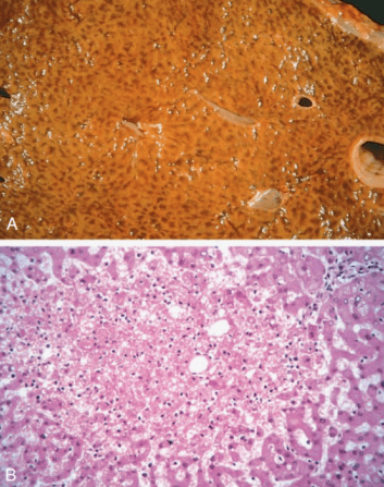
Liver with Chronic Passive Congestion and Hemorrhagic Necrosis
Edema and Fluid Dynamics
Key Definitions: Extravascular Fluid Accumulations
- Edema: Excessive collection of free liquid within the extravascular interstitial tissue compartments.
- Effusion: Pathological accumulation of liquid inside preformed mesothelial-lined serous cavities.
- Anasarca: Severe, profound, generalized subcutaneous edema extending throughout the entire body, accompanied by simultaneous multi-cavity effusions.
| Anatomical Cavity Location | Designated Effusion Terminology |
|---|---|
| Pleural Space | Hydrothorax / Pleural effusion |
| Pericardial Sac | Hydropericardium / Pericardial effusion |
| Peritoneal Cavity | Hydroperitoneum / Ascites |
| Synovial Joint Capsule | Hydroarthrosis / Joint effusion |
Total Body Water Distribution: Water comprises roughly 60% of lean body weight. This volume is partitioned physiologically into two main compartments:
- Intracellular Fluid (ICF): Contains approximately two-thirds (66%) of all body water.
- Extracellular Fluid (ECF): Accounts for the remaining one-third (33%), distributed across interstitial spaces, lymphatic channels, and intravascular plasma (plasma represents only 5% of total body water).
Starling Equilibrium of Microvascular Exchange
Fluid movement across capillary endothelia is dictated by four interdependent forces, summarized by Starling's equation:
- Capillary Hydrostatic Pressure (Pc): Physical outward fluid propulsion driven by cardiac output and vascular resistance. Highest at the arteriolar end (~35 mmHg) and lowest at the venular limb (~15 mmHg).
- Plasma Colloid Osmotic / Oncotic Pressure (πc): Inward osmotic pulling force generated by nondiffusible plasma proteins (primarily serum albumin), maintaining an opposing pressure (~25–28 mmHg).
- Interstitial Hydrostatic Pressure (Pif): Fluid pressure within tissue interstices (close to 0 mmHg under baseline states).
- Interstitial Colloid Osmotic Pressure (πif): Osmotic draw from minute interstitial proteins (~1–3 mmHg).
At the arteriolar extremity, net outward hydrostatic driving forces exceed inward oncotic forces, driving interstitial filtration. At the venular end, fallen hydrostatic pressure allows inward oncotic suction to recover most filtered volume. The residual filtered fluid and extravasated proteins enter terminal lymphatic capillaries to be returned to the venous circulation via the thoracic duct. Edema develops whenever microvascular transudation outpaces lymphatic return capacity.
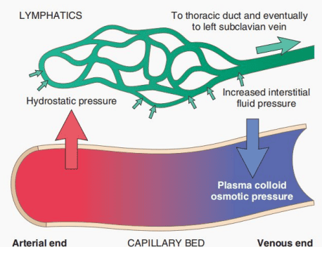
Starling Equilibrium and Capillary Microvascular Fluid Exchange
Pathological Classification: Transudates vs. Exudates
| Characteristic | Transudate Fluid | Exudate Fluid |
|---|---|---|
| Primary Mechanism | Hydrostatic elevation or reduced intravascular colloid oncotic pressure | Increased microvascular endothelial permeability from inflammation |
| Endothelial Barrier Integrity | Intact; inter-endothelial junctions remain preserved | Disrupted; widened inter-endothelial gaps and endothelial injury |
| Protein Concentration | Low (< 3.0 g/dL) | High (> 3.0 g/dL, rich in fibrinogen and globulins) |
| Specific Gravity | Low (< 1.012) | High (> 1.020) |
| Cellular Constituents | Acellular or sparse exfoliated mesothelial cells | Abundant inflammatory cells (neutrophils, histiocytes) |
| Clarity & Appearance | Clear, straw-colored, watery | Cloudy, purulent, turbid, or blood-tinged |
| Clinical Settings | Congestive heart failure, hepatic cirrhosis, nephrotic syndrome | Pneumonia, acute peritonitis, suppurative infections, pleuritis |
Five Principal Etiological Mechanisms of Edema
1. Elevated Capillary Hydrostatic Pressure
Hydrostatic hypertension pushes fluid out of intravascular spaces into interstitial tissues, stemming from localized drainage failure or generalized venous backpressure:
- Localized: Intravascular luminal occlusion by deep-vein thrombosis (DVT) elevates distal capillary pressure, resulting in uncompensated unilateral limb swelling and pain below the obstruction. External compressive masses or pregnancy-related pelvic compression cause similar localized shifts.
- Generalized: Congestive heart failure causes diminished cardiac forward stroke output while increasing end-diastolic filling pressures. This leads to blood pooling upstream in pulmonary capillaries (left failure) or systemic venous networks (right failure). Decreased renal perfusion triggers compensatory renin-angiotensin-aldosterone axis activation, promoting secondary renal salt and water retention that exacerbates hydrostatic pressure.
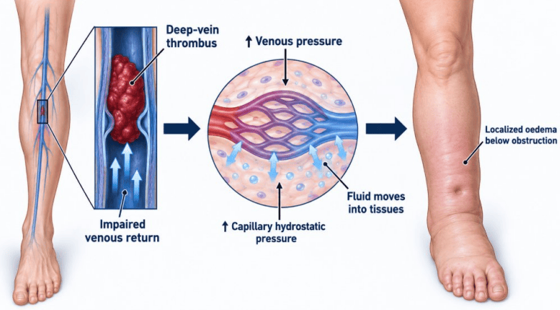
Localized Hydrostatic Edema Generated by Deep Vein Thrombosis
2. Reduced Plasma Colloid Osmotic Pressure (Hypoalbuminemia)
Albumin provides the bulk of plasma oncotic pressure. When total serum albumin drops below critical thresholds (< 2.5 g/dL), water retention within the vascular compartment is compromised, allowing fluid transudation into interstitial spaces and causing systemic hypovolemia:
- Excessive Protein Loss: Nephrotic syndrome produces heavy proteinuria (> 3.5 g/24 hr) secondary to damaged glomerular basement membrane podocytes. Protein-losing gastroenteropathy, severe extensive thermal burns, and exfoliating skin diseases cause direct trans-epithelial loss of serum proteins.
- Decreased Protein Synthesis: Advanced hepatic cirrhosis impairs hepatocyte protein synthesis, while severe protein malnutrition (kwashiorkor) deprives the liver of essential amino acid precursors.
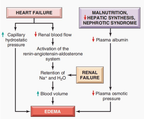
Interconnected Pathways Leading to Systemic Edema
3. Lymphatic Obstruction (Lymphedema)
Mechanical blockage of lymphatic drainage channels prevents the reabsorption of interstitial proteins and water, resulting in localized non-pitting lymphedema:
- Inflammatory and Fibrotic: Radiation therapy induces perivascular scar tissue that obliterates delicate lymphatic channels.
- Infectious Parasitism: Filariasis (Wuchereria bancrofti) causes chronic lymphatic inflammation, fibrosis, and massive dependent lymphedema (elephantiasis).
- Surgical Ablation: Radical mastectomy with axillary lymph node dissection disrupts upper-extremity lymphatic clearance, leading to chronic unilateral arm lymphedema.
- Malignant Infiltration: Metastatic breast carcinoma invading dermal lymphatics produces skin tethering around hair follicles, creating a textured surface resembling an orange peel (peau d'orange).
4. Sodium and Water Retention
Expanded sodium mass retains stoichiometric quantities of water, increasing intravascular volume, raising hydrostatic pressures, and diluting plasma proteins:
- Primary Renal Hypo-excretion: Acute glomerulonephritis and end-stage chronic kidney disease reduce the glomerular filtration rate (GFR), preventing adequate water and solute clearance.
- Secondary Hyperaldosteronism: Circulatory hypoperfusion in congestive heart failure and cirrhotic splanchnic arterial vasodilation triggers continuous renin secretion, promoting renal salt-water retention.
5. Increased Vascular Permeability (Inflammation)
Inflammatory mediators (histamine, bradykinin, leukotrienes C4/D4, platelet-activating factor) induce endothelial cell contraction and gap formation in postcapillary venules. The escape of protein-rich fluid into the extravascular compartment produces inflammatory exudative edema.
Morphological Presentations of Edema
Subcutaneous Edema Patterns
- Dependent Edema: Distribution guided by gravity; prominent across the ankles and pretibial regions in ambulatory individuals, and over the presacral cushion in bedridden patients. Characteristic of congestive heart failure.
- Pitting Edema: Finger compression displaces interstitial water from the intercellular matrix, leaving an indentation that persists for seconds to minutes.
- Periorbital Edema: Fluid accumulation around the eyes due to the low baseline turgor of loose peri-orbital connective tissue. Characteristically prominent upon morning awakening in nephrotic syndrome.
Pulmonary Edema: Lungs become heavy, soggy, and two to three times their normal weight. Sectioning express an abundance of foamy, blood-tinged liquid mixed with air. Histologically, alveolar capillaries appear distended with red blood cells, septa are widened by fluid, and alveoli contain pink, amorphous, proteinaceous material. This fluid barrier reduces pulmonary compliance, limits ventilation, and impairs gas exchange, producing acute respiratory failure.
Cerebral Edema: May be localized (surrounding neoplasms, abscesses, or trauma) or generalized (following anoxic-ischemic encephalopathy, hypertensive crisis, or severe trauma). The brain swells against the rigid cranium; gyri become widened and flattened, while sulci are narrowed. As intracranial pressure (ICP) rises, expansion can lead to transtentorial or tonsillar herniation, brainstem compression, and fatal respiratory arrest.
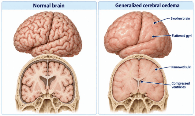
Morphological Features of Generalized Cerebral Edema
Pediatric Pitting Edema and Proteinuria
Clinical Scenario: A young child presents with bilateral dependent pitting lower extremity edema, facial puffiness, and heavy proteinuria.
- Mechanism: Massive renal loss of low-molecular-weight plasma proteins, predominantly albumin, leads to profound hypoalbuminemia.
- Systemic Distribution: Because plasma oncotic pressure falls uniformly throughout the circulation, transudation is generalized.
- Sodium Exacerbation: Hypovolemia triggers renin-angiotensin-aldosterone axis activation. The resulting renal sodium retention expands plasma volume and raises capillary hydrostatic pressure, worsening fluid extravasation.
- Morning Periorbital Distribution: Supine overnight positioning redistributes hydrostatic forces toward the head and periorbital tissue, causing prominent morning facial puffiness.
Hemorrhage
Key Definition: Hemorrhage
Hemorrhage: The extravasation of whole blood components from the cardiovascular compartment into the surrounding tissues, preformed body cavities, or external environment.
Basic Mechanisms of Hemorrhage:
- Physical Vascular Disruption: Macro-rupture from mechanical trauma, penetrating injury, atherosclerotic aneurysmal rupture, or invasive neoplastic erosion.
- Microvascular Diapedesis: Capillary leakage occurring without physical macro-rupture, driven by severe congestion, endothelial toxins, or extreme hypoxia.
- Hemostatic Failure: Qualitative or quantitative platelet defects and inherited or acquired coagulation factor deficiencies.
Categorization by Relationship to Exterior:
- External Hemorrhage: Blood directly leaves the body (e.g., lacerations, epistaxis, menorrhagia).
- Internal Hemorrhage: Blood is contained within organ tissues or enclosed spaces (e.g., intracranial, retroperitoneal).
- Concealed Internal Hemorrhage: Hidden, potentially massive collections that may lead to hemorrhagic shock before outward signs appear (e.g., ruptured ectopic tubal gestation, traumatic splenic laceration, aortic dissection).
Classification by Size and Appearance
| Hemorrhage Subtype | Dimension | Morphological Characteristics | Primary Clinical Etiologies |
|---|---|---|---|
| Petechiae | 1 to 2 mm | Pinpoint, punctate mucosal or cuticular hemorrhages; non-blanching | Severe thrombocytopenia, platelet dysfunction, scurvy, sudden hydrostatic elevation |
| Purpura | 3 to 5 mm | Slightly larger, circumscribed purpuric patches on cutaneous or serosal surfaces | Immune thrombocytopenic purpura (ITP), IgA vasculitis (Henoch-Schönlein), direct trauma, vessel fragility |
| Ecchymoses | 1 to 2 cm or larger | Broad subcutaneous hematomas; classic soft tissue "bruises" | Blunt tissue trauma, severe coagulopathies, anticoagulant therapy |
| Hematoma | Variable (millimeters to massive) | Localized mass-forming collection of clotted blood within a distinct space | Aortic rupture, intracranial hematomas, intramuscular hemorrhage, retroperitoneal injury |
Chromatographic Degradation of an Ecchymosis (Bruise)
A healing bruise undergoes a characteristic sequence of enzymatic color changes:
- Red-Blue: Intravasated erythrocytes containing intact oxygenated/deoxygenated hemoglobin.
- Blue-Green: Macrophage heme oxygenase converts red-blue heme into green biliverdin.
- Yellow-Gold: Biliverdin is reduced enzymatically to yellow bilirubin.
- Golden-Brown: Unbound ferric iron aggregates into ferritin clusters, forming brown hemosiderin before final resolution.
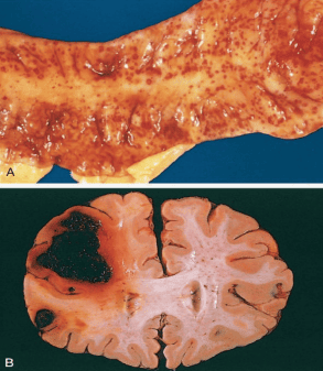
Hemorrhage Patterns from Mucosal Petechiae to Fatal Hematoma
Internal Cavitary Hemorrhages
- Hemothorax: Blood pooling in the pleural cavity from thoracic trauma, vascular rupture, or invasive neoplasia.
- Hemopericardium: Blood collecting inside the noncompliant pericardial sac. Rapid entry of as little as 150–250 mL of blood can cause cardiac tamponade, compressing thin-walled right chambers and preventing diastolic filling.
- Hemoperitoneum: Blood inside the peritoneal cavity, seen in ruptured ectopic gestation, splenic laceration, or abdominal aortic rupture.
- Hemarthrosis: Blood collecting within synovial joints, common after traumatic injuries and characteristically recurrent in severe inherited factor deficiencies like hemophilia A or B.
Normal Hemostasis
Key Definition: Physiological Hemostasis
Hemostasis: A regulated process that forms a localized hemostatic plug at sites of vascular injury while maintaining blood in a fluid, clot-free state within intact vessels.
The Four Sequential Phases of Hemostasis ("V-P-C-F"):
- V – Vasoconstriction: Immediate, transient arteriolar smooth muscle contraction.
- P – Platelet Plug Formation (Primary Hemostasis): Adhesion, activation, and aggregation of platelets.
- C – Coagulation Cascade (Secondary Hemostasis): Fibrin formation cross-linking the platelet plug.
- F – Fibrinolysis & Anticoagulation: Limitation and eventual breakdown of the hemostatic plug during tissue repair.
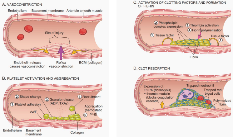
Sequential Stages of Normal Hemostasis
Primary Hemostasis: The Platelet Response
Platelets are disc-shaped anucleate cell fragments derived from bone marrow megakaryocytes. Following endothelial injury, primary hemostasis proceeds through three steps ("A-A-A"): Adhesion, Activation, and Aggregation.
1. Platelet Adhesion
Endothelial damage exposes subendothelial type IV collagen and extracellular matrix. Von Willebrand factor (vWF), produced by endothelial Weibel-Palade bodies and platelet alpha granules, binds directly to exposed collagen fibrils. Platelets adhere by binding their membrane glycoprotein receptor, GPIb, to immobilized vWF.
Platelet Adhesion vs. Aggregation Receptors
GPIb = Adhesion (Binds vWF): Deficient in Bernard-Soulier syndrome ("Bernard-Soulier has Bad Binding"). Presents with impaired adhesion and abnormally large platelets.
GPIIb/IIIa = Aggregation (Binds Fibrinogen): Deficient in Glanzmann thrombasthenia ("Glanzmann lacks Aggregation"). Adhesion is intact, but platelets cannot form inter-platelet cross-links.
2. Platelet Activation and Secretion
Adhesion triggers a conformational shape change: platelets transition from smooth discs into irregular spheres extending long, spiky pseudopods, which maximizes their surface area. Activated platelets flip their plasma membranes to expose negatively charged phosphatidylserine, providing a critical catalytic surface for coagulation factor assembly.
Granule Exocytosis:
- Alpha (α) Granules: Secrete coagulation factor V, fibrinogen, vWF, fibronectin, platelet factor 4 (PF4), platelet-derived growth factor (PDGF), and transforming growth factor-beta (TGF-β).
- Dense (δ) Granules: Secrete adenosine diphosphate (ADP), ionized calcium (Ca2+), adenosine triphosphate (ATP), serotonin, and epinephrine.
Recruitment Amplification: Extracellular ADP binds platelet P2Y1 and P2Y12 receptors, promoting platelet activation. Simultaneously, platelet cyclooxygenase (COX) converts arachidonic acid into thromboxane A2 (TXA2), a potent platelet aggregator and vasoconstrictor.
Aspirin Mechanism of Action
Aspirin: Irreversibly acetylates and inhibits cyclooxygenase (COX) enzymes, blocking the production of thromboxane A2. Because anucleate platelets cannot synthesize new enzymes, this inhibitory defect persists for the platelet's entire 7- to 10-day lifespan.
3. Platelet Aggregation & Primary Plug Formation
Activation induces a conformational change in the GPIIb/IIIa receptor complex, exposing binding sites for multivalent circulating fibrinogen. Fibrinogen molecules bridge adjacent platelets, generating an expanding aggregate that forms the reversible primary hemostatic plug.
Secondary Hemostasis: The Coagulation Cascade
Secondary hemostasis stabilizes the primary platelet mass through a sequential series of proenzyme activations that culminates in thrombin generation and insoluble, cross-linked fibrin deposition.
Enzymatic Complex Assembly: Efficient cascade progression requires an active enzyme, a proenzyme substrate, an activating cofactor, and ionized calcium (Ca2+) assembled upon the negatively charged phosphatidylserine surface of activated platelets.
Vitamin K-Dependent Coagulation Factors ("1972")
Hepatic gamma-glutamyl carboxylase requires reduced vitamin K as a cofactor to add carboxyl groups to glutamic acid residues on specific clotting proteins. This post-translational carboxylation enables calcium-dependent binding to platelet phospholipids:
- Procoagulant factors: Factor X, Factor IX, Factor VII, Factor II (Prothrombin) (Mnemonic: 1972).
- Anticoagulant regulatory proteins: Protein C and Protein S.
Laboratory Pathways vs. In Vivo Hemostasis
In Vivo Physiology: Physiological clotting is initiated by the Tissue Factor (TF) pathway. Exposure of subendothelial Tissue Factor at the injury site binds circulating Factor VIIa. The TF-VIIa complex directly cleaves Factor X to Xa and Factor IX to IXa. Small initial amounts of thrombin generated by Factor Xa provide positive feedback by activating Factors V, VIII, and XI, which amplifies subsequent thrombin generation.
In Vitro Testing Pathways:
- Extrinsic Pathway (Prothrombin Time / PT): Evaluates factors VII, X, V, prothrombin (II), and fibrinogen. Thromboplastin (tissue factor), phospholipids, and calcium are added to citrated plasma. Standardized clinically as the International Normalized Ratio (INR) to monitor warfarin therapy.
- Intrinsic Pathway (Activated Partial Thromboplastin Time / aPTT): Evaluates factors XII, XI, IX, VIII, X, V, II, and fibrinogen. Clotting is initiated in vitro by adding contact activators (silica, celite, or kaolin) alongside phospholipids and calcium. Used to monitor unfractionated heparin therapy.
| Laboratory Assay | Target Coagulation Pathway | Coagulation Factors Evaluated | Therapeutic Drug Monitoring |
|---|---|---|---|
| Prothrombin Time (PT / INR) | Extrinsic and Common pathways | Factors VII, X, V, Prothrombin (II), Fibrinogen | Warfarin (Vitamin K antagonists) |
| Partial Thromboplastin Time (aPTT) | Intrinsic and Common pathways | Factors XII, XI, IX, VIII, X, V, II, Fibrinogen | Unfractionated Heparin |
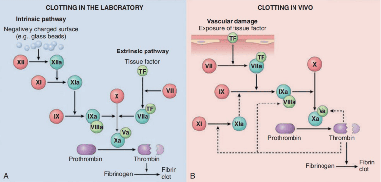
The Coagulation Cascade in the Laboratory and In Vivo
Thrombin: The Central Regulatory Hub of Hemostasis
Thrombin (Factor IIa) serves multiple catalytic, regulatory, and inflammatory functions:
- Fibrin Polymerization: Cleaves soluble fibrinogen to yield insoluble fibrin monomers that polymerize spontaneously into fibrils.
- Fibrin Stabilization: Directly activates Factor XIII (fibrin-stabilizing factor), a transglutaminase that forms covalent cross-links between adjacent fibrin strands.
- Feedback Amplification: Activates upstream cofactors V and VIII, as well as Factor XI, driving rapid burst-like thrombin production.
- Platelet Aggregation: Activates platelets via cleavage of Protease-Activated Receptors (PAR-1 and PAR-4).
- Pro-inflammatory Signaling: Stimulates endothelial cells and leukocytes via PARs to synthesize selectins, cytokines (IL-1, IL-6), chemokines, and nitric oxide.
- Anticoagulant Switch: Upon reaching intact downstream endothelium, thrombin binds membrane-bound thrombomodulin. This changes its substrate specificity: thrombin ceases cleavage of fibrinogen and instead activates Protein C, which limits further clot expansion.
Limitation of Clotting and Fibrinolysis
To prevent thrombosis from extending throughout the vascular tree, hemostatic activation is restricted to the site of injury by several regulatory systems:
- Hemodynamic Dilution: Flowing blood washes activated factors downstream, where they are diluted and cleared by hepatic Kupffer cells.
- Antithrombin III (ATIII): A circulating serine protease inhibitor that complexes with and inactivates thrombin, Factor Xa, Factor IXa, Factor XIa, and Factor XIIa. This activity is enhanced over 1,000-fold in the presence of heparin-like glycosaminoglycans on endothelial membranes.
- Thrombomodulin-Protein C-Protein S System: Thrombin-thrombomodulin complexes cleave Protein C to form Activated Protein C (APC). Along with its cofactor, Protein S, APC cleaves and inactivates active cofactors Factor Va and Factor VIIIa.
- Tissue Factor Pathway Inhibitor (TFPI): Endothelial-derived protein that binds and neutralizes Factor Xa and the initial TF-VIIa catalytic complexes.
- The Fibrinolytic System: Fibrinolysis degrades polymerized fibrin deposits to maintain vessel patency. Endothelial cells synthesize tissue plasminogen activator (t-PA), which cleaves proenzyme plasminogen into active plasmin. Plasmin cleaves fibrin into soluble fragments called fibrin degradation products (FDPs), including D-dimers.
Clinical Utility of D-Dimer Assays
D-Dimers: Specific cleavage products generated exclusively when plasmin breaks down covalently cross-linked fibrin (cross-linked by Factor XIIIa). Elevated serum D-dimer levels indicate recent intravascular coagulation followed by secondary fibrinolysis (e.g., deep-vein thrombosis, pulmonary embolism, disseminated intravascular coagulation).
Thrombosis
Key Definition: Thrombosis
Thrombosis: The pathological, non-physiological formation of an intravascular solid or semisolid aggregate composed of platelets, fibrin, and entrapped cellular elements inside an intact or minimally injured vessel during life.
| Characteristic | Antemortem Thrombus | Postmortem Clot |
|---|---|---|
| Timing of Formation | Formed during life within flowing blood | Formed after death in stagnant blood |
| Vascular Attachment | Firmly adherent to the underlying endothelial surface | Non-adherent; easily lifted out of the vessel lumen |
| Consistency & Texture | Firm, dry, granular, and friable | Soft, rubbery, moist, and gelatinous |
| Internal Laminations | Lines of Zahn present (pale platelet-fibrin layers alternating with dark erythrocyte-rich layers) | Absence of true lines of Zahn; demonstrates gravitational layering ("currant jelly" red base under a yellow "chicken fat" supernatant) |
Pathogenesis: Virchow's Triad ("E-A-H")
Thrombus generation is driven by three primary abnormalities: E – Endothelial Injury, A – Abnormal Blood Flow, and H – Hypercoagulability.
1. Endothelial Injury
Loss or dysfunction of the non-thrombogenic endothelial lining exposes subendothelial vWF and collagen to platelets, releases Tissue Factor, and downregulates endothelial thrombomodulin, protein C receptors, and prostacyclin (PGI2). Causes include ulcerated atherosclerotic plaques, vasculitis, hypertension, cigarette smoke toxins, and endocardial damage following myocardial infarction.
2. Abnormal Blood Flow: Turbulence and Stasis
- Turbulence (Arterial/Cardiac): Disordered chaotic flow damages endothelial cells and creates local recirculating pockets of stasis. Commonly caused by arterial aneurysms, atherosclerotic plaques, and stenotic heart valves.
- Stasis (Venous): Sluggish or halted flow disrupts normal laminar streaming, in which cellular elements travel in the central axial stream separated from the endothelium by a clear plasma zone. Stasis allows platelets to make contact with the vessel wall, delays hepatic clearance of activated clotting factors, and prevents local delivery of circulating anticoagulant proteins.
3. Hypercoagulability (Thrombophilia)
Refers to inherited or acquired alterations in the coagulation cascade that predispose to thrombosis:
| Category | Specific Condition | Molecular / Clinical Mechanism |
|---|---|---|
| Primary (Inherited) | Factor V Leiden Mutation | Point mutation (Arg506Gln substitution) making Factor Va resistant to cleavage by Activated Protein C (APC). The most common inherited thrombophilia in Caucasian populations. |
| Primary (Inherited) | Prothrombin G20210A Mutation | Single nucleotide substitution in the 3'-untranslated region that increases prothrombin gene expression, elevating circulating prothrombin levels 2- to 3-fold. |
| Primary (Inherited) | Antithrombin III Deficiency | Impaired neutralization of thrombin and Factor Xa; patients do not exhibit the expected elongation in aPTT when administered heparin. |
| Primary (Inherited) | Protein C or S Deficiency | Impaired proteolytic degradation of Factors Va and VIIIa. Severe homozygous deficiency in neonates presents as fatal purpura fulminans. |
| Secondary (Acquired) | Antiphospholipid Syndrome (APS) | Autoantibodies against phospholipid-binding proteins (anti-cardiolipin, anti-β2-glycoprotein I, lupus anticoagulant) that trigger widespread endothelial activation, recurrent venous/arterial thrombosis, and pregnancy loss. |
| Secondary (Acquired) | Heparin-Induced Thrombocytopenia (HIT) | IgG autoantibodies develop against platelet factor 4 (PF4)-heparin complexes, activating platelets and producing widespread paradoxical thrombosis alongside a falling platelet count. |
Morphology and Fate of Thrombi
Arterial Thrombi: Typically develop at sites of endothelial injury or plaque rupture. They grow in a retrograde direction (toward the heart, against the flow of arterial blood). Composed predominantly of platelet-rich layers, earning the designation "white thrombi." They are frequently occlusive, producing acute downstream tissue ischemia or infarction (e.g., myocardial infarction, ischemic stroke).
Venous Thrombi (Phlebothrombosis): Form in low-pressure, stagnant environments, characteristically in the deep calf and thigh veins. They propagate anterograde (in the direction of venous blood flow, toward the heart). Formed largely of dense erythrocyte networks trapped in fibrin meshes, earning the designation "red thrombi." The loosely attached, propagating tail is prone to fragmentation and embolization to the lungs.
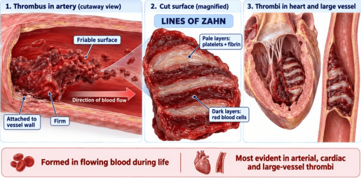
Morphological Architecture and Lines of Zahn in Thrombi
The Four Potential Fates of a Thrombus
- Propagation: The thrombus accumulates additional platelets and fibrin, extending along the vessel and increasing the likelihood of complete vascular occlusion.
- Embolization: The thrombus (or a portion of it) dislodges from the vessel wall, travels through the circulation, and impacts within distant luminal branches.
- Dissolution: Early-stage thrombi can be dissolved through endogenous fibrinolysis (plasmin activation). Older, cross-linked thrombi become resistant to lysis, which is why clinical thrombolytic drugs (e.g., t-PA) must be administered early in acute ischemic events.
- Organization and Recanalization: Ingrowth of endothelial cells, smooth muscle cells, and fibroblasts converts the thrombus into vascularized connective tissue. Over time, small endothelial-lined lumens (recanalized channels) form through the mass to partially restore blood flow.
Disseminated Intravascular Coagulation (DIC)
Pathophysiology: DIC is an acquired, secondary thrombohemorrhagic disorder that complicates various severe systemic diseases (e.g., Gram-negative or Gram-positive sepsis, amniotic-fluid embolism, acute promyelocytic leukemia, extensive burns). Widespread release of Tissue Factor or diffuse endothelial injury triggers systemic, microcirculatory coagulation.
The Thrombohemorrhagic Paradox:
- Diffuse microvascular fibrin-platelet thrombi obstruct regional blood flow, producing multi-organ ischemia, end-organ dysfunction, and microangiopathic hemolytic anemia (schistocytes).
- Widespread thrombus generation rapidly consumes available platelets, fibrinogen, Factor V, and Factor VIII.
- Secondary activation of the fibrinolytic system produces elevated levels of fibrin degradation products (FDPs and D-dimers), which interfere with remaining platelet function and fibrin polymerization.
- This combination of clotting factor consumption and fibrinolysis leads to severe spontaneous hemorrhage (e.g., oozing from intravenous puncture sites, petechiae, gastrointestinal bleeding).
| Diagnostic Laboratory Assay | Observed Result in Active DIC | Underlying Mechanism |
|---|---|---|
| Platelet Count | Markedly decreased (↓) | Accelerated consumption into widespread microthrombi |
| Prothrombin Time (PT / INR) | Prolonged (↑) | Depletion of extrinsic and common pathway coagulation factors |
| Partial Thromboplastin Time (aPTT) | Prolonged (↑) | Consumption of intrinsic and common pathway factors |
| Fibrinogen Level | Severely depleted (↓) | Consumption via widespread conversion into fibrin networks |
| D-Dimer and FDP Levels | Markedly elevated (↑↑) | Secondary fibrinolysis of cross-linked fibrin clots |
| Peripheral Blood Smear | Presence of Schistocytes | Mechanical fragmentation of erythrocytes sheared across microvascular fibrin strands |
Embolism
Key Definition: Embolus and Embolism
Embolus: A detached, traveling intravascular solid, liquid, or gaseous mass carried by the bloodstream from its point of origin to a distant site, where it can cause downstream vascular obstruction, tissue ischemia, or infarction.
Constituent Classification of Emboli:
- Solid Emboli: Dislodged thromboemboli (accounting for > 95% of cases), ulcerated atherosclerotic plaque debris (cholesterol emboli), tumor fragments, or infective bacterial/fungal vegetations.
- Liquid Emboli: Bone marrow fat droplets (following skeletal fractures) or amniotic fluid components.
- Gaseous Emboli: Atmospheric air bubbles introduced during trauma/surgery or insoluble nitrogen bubbles in decompression sickness.
1. Pulmonary Thromboembolism (PTE)
In over 95% of cases, pulmonary emboli originate from deep-vein thrombi (DVT) of the lower extremities proximal to or at the popliteal vein (popliteal, femoral, and iliac veins). Dislodged fragments travel through the inferior vena cava, navigate the right atrium and right ventricle, and impact within the main pulmonary arterial tree or its distal branches.
- Massive / Saddle Embolus: A large embolus that occludes the main bifurcation of the pulmonary artery. This causes acute right ventricular pressure overload, acute cor pulmonale, electromechanical dissociation, cardiogenic/obstructive shock, and sudden death.
- Medium-Sized Emboli: Travel into peripheral segmental pulmonary branches. In patients with compromised baseline cardiopulmonary function, this can lead to wedge-shaped hemorrhagic pulmonary infarction, pleuritic chest pain, tachypnea, and hemoptysis.
- Small / Micro-Emboli: Often clinically silent because the lung receives a dual blood supply from the pulmonary and bronchial arterial systems. Recurrent micro-emboli over time can cause progressive obliteration of the pulmonary vascular bed, leading to pulmonary hypertension and chronic right heart failure.
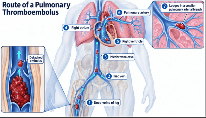
Migration Pathway of a Pulmonary Thromboembolus
2. Systemic Thromboembolism
Systemic emboli travel through the arterial circulation. Roughly 80% originate from intracardiac mural thrombi (associated with left ventricular myocardial infarctions or left atrial dilation secondary to mitral stenosis and atrial fibrillation). The remaining 20% arise from aortic aneurysms, ulcerated atherosclerotic plaques, or infected heart valve vegetations. Common sites of arrest include the lower extremities (75%), brain (10%), kidneys, spleen, and mesenteric vasculature.
3. Paradoxical Embolism
Occurs when an embolus of venous origin bypasses the pulmonary capillary bed by crossing an abnormal right-to-left intracardiac communication (e.g., patent foramen ovale [PFO], atrial septal defect [ASD], or ventricular septal defect [VSD]). This allows a venous thrombus to enter the arterial circulation and cause an ischemic stroke or systemic arterial infarction.
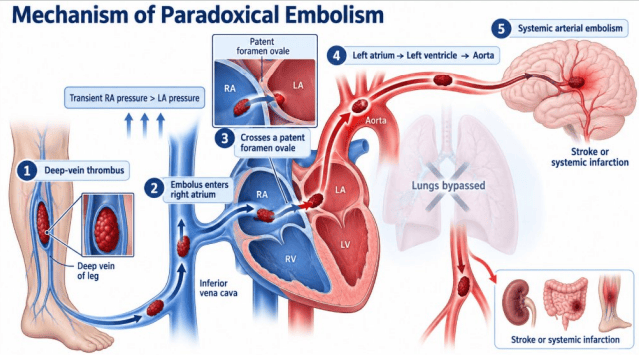
Hemodynamic Pathway of Paradoxical Embolism
4. Fat Embolism Syndrome (FES)
Following traumatic fractures of long bones (e.g., femur, tibia) or severe pelvic trauma, ruptured medullary fat globules enter exposed bone marrow venules. The clinical syndrome characteristically presents 24 to 72 hours after trauma with a classic triad: acute respiratory insufficiency (dyspnea, hypoxemia), neurological deficits (confusion, delirium, seizures), and a petechial rash localized across the anterior neck, chest, and conjunctivae. Pathogenesis involves mechanical microvascular plugging alongside biochemical endothelial injury driven by free fatty acids released from lipolyzed globules.
5. Air and Gas Embolism
Exogenous atmospheric air (> 100 mL) can be drawn into the venous circulation during central venous catheter insertion/removal, head and neck surgery, or thoracic trauma. In Decompression Sickness, deep-sea divers breathing high-pressure air accumulate dissolved atmospheric nitrogen in their blood and lipid-rich tissues. If depressurization during ascent occurs too rapidly, nitrogen comes out of solution as microscopic gas bubbles. These bubbles obstruct microvessels, producing painful periarticular ischemia ("the bends"), acute pulmonary edema with respiratory distress ("the chokes"), and ischemic necrosis of skeletal bones (caisson disease).
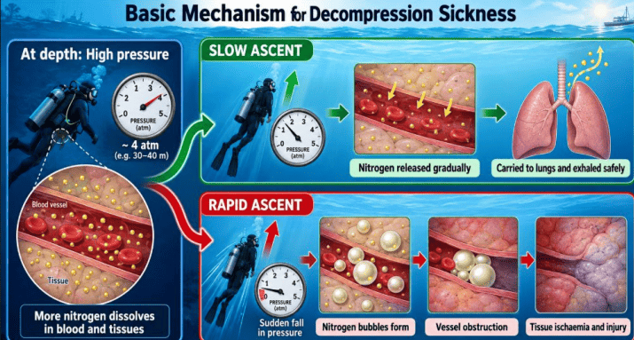
Pathogenic Mechanism of Nitrogen Gas Embolism in Decompression Sickness
6. Amniotic Fluid Embolism (AFE)
A rare, catastrophic obstetric emergency caused by breaches in the utero-placental membrane barrier during labor, delivery, or the immediate postpartum period. Amniotic fluid containing fetal elements (squamous squames, lanugo hair, vernix, mucin, and procoagulant tissue factor) enters the maternal venous circulation. This triggers severe pulmonary vasoconstriction, acute right ventricular failure, cardiogenic shock, and consumption coagulopathy (DIC). Microscopic examination of the maternal pulmonary microvasculature confirms the diagnosis by revealing laminated swirls of fetal squamous cells and mucin within alveolar arterioles.
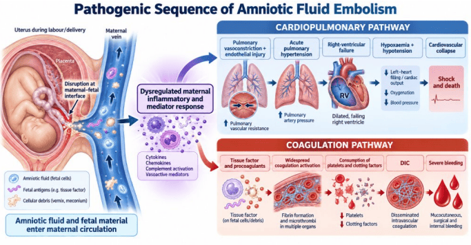
Pathogenic Sequence of Amniotic Fluid Embolism
Infarction
Key Definition: Infarction and Ischemia
- Ischemia: Inadequate arterial perfusion to a tissue bed, causing a simultaneous deficit of oxygen and metabolic substrates alongside the accumulation of toxic metabolic wastes.
- Infarct: A circumscribed, localized area of ischemic tissue necrosis resulting from occlusion of the arterial blood supply or, less commonly, venous outflow obstruction.
Ischemia vs. Hypoxia: Ischemia is more damaging than simple hypoxemia (low dissolved arterial PO2). While hypoxemic tissues continue to receive glucose and have metabolic wastes removed via intact perfusion, ischemic tissues lose substrate delivery and experience toxic metabolite buildup, accelerating cell death.
Classification: White (Anemic) vs. Red (Hemorrhagic) Infarcts
| Diagnostic Feature | White (Anemic) Infarct | Red (Hemorrhagic) Infarct |
|---|---|---|
| Primary Vascular Mechanism | Arterial occlusion in solid compact organs with end-arterial circulations | Venous occlusion, loose spongy tissues, tissues with dual circulations, or reperfusion |
| Erythrocyte Extravasation | Minimal | Abundant intra-parenchymal erythrocyte pooling |
| Gross Tissue Chromaticity | Pale, tan, or yellow-white with an erythematous border | Dark red, purple, or black |
| Representative Organs | Heart, Kidney, Spleen | Lungs, Small and Large Intestine, Testis, Ovary |
| Reperfusion Vulnerability | Secondary hemorrhage can occur if arterial patency is restored | Inherent to the primary pathology |
Five Conditions Favoring Red (Hemorrhagic) Infarcts:
- Venous Occlusion: Torsion of the spermatic cord or ovary blocks thin-walled venous drainage while high-pressure arterial inflow continues transiently, forcing blood into the necrotic tissue.
- Loose, Spongy Tissues: The lung has an expansive alveolar network that readily fills with blood escaping from injured capillaries.
- Dual Blood Supplies: Tissues with collateral vascular supplies (e.g., lungs via pulmonary and bronchial arteries; liver via hepatic artery and portal vein; small intestine via arcades) bleed into ischemic territories from patent alternate vessels.
- Prior Sluggish Congestion: Tissue beds engorged with venous blood prior to occlusion extravasate large volumes of erythrocytes upon necrosis.
- Reperfusion of Ischemic Zones: Re-establishing arterial flow into mechanically fragile, necrotic microvasculature causes secondary trans-endothelial hemorrhage.
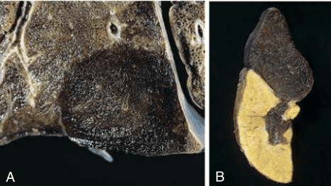
Gross Morphology of Red (Hemorrhagic) and White (Anemic) Infarcts
Morphology and Resolution of Infarctions
Geometric Configuration: Most arterial infarcts assume a wedge shape. The apex points toward the occluded vessel, while the broad base aligns with the organ's subcapsular serosal surface. Overlying serosa often displays reactive fibrinous exudates.
Microscopic Characteristics: In most tissues (heart, kidney, spleen), ischemia causes coagulative necrosis. The basic cellular architecture remains identifiable for several days, but cells lose their nuclei, exhibit karyolysis, and develop increased cytoplasmic eosinophilia. The critical exception is the central nervous system: cerebral ischemic infarction results in liquefactive necrosis, in which hydrolytic enzymes digest the lipid-rich brain tissue into a fluid-filled cystic cavity lined by reactive astrocytes (gliosis).
Shock
Key Definition: Systemic Shock
Shock: A profound state of systemic tissue hypoperfusion caused by reduced cardiac output or decreased effective circulating blood volume, resulting in cellular hypoxia, energy depletion, and multiorgan failure.
General Pathogenic Progression:
Classification of Shock Syndromes
| Shock Category | Primary Hemodynamic Defect | Clinical Causes | Early Physical Findings |
|---|---|---|---|
| Cardiogenic Shock | Myocardial pump failure leading to decreased cardiac output and stroke volume | Extensive myocardial infarction (> 40% LV loss), ventricular rupture, papillary muscle rupture, sustained arrhythmias | Hypotension, tachycardia, weak pulse, cold, clammy, cyanotic skin, pulmonary edema (crackles) |
| Hypovolemic Shock | Severe loss of intravascular whole blood or plasma volume | Massive hemorrhage, extensive thermal burns, severe vomiting or diarrhea, dehydration | Hypotension, narrow pulse pressure, tachycardia, cool, pale extremities, oliguria, intense thirst |
| Septic Shock | Systemic vasodilation, microvascular pooling, and endothelial activation from infection | Gram-negative septicemia (endotoxins/LPS), Gram-positive sepsis (superantigens), invasive fungal infections | Early phase: warm, flushed skin and bounding pulses; Late phase: cold extremities, profound refractory hypotension, DIC |
| Obstructive Shock | Physical mechanical barrier impairing cardiac filling or ventricular outflow | Massive pulmonary saddle embolism, cardiac tamponade, tension pneumothorax | Distended neck veins (elevated JVD), pulsus paradoxus, hypotension, cold extremities |
| Anaphylactic Shock | IgE-mediated systemic hypersensitivity causing acute vasodilation and increased permeability | Hymenoptera stings, medications (penicillin), food allergies (peanuts) | Urticaria, pruritus, laryngeal edema, bronchospasm, flushing, acute hypotension |
| Neurogenic Shock | Sudden loss of sympathetic vasomotor vascular tone | High cervical spinal cord transection, severe traumatic brain injury, high spinal anesthesia | Hypotension accompanied paradoxically by bradycardia and warm, dry skin (loss of sympathetic input) |
Pathogenesis of Septic Shock
Microbial Pathogen-Associated Molecular Patterns (PAMPs, such as Gram-negative bacterial lipopolysaccharide) bind Toll-like receptors on monocytes, macrophages, and neutrophils. This triggers high-level release of pro-inflammatory cytokines (TNF-α, IL-1, IL-6) and secondary mediators (nitric oxide, platelet-activating factor). These factors produce systemic endothelial activation, generalized precapillary arteriolar vasodilation, capillary leakage, and widespread Tissue Factor expression, which drives microvascular thrombosis (DIC). Concurrent metabolic abnormalities include insulin resistance, cellular hyperglycemia, impaired mitochondrial oxidative phosphorylation, and tissue lactic acidosis.
Sepsis-3 Clinical Criteria for Septic Shock
A clinical subset of sepsis characterized by significant circulatory, cellular, and metabolic abnormalities:
- Persistent hypotension requiring vasopressor therapy to maintain a Mean Arterial Pressure (MAP) ≥ 65 mmHg, and
- A serum lactate concentration > 2.0 mmol/L (18 mg/dL) despite adequate volume resuscitation.
The Three Sequential Stages of Shock
1. Non-Progressive (Compensated) Stage
Neurohormonal compensatory mechanisms maintain central organ perfusion without irreversible injury. Carotid sinus and aortic arch baroreceptors detect drops in arterial stretch, firing the sympathetic nervous system to release catecholamines (epinephrine and norepinephrine). This produces tachycardia, increased cardiac contractility, and peripheral arteriolar vasoconstriction, preserving blood flow to the brain and heart. Renal hypoperfusion triggers the renin-angiotensin-aldosterone axis, releasing aldosterone and antidiuretic hormone (ADH) to preserve sodium and water, which causes compensatory oliguria.
2. Progressive (Decompensated) Stage
If the inciting cause is not corrected, systemic hypoperfusion persists. Cells turn to anaerobic glycolysis, producing lactic acid that triggers systemic metabolic acidosis. Acidosis blunts arteriolar responsiveness to catecholamines; precapillary sphincters relax while postcapillary venules remain constricted, causing peripheral blood pooling. This decreases venous return, reduces cardiac output, and initiates a self-reinforcing downward spiral of tissue hypoperfusion.
3. Irreversible Stage
Widespread cellular injury becomes permanent. Extreme ATP depletion disables ATP-dependent Na+/K+ membrane pumps, causing intracellular sodium and water accumulation and cell swelling. Lysosomal membranes rupture, releasing autolytic acid hydrolases into the cytoplasm. Ischemic bowel injury compromises the mucosal barrier, allowing intestinal bacteria and endotoxins to enter the bloodstream (superimposed endotoxic shock). Renal acute tubular necrosis causes acute renal failure, and severe myocardial ischemia limits cardiac pump function. Even if systemic arterial pressure is restored with fluids and inotropes, cellular injury has progressed beyond recovery, and death is inevitable.
Summary Mnemonics for Shock
- Three Major Shock Classes ("H-C-S"): Hypovolemic, Cardiogenic, Septic.
- Chronological Stages of Shock ("C-P-I"): Compensated (Non-progressive), Progressive, Irreversible.
- Four Key Drivers of Septic Shock ("V-C-M-I"): Vascular leakage and vasodilation, Coagulation activation (DIC), Metabolic failure and myocardial depression, Immune dysregulation.