Environmental and Nutritional Pathology
Scope of Environmental & Nutritional Pathology
Pathology encompasses cellular and systemic injuries arising from both exogenous ecological exposures and disruptions in metabolic fuel homeostasis. This comprehensive syllabus explores physical agents, chemical xenobiotics, atmospheric pollutants, occupational carcinogens, recreational toxins, thermal extremes, ionizing radiation, and the full spectrum of undernutrition, vitamin deficiencies, and trace element syndromes.
1. Introduction to Environmental Diseases
Environmental diseases represent pathological alterations provoked or modulated by deleterious chemical, biological, or physical agents encountered across outdoor surroundings, enclosed living spaces, workplaces, and personal behavioral environments. The concept of the human "environment" extends substantially beyond ambient outdoor air.
| Environmental Domain | Representative Exposure Sources | Primary Pathological Impact |
|---|---|---|
| Outdoor Environment | Ambient air particulate pollution, polluted waterways, excessive climatic heat, and extreme freezing conditions | Direct pulmonary irritation, systemic vascular strain, waterborne gastrointestinal infection, and tissue freezing |
| Indoor Environment | Biomass combustion smoke, passive environmental tobacco smoke, poorly ventilated architectural spaces, and radon gas | Chronic airway inflammation, carbon monoxide hypoxia, and delayed mucosal carcinogenesis |
| Occupational Environment | Industrial reagents, heavy metals, synthetic organic solvents, mineral dusts, pesticides, and physical radiation | Pneumoconioses, acute tubular necrosis, chemical hepatitis, marrow aplasia, and occupational neoplasms |
| Personal Environment | Voluntary tobacco use, ethanol intake, non-prescribed substances of abuse, prescription therapeutics, and diet | Atherosclerosis, direct organ necrosis, mucosal ulceration, neurochemical dependence, and metabolic imbalance |
Tissue damage following environmental insult may manifest secondary to a single high-intensity acute exposure (such as an industrial toxic spill or acute carbon monoxide inhalation) or as an insidious, progressive pathology driven by chronic low-dose repetitive exposure over years (such as occupational coal dust inhalation or sustained heavy metal accumulation).
Determinants of Toxic Harm
Evaluating chemical and physical injury requires systematic assessment of five fundamental variables:
- Agent Identity: Different toxic agents target unique cellular organelles, enzymes, or molecular pathways.
- Absorbed Dose: The quantity of chemical entering biological compartments dictates the degree of cellular damage (the cornerstone of classical toxicology).
- Exposure Duration: Acute peak exposures provoke catastrophic necrosis or sudden functional arrest, whereas prolonged low-dose contact facilitates chronic fibrosis, metaplasia, or neoplastic transformation.
- Port of Entry: Absorption kinetics and systemic distribution diverge markedly depending on whether contact occurs via transdermal absorption, ingestion, or inhalation.
- Host Susceptibility: Individual resilience is modified by chronological age, genetic polymorphisms in biotransformation enzymes, pre-existing organ failure, and immune status.
2. Climate Change as an Emerging Health Problem
Global climatic instability involves progressive long-term alterations in baseline temperatures and meteorological patterns. The underlying driver is the greenhouse effect, wherein atmospheric trace gases absorb thermal energy radiating from Earth's crust, limiting heat dissipation into space. As greenhouse gas concentrations escalate, progressive global warming results.
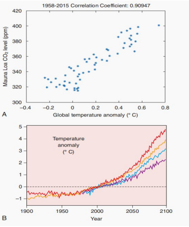
Atmospheric Carbon Dioxide Accumulation and Global Temperature Anomalies
From a pathological perspective, climate change modifies human disease burdens through multiple biological and environmental pathways identified by the World Health Organization (WHO):
| Climatic Shift / Exposure | Primary Pathophysiological Mechanism | Direct Clinical & Pathological Consequence |
|---|---|---|
| Excessive Thermal Heatwaves | Evaporative fluid depletion, extreme cutaneous vasodilation, systemic inflammatory cascade, and protein denaturation | Hypovolemic circulatory shock, heat stroke, acute tubular necrosis, and decompensation of underlying cardiovascular illness |
| Severe Floods & Droughts | Compromise of municipal sewage containment and depletion of sanitary potable drinking water supplies | Outbreaks of acute enteritis, infectious dysentery, secretory diarrhea, and rapid hypovolemic dehydration |
| Altered Precipitation & Thermal Zones | Expansion of geographical ranges and breeding seasonality suitable for arthropod reproduction and survival | Shifting geographic prevalence of malaria (Anopheles vector) and dengue fever (Aedes vector) |
| Agricultural Disruption & Crop Failure | Drought- and flood-induced agricultural collapse triggering severe regional food supply deficits | Somatic protein-energy wasting, stunted linear growth, immune suppression, and widespread micronutrient deficiencies |
| Extreme Weather Events | Physical impacts from collapsing architecture, landslides, high winds, and flying blunt debris | Blunt mechanical trauma, extensive crush lacerations, traumatic bone fractures, and drowning |
| Healthcare System Disruption | Structural damage to clinics, supply chain failures, and loss of chronic pharmacotherapy access | Accelerated morbidity and mortality from untreated chronic non-communicable diseases |
3. Toxicology of Chemical and Physical Agents
Toxicology is the quantitative biological science investigating how harmful substances enter the host organism, undergo physiological distribution and biotransformation, and execute molecular cellular injury. Agents include synthetic organic chemicals, environmental poisons, and physical energies such as heat and radiation.
Xenobiotics: Nature and Entry Routes
A xenobiotic is any exogenous chemical substance originating outside the organism that enters biological tissues. This includes pharmacological drugs, agricultural pesticides, industrial solvents, food additives, and environmental pollutants.
- Inhalation Route: Volatile organic vapors, toxic gases, toxic fumes, and suspended respirable particulates reach alveolar membranes, achieving rapid entry into the systemic circulation.
- Ingestion Route: Contaminated nutrition, adulterated water, and orally consumed pharmaceuticals penetrate gastric or intestinal mucosa, passing directly into the portal venous circulation.
- Dermal Contact Route: Lipid-soluble agents penetrate the stratum corneum barrier, entering subcutaneous capillaries or inducing local allergic dermatitis.
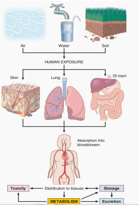
Biological Fate of Environmental Xenobiotics
Physiological Processing: Absorption, Distribution, Storage, and Excretion
Following barrier penetration, the chemical's internal pharmacological fate governs the anatomical location, onset latency, and clinical duration of tissue pathology:
- Distribution: The systemic arterial circulation conveys the dissolved or protein-bound xenobiotic to peripheral target tissues.
- Storage in Reservoir Tissues: Highly lipophilic agents accumulate within neutral adipocyte fat stores, while bone-seeking heavy metals substitute for calcium ions in mineralized osteoid, persisting for decades.
- Enzymatic Metabolism: Hepatic and extrahepatic enzyme systems chemically transform the parent compound, fundamentally modifying its polarity and biological reactivity.
- Excretion: Polar, hydrophilic metabolites are filtered and eliminated through renal urine, hepatic biliary excretion into feces, or pulmonary exhalation of volatile compounds.
Clinical Teaching Axiom: The organ suffering structural injury does not necessarily represent the anatomical port of entry. For instance, skin absorption of carbon tetrachloride produces severe centrilobular liver necrosis, while chronic oral lead ingestion damages the central nervous system and proximal renal tubules.
Biotransformation: Detoxification versus Bioactivation
Metabolism does not universally mitigate toxicity; enzymatic alteration represents a dual-edged biological mechanism:
| Metabolic Outcome | Biochemical Mechanism | Cellular Consequence |
|---|---|---|
| Detoxification | Conversion of a lipophilic or chemically active compound into an inert, water-soluble product | Facilitates safe renal or biliary elimination, preventing pathological cell injury |
| Bioactivation | Enzymatic processing converts an innocuous parent compound into an electrophilic, highly reactive intermediate | Direct covalent binding to cellular DNA, membrane phospholipid peroxidation, and enzyme inactivation |
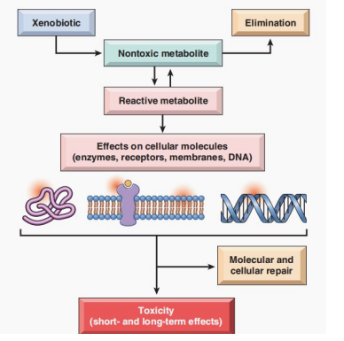
Pathways of Xenobiotic Detoxification and Bioactivation
Phase I and Phase II Reactions
Biotransformation is classified into two sequential, highly coordinated reaction phases:
- Phase I Functionalization: Chemical introduction or unmasking of functional groups (-OH, -NH2, -SH, -COOH) through oxidation, reduction, or hydrolysis. While this increases polarity, Phase I frequently yields unstable, highly toxic reactive intermediates.
- Phase II Conjugation: Covalent linkage of endogenous polar hydrophilic moieties to the parent compound or its Phase I metabolite. Major pathways include glucuronidation, sulfation, and glutathione (GSH) conjugation, typically neutralizing toxicity and ensuring prompt urinary or fecal excretion.
The Cytochrome P450 Enzyme System
The Cytochrome P450 (CYP) heme-protein superfamily, situated within the smooth endoplasmic reticulum (microsomal fraction) of hepatocytes and enterocytes, represents the principal catalytic driver of Phase I oxidation reactions.
While clearing innumerable xenobiotics, CYP metabolism can:
- Accidentally bioactivate non-toxic pro-toxins into cellular mutagens or destructive electrophiles.
- Generate dangerous reactive oxygen species (ROS) via uncoupled catalytic cycling, triggering secondary oxidative membrane injury.
- Exhibit wide interpersonal variability due to genetic polymorphisms (e.g., CYP2D6, CYP2E1) and drug-drug competitive inhibition or enzyme induction.
4. Environmental Air Pollution
Air pollution constitutes the dispersion of toxic gases, volatile mists, or microscopic suspended particulate matter within atmospheric or indoor environments. The human respiratory tree represents the primary target of injury due to direct contact, but systemic penetration frequently compromises cardiovascular, renal, and central nervous systems.
| Air Pollutant | Vulnerable Patient Groups | Clinical Pathological Effects |
|---|---|---|
| Ozone (O3) | Healthy children, exercising adults, athletes, and patients with asthma | Direct oxidative epithelial damage, upper airway inflammation, decreased FEV1, and asthma exacerbations |
| Nitrogen Dioxide (NO2) | Infants, children, and chronic asthmatic patients | Airway mucosal irritation, increased bronchial reactivity, and heightened vulnerability to respiratory infections |
| Sulfur Dioxide (SO2) | Patients with chronic bronchitis, emphysema, and bronchial asthma | Severe reflex bronchoconstriction, acid-induced mucosal injury, and increased cardiopulmonary mortality |
| Acid Aerosols | Children and individuals with chronic respiratory disease | Paralysis of mucociliary clearance, deep alveolar irritation, and increased emergency hospitalizations |
| Particulate Matter (PM) | Elderly adults, children, patients with coronary artery disease or COPD | Macrophage inflammasome activation, systemic pro-thrombotic inflammation, myocardial infarction, and excess mortality |
Detailed Outdoor Pollutants
- Ground-Level Ozone (O3): Forms at ground level when sunlight and atmospheric heat drive photochemical reactions between nitrogen oxides (NOx) and volatile organic compounds derived from vehicle emissions. It produces free radical-mediated lipid peroxidation of respiratory epithelial membranes.
Teaching distinction: Ground-level tropospheric ozone is a dangerous oxidant pollutant, whereas stratospheric ozone is beneficial, absorbing harmful solar ultraviolet (UV) radiation. - Nitrogen Dioxide (NO2): Produced by industrial combustion and vehicle engines; dissolves into respiratory secretions to generate nitric acid, inducing deep mucosal necrosis.
- Sulfur Dioxide (SO2) and Acid Aerosols: Emitted during combustion of sulfur-rich fossil fuels (coal, crude oil). In the atmosphere, it converts into sulfuric acid, disrupting epithelial defenses and stimulating sensory receptors to provoke acute bronchospasm.
- Particulate Matter (PM): Heterogeneous collection of microscopic airborne solids and liquid droplets, including carbonaceous soot, heavy metal fragments, and organic hydrocarbons. Particles under 10 μm (PM10) reach the bronchioles, while fine particulates under 2.5 μm (PM2.5) penetrate directly into terminal alveoli.
Alveolar macrophages phagocytose PM, triggering the NLRP3 inflammasome, releasing Interleukin-1 (IL-1) and TNF, and recruiting destructive neutrophilic infiltrates that accelerate chronic emphysematous tissue destruction.
5. Carbon Monoxide Poisoning
Carbon monoxide (CO) is an insidious, non-irritating, completely odorless, colorless, and tasteless gas formed during incomplete combustion of carbonaceous organic materials. Unlike other air contaminants that irritate the mucosal lining, carbon monoxide acts as an acute systemic asphyxiant.
Molecular Mechanisms of Carbon Monoxide Toxicity
- Extreme Hemoglobin Affinity: Carbon monoxide binds to the ferrous (Fe2+) iron of hemoglobin with roughly 200 times greater affinity than molecular oxygen, synthesizing carboxyhemoglobin (COHb) and completely abolishing oxygen transport across occupied binding sites.
- Haldane Allosteric Shift: The binding of CO to one heme site alters the quaternary structure of the hemoglobin tetramer, shifting the oxygen-hemoglobin dissociation curve steeply to the left. Consequently, oxygen remaining on neighboring heme subunits is held tightly and cannot be released to hypoxic peripheral tissues.
- Inhibition of Cellular Respiration: Dissolved carbon monoxide crosses cellular membranes and binds directly to the iron-copper center of mitochondrial cytochrome c oxidase (Complex IV), halting aerobic ATP generation.
Environmental Sources: Motor vehicle combustion exhaust, malfunctioning indoor kerosene heaters, gas-powered generators operated in enclosed spaces, indoor charcoal stoves ("sigiri"), building fires, and industrial furnaces.
Clinical Manifestations and Diagnostic Caveats
- Early/Mild Poisoning: Frontal headache, lightheadedness, nausea, progressive weakness, and cognitive confusion. Impaired judgment frequently prevents exposed individuals from escaping the lethal environment.
- Severe/Acute Toxicity: Loss of consciousness, generalized tonic-clonic convulsions, ventricular arrhythmias, myocardial ischemia, pulmonary edema, coma, and cardiorespiratory arrest.
- Diagnostic Trap: Standard non-invasive pulse oximeters cannot distinguish carboxyhemoglobin from oxyhemoglobin because both absorb light at 660 nm. A patient may present with fatal tissue anoxia while showing a deceptive "99% SpO2" reading. Confirmation demands blood co-oximetry to quantify exact carboxyhemoglobin percentages.
Pathological and Post-Mortem Findings
- Gross Tissue Appearance: Blood, musculature, and visceral organs exhibit a pathognomonic bright cherry-red coloration due to high concentrations of circulating carboxyhemoglobin.
- Fulminant Rapid Death: Individuals who die within minutes show minimal structural microscopic cellular changes because demise precedes reactive morphological adaptations.
- Prolonged Survival After Severe Anoxia: Patients maintained on life support following severe poisoning exhibit marked cerebral edema, petechial hemorrhages, laminar cortical neuronal necrosis, and bilateral, selective ischemic necrosis of the globus pallidus within the lenticular nucleus.
6. Indoor Air Pollution and Environmental Carcinogens
Enclosed domestic environments can accumulate high concentrations of airborne contaminants due to limited air exchange:
- Biomass and Organic Fuel Smoke: Millions of households globally burn wood, crop residues, dried dung, and charcoal in unvented indoor hearths for domestic cooking and heating. This creates dense exposures to irritating aldehydes, carbon monoxide, respirable soot, and polycyclic aromatic hydrocarbons (PAHs), predisposing to chronic obstructive pulmonary disease, recurrent lower respiratory tract infections in infants, and upper airway neoplasms.
- Radon Gas: A colorless, odorless, naturally occurring radioactive noble gas produced by the radioactive decay chain of uranium-238 in granite, soil, and rock. Radon seeps through foundation cracks, accumulating in basements. Inhaled alpha-emitting decay products deposit along bronchial epithelium, inflicting double-strand DNA fractures. Radon represents the leading environmental cause of lung carcinoma in non-smokers and exhibits a potent multiplicative synergistic risk in active cigarette smokers.
- Bioaerosols: Suspended airborne organic particulates comprising bacterial endotoxins, respiratory viruses, fungal/mold spores (e.g., Aspergillus, Stachybotrys), dust-mite antigens, and animal dander, triggering allergic rhinitis, hypersensitivity pneumonitis, and severe extrinsic asthma.
7. Toxic Heavy Metals as Environmental Pollutants
The transition elements and heavy metals lead, mercury, arsenic, and cadmium exert severe clinical toxicity by binding to structural nucleophilic sites, displacing essential physiological cations, and producing uninhibited oxidative damage.
1. Lead Poisoning (Plumbism)
Exposure Sources: Ingestion of flaking lead-based household paint, inhalation of contaminated demolition dust, contaminated municipal water delivered via lead-soldered plumbing, lead-acid battery manufacturing, non-ferrous foundry recycling, and traditional folk cosmetics/medicines.
Pediatric Vulnerability: Children absorb up to 50% of ingested lead (compared to roughly 10% in adults), possess an immature, hyper-permeable blood-brain barrier, display physiological exploratory hand-to-mouth behaviors (pica), and sustain irreversible neurocognitive deficits at trace blood concentrations. There is no established safe blood lead concentration in children.
Biochemical Mechanisms and Organ Pathologies of Lead
- Binding to Protein Sulfhydryl Groups: Inactivates essential enzymes throughout cellular metabolism.
- Calcium Cation Mimicry: Competes with Ca2+ in neurotransmitter release mechanisms and deposits into the mineral phase of skeleton. Over 80% to 90% of absorbed lead is sequestered in bones and teeth, serving as an endogenous reservoir with a half-life of 20 to 30 years.
- Inhibition of Heme Biosynthesis: Selectively blocks two critical enzymes in the heme synthetic pathway:
- δ-Aminolevulinic Acid Dehydratase (δ-ALAD): Catalyzes pyrrole ring formation. Inhibition leads to elevated blood levels of toxic δ-ALA.
- Ferrochelatase: Catalyzes the insertion of Fe2+ into protoporphyrin IX. Its blockade halts heme completion, driving high levels of free erythrocyte protoporphyrin (zinc protoporphyrin).
- Hematological Morphology: Produces a microcytic, hypochromic anemia. Peripheral blood smears demonstrate characteristic basophilic stippling within erythrocytes (representing abnormal aggregates of degraded ribosomal RNA caused by lead-induced inhibition of erythrocyte 5'-nucleotidase).
- Central and Peripheral Nervous Systems: In children, lead damages cerebellar Purkinje cells and cerebral cortical gray matter, causing cerebral edema, encephalopathy, ataxia, IQ deficits, and aggressive behavioral disorders. In adults, toxicity presents as a painless motor peripheral neuropathy characterized by bilateral extensor weakness—manifesting classically as wrist drop (radial nerve palsy) and foot drop (peroneal nerve palsy).
- Gastrointestinal Plumbism: Severe, intensely painful, poorly localized intestinal smooth muscle spasms designated as lead colic.
- Renal Tubulopathy: Acute exposure induces proximal tubular epithelial dysfunction with pathognomonic acid-fast intranuclear lead-protein inclusion bodies. Chronic plumbism leads to progressive tubulointerstitial fibrosis, nephrosclerosis, and secondary saturation gout ("saturnine gout").
- Bones and Gingiva: In growing children, lead interferes with the normal osteoclast resorption and remodeling of calcified provisional cartilage at the epiphyseal plates, producing dense, transverse radio-opaque bands on plain radiographs known as lead lines (reflecting hypermineralized, retained calcified cartilage rather than metallic lead). In the oral cavity, bacterial production of hydrogen sulfide reacts with lead in saliva to form a dark blue-black line along the gingival margin (Burton's line).
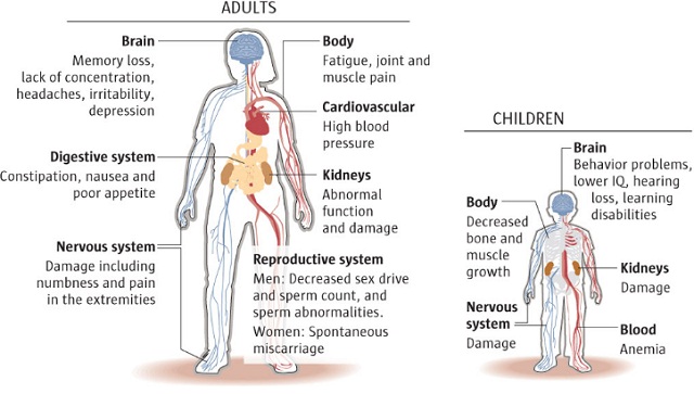
Systemic Pathological Features of Lead Poisoning
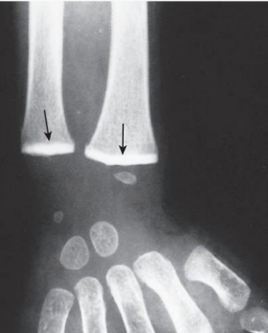
Dense Metaphyseal "Lead Lines" at the Distal Radius and Ulna
2. Mercury Poisoning (Hydrargyria)
Mercury presents in three physical and chemical configurations, each exhibiting divergent absorption profiles, biological affinities, and pathological consequences:
| Chemical State | Primary Exposure Source | Critical Target Organs & Pathology |
|---|---|---|
| Elemental Mercury (Hg0) | Inhalation of volatile mercury vapors in artisanal gold mining, dental amalgams, broken thermometers | Central nervous system and kidneys. Inhaled vapor crosses the blood-brain barrier causing intention tremors, gingivitis, and psychiatric irritability ("mad hatter syndrome" / erethism) |
| Inorganic Mercury Salts (e.g., Mercuric Chloride) | Ingestion of industrial chemical salts, older mercurial skin-lightening soaps, and laboratory reagents | Gastrointestinal mucosa and renal cortex. Direct chemical corrosiveness produces bloody mucosal ulceration, followed by acute tubular necrosis (ATN) and anuric renal failure |
| Organic Mercury (Methylmercury) | Bioaccumulation and biomagnification in the aquatic food web; consumption of large apex predatory marine fish | Central nervous system, particularly the developing fetal brain. Lipophilic methylmercury readily crosses the placenta and blood-brain barrier, causing severe microcephaly, cerebral palsy, visual field constriction, and sensory ataxia (Minamata disease) |
3. Arsenic Poisoning
Environmental Exposure: Arsenic leaches into alluvial drinking water wells from mineral-rich ground aquifers (a major crisis in South Asia), or contaminates populations via mining runoff, wood preservatives, and non-ferrous metal smelting.
Cellular Mechanisms: Trivalent inorganic arsenic (arsenite, As3+) binds sulfhydryl groups, disrupting pyruvate dehydrogenase and halting the Krebs cycle, while pentavalent arsenate (As5+) acts as a phosphate analog, uncoupling mitochondrial oxidative phosphorylation and depleting ATP pools.
Clinical & Pathological Spectrum:
- Acute Arsenic Toxicity: Catastrophic hemorrhagic gastroenteritis ("rice-water" diarrhea with shredding mucosal sheets), generalized vascular collapse, QTc prolongation, torsades de pointes, and circulatory shock.
- Chronic Cutaneous Plaque Formation: Symmetrical palmar and plantar hyperkeratosis (thickened, nodular, verrucous lesions) accompanied by diffuse mottled "raindrop" hyperpigmentation of the trunk.
- Malignant Transformation: Arsenic is a potent human carcinogen, uniquely predisposing to multiple cutaneous basal cell carcinomas, squamous cell carcinomas, pulmonary squamous carcinomas, and hepatic angiosarcomas.
8. Industrial and Agricultural Exposures
Chemical exposures in occupational settings occur primarily via transdermal absorption, direct inhalation of aerosols, or inadvertent ingestion, manifesting across numerous anatomical systems:
| Organ / Physiological System | Specific Pathological Effect | Primary Industrial / Chemical Toxicant |
|---|---|---|
| Cardiovascular System | Arrhythmias, myocardial infarction, coronary vasoconstriction | Carbon monoxide, lead, organic fluorocarbons, cadmium |
| Respiratory Tract | Sinonasal carcinoma Bronchogenic carcinoma Pneumoconioses (pulmonary fibrosis) Hypersensitivity lung disease |
Wood dust, isopropyl alcohol Asbestos, radon, arsenic, chromium, nickel Crystalline silica, coal worker's dust, asbestos Beryllium, toluene diisocyanate |
| Nervous System | Peripheral sensorimotor neuropathy Ataxic gait disturbance Acute central depression / coma |
Lead, arsenic, acrylamide, carbon disulfide, n-hexane Chlordane, methylmercury, toluene Halogenated hydrocarbons, industrial alcohols |
| Urinary System | Acute tubular necrosis Transitional cell urothelial carcinoma |
Mercuric chloride, ethylene glycol, carbon tetrachloride 2-Naphthylamine, 4-aminobiphenyl, benzidine dyes |
| Hematopoietic System | Aplastic anemia and Acute Myeloid Leukemia (AML) | Benzene, ionizing radiation |
| Hepatic Parenchyma | Centrilobular necrosis & fatty steatosis Hepatic Angiosarcoma |
Carbon tetrachloride (CCl4), chloroform Monomeric vinyl chloride, Thorotrast, arsenic |
| Integumentary System | Chloracne, follicular cysts, cutaneous hyperpigmentation | Polychlorinated biphenyls (PCBs), 2,3,7,8-TCDD (dioxin) |
Detailed Industrial Reagents and Pesticides
- Organic Solvents: Chloroform, carbon tetrachloride, and hexane are volatile lipid-clearing agents used in commercial degreasing and dry cleaning.
- Carbon Tetrachloride (CCl4): In the liver, microsomal CYP2E1 metabolizes CCl4 via homolytic cleavage into the highly reactive trichloromethyl free radical (•CCl3). This radical initiates rapid auto-catalytic lipid peroxidation of endoplasmic reticulum membranes, halting apolipoprotein B-100 synthesis within 30 minutes. Triglycerides cannot be exported as VLDL, resulting in profound macrovesicular hepatic steatosis, followed by severe ballooning degeneration and massive centrilobular necrosis.
- Benzene: Found in light oil distillates, petrochemicals, and rubber fabrication. Hepatic biotransformation yields toxic quinone metabolites that migrate into bone marrow stroma, damaging hematopoietic stem cells to cause marrow aplasia (aplastic anemia) and cytogenetic translocations driving acute myeloid leukemia (AML).
- Polycyclic Aromatic Hydrocarbons (PAHs): Benzo[a]pyrene and related compounds generated by incomplete combustion of coal, furnace tar, and industrial soot. In the liver, CYP1A1 oxidizes PAHs into electrophilic epoxides, which form covalent adducts with guanine bases in DNA, inactivating tumor suppressors such as TP53. Historically, Sir Percivall Pott identified PAHs in soot as the etiology of scrotal squamous cell carcinomas in chimney sweeps.
- Organochlorine Compounds: Halogenated agricultural insecticides including DDT (dichlorodiphenyltrichloroethane), lindane, aldrin, and industrial polychlorinated biphenyls (PCBs) and dioxins (TCDD). These stable compounds bioaccumulate in adipose tissue and aquatic food chains. Acute toxic exposure produces tremors and convulsions. Chronic exposure triggers chloracne—characterized by dense, disfiguring facial comedones, keratinizing follicular cysts, and hyperpigmentation—along with endocrine disruption via estrogen receptor antagonism or mimicry.
- Crucial Pesticide Distinction: Organochlorines must not be conflated with organophosphate insecticides (such as parathion, malathion, and chlorpyrifos). Organophosphates are non-chlorinated compounds that irreversibly phosphorylate and inactivate the enzyme acetylcholinesterase (AChE) at synaptic junctions, producing acute cholinergic toxidromes (salivation, lacrimation, urination, defecation, bronchospasm, and muscular fasciculations).
- Bisphenol A (BPA): An organic synthetic compound polymerized to produce rigid polycarbonate food containers, beverage bottles, and epoxy resins lining metal cans. It leaches into dietary products upon heating and functions as a synthetic xenoestrogen, binding nuclear estrogen receptors and interfering with endocrine and reproductive physiology.
- Vinyl Chloride: Monomeric gas polymerized in industry to yield polyvinyl chloride (PVC) plastics. Chronic occupational inhalation exhibits an absolute pathological specificity for angiosarcoma of the liver, a rare, aggressive malignant neoplasm arising from endothelial cells lining the hepatic sinusoids (distinct from hepatocellular carcinoma).
- Mineral Dusts and Pneumoconioses: Chronic inhalation of inorganic mineral dusts:
- Coal Workers' Pneumoconiosis: Ranges from simple anthracosis (asymptomatic soot accumulation along peribronchial lymphatics) to progressive massive fibrosis (dense black collagenous nodules with ischemic cavitation).
- Silicosis: Inhalation of crystalline silicon dioxide (quartz) in quarrying and sandblasting. Phagocytosed silica crystals rupture macrophage phagolysosomes, triggering severe inflammasome activation and concentric "whorled" hyalinized fibrotic nodules that increase susceptibility to pulmonary tuberculosis.
- Asbestosis: Serpentines and amphiboles provoke diffuse interstitial pulmonary fibrosis, pleural plaques, bronchogenic carcinoma, and malignant pleural mesothelioma.
9. Pathology of Tobacco Consumption
Tobacco usage remains the leading preventable cause of human morbidity and premature mortality worldwide. Tobacco smoke is a complex heterogeneous aerosol containing over 4,000 individual chemical compounds, including gas-phase irritants, cellular asphyxiants, and proven chemical carcinogens.
| Smoke Component | Primary Biological Mechanism | Major Pathological Disease Association |
|---|---|---|
| Nicotine | Crosses the blood-brain barrier within seconds; binds central nicotinic acetylcholine receptors (nAChR), stimulating dopamine release in reward pathways; stimulates autonomic ganglia to release systemic catecholamines | Psychological and physical dependence; acute tachycardia, peripheral vasoconstriction, elevated blood pressure, and increased myocardial oxygen demand |
| Polycyclic Aromatic Hydrocarbons (PAHs) | Metabolically converted by host CYP enzymes into reactive diol epoxides that form covalent DNA adducts and induce G-to-T transversions in TP53 and KRAS | Bronchogenic squamous cell and adenocarcinoma, laryngeal carcinoma |
| Nitrosamines (NNK, NNN) | Tobacco-specific nitrosamines produced during leaf curing; induce point mutations in bronchial and pancreatic progenitor cells | Adenocarcinoma of the lung, pancreatic carcinoma |
| Aromatic Amines (e.g., 2-Naphthylamine) | Absorbed into circulation, conjugated in the liver, filtered into urine, and cleaved by acidic urinary enzymes into active mutagens | Transitional cell (urothelial) carcinoma of the urinary bladder |
| Carbon Monoxide | Synthesizes carboxyhemoglobin, reducing oxygen carrying capacity and impairing peripheral oxygen unloading | Subclinical tissue hypoxia, vascular endothelial injury, accelerated atherogenesis, and intrauterine growth restriction |
| Formaldehyde & Oxides of Nitrogen | Direct chemical toxicity to airway cilia, causing widespread ciliostasis and mucosal epithelial death | Impaired mucociliary clearance, chronic mucosal inflammation, and recurrent lower respiratory tract infections |
| Tar | Particulate residue concentrating thousands of condensed organic carcinogens and mutagens | Extensive mucosal staining, local contact mutagenesis along the aerodigestive tract |
Chronic Respiratory Diseases: Bronchitis and Emphysema
1. Chronic Bronchitis: Defined clinically as persistent cough with sputum production for at least 3 consecutive months in 2 consecutive years. Repeated smoke inhalation causes hyperplasia of bronchial submucosal glands, marked proliferation of surface goblet cells, and loss of ciliary function. Mucus plugs lodge within bronchioles, accompanied by chronic lymphocytic infiltration and peribronchiolar fibrosis.
2. Emphysema: Characterized by permanent, abnormal enlargement of airspaces distal to the terminal bronchioles, accompanied by destruction of alveolar walls without gross fibrosis. Cigarette smoke recruits alveolar macrophages and neutrophils, which release excess proteolytic enzymes, predominantly neutrophil elastase. Simultaneously, oxidants in tobacco smoke inactivate endogenous α1-antitrypsin. The resulting protease-antiprotease imbalance leads to enzymatic breakdown of alveolar elastin, loss of pulmonary elastic recoil, and expiratory airway collapse with severe air trapping.
Malignant Transformations Induced by Smoking
Repeated exposure of normal respiratory pseudo-stratified ciliated columnar epithelium to carcinogenic smoke constituents induces an orderly progression of morphological adaptations:
Normal Respiratory Epithelium → Squamous Metaplasia → Squamous Dysplasia → Carcinoma in Situ → Invasive Bronchogenic Carcinoma
Tobacco smoke directly provokes malignancies beyond the respiratory tree, including carcinomas of the oral cavity, pharynx, larynx, esophagus, pancreas, cervix, and urinary bladder.
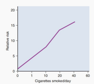
Impact of Cigarette Consumption on Lung Cancer Relative Risk
Quantification of Exposure: Pack-Years
Cumulative lifetime smoking is standardized using the clinical metric Pack-Years:
Pack-Years = (Packs Smoked Per Day) × (Years of Active Smoking)
- Example 1: Smoking 20 cigarettes (1 pack) daily for 20 years = 1 × 20 = 20 pack-years.
- Example 2: Smoking 10 cigarettes (0.5 pack) daily for 20 years = 0.5 × 20 = 10 pack-years.
Clinical Caution: The pack-year formula quantifies aggregate burden for screening criteria (e.g., low-dose chest CT), but there is no safe threshold of tobacco exposure.
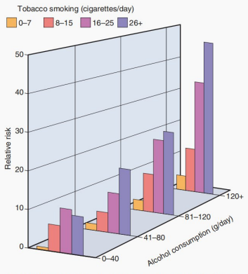
Synergistic Carcinogenesis of Concurrent Tobacco and Alcohol Exposure
Synergistic Interactions and Cardiovascular Consequences
- Asbestos and Tobacco Synergism: Asbestos exposure alone elevates lung cancer risk approximately 5-fold; cigarette smoking alone raises it 10-fold. Concomitant exposure acts multiplicatively, driving lung cancer risk up to 50-fold.
Crucial Distinction: Tobacco smoking synergizes with asbestos to multiply the incidence of bronchogenic carcinoma, but smoking is not an etiological driver of malignant pleural mesothelioma. - Cardiovascular Pathology: Smoking drives atherogenesis by inflicting direct endothelial cell injury, promoting circulating LDL oxidation, triggering chronic vascular inflammation, and stimulating platelet aggregation via thromboxane release. Nicotine accelerates myocardial workload via adrenergic stimulation, while carbon monoxide reduces oxygen delivery, drastically elevating risks of acute myocardial infarction, sudden cardiac death, ischemic stroke, and peripheral arterial disease.
- Obstetrical Complications: Maternal smoking and passive smoke exposure during gestation cause fetal hypoxia secondary to carboxyhemoglobin formation and nicotine-mediated placental vasoconstriction. This leads to fetal growth restriction (IUGR), premature labor, placental abruption, low birth weight, and sudden infant death syndrome (SIDS).
- Passive (Secondhand) Smoke: Environmental tobacco smoke exhaled by smokers or released from smoldering tips causes coronary heart disease, lung cancer, and exacerbated pediatric asthma in non-smokers. The WHO affirms there is no safe lower limit of passive smoke exposure.
- Benefits of Cessation: Cardiovascular and thrombotic risks decline rapidly within years of quitting. However, destructive architectural changes of panacinar or centriacinar emphysema are irreversible, and lung cancer risk remains elevated above baseline non-smokers for decades.
10. Pathology of Alcohol (Ethanol) Consumption
Ethanol is a small, water- and lipid-soluble aliphatic alcohol rapidly absorbed intact from the stomach (20%) and small intestine (80%), rapidly distributing throughout total body water. Over 90% to 95% of absorbed ethanol is oxidized within hepatocytes, with less than 5% to 10% excreted unchanged through expired alveolar air, urine, and sweat.
Hepatic Biochemical Pathways of Ethanol Oxidation
The core oxidative pathway converts Ethanol → Acetaldehyde → Acetate via three enzymatic cascades:
1. Alcohol Dehydrogenase (ADH) Pathway
The principal hepatic pathway located within the cytosol of hepatocytes. ADH oxidizes ethanol to acetaldehyde while reducing oxidized nicotinamide adenine dinucleotide (NAD+) to its reduced form (NADH):
Ethanol + NAD+ → Acetaldehyde + NADH + H+
This reaction shifts the hepatic redox potential, yielding an elevated NADH:NAD+ ratio that disrupts multiple intermediary metabolic pathways in the liver.
2. Microsomal Ethanol-Oxidizing System (MEOS)
Situated within the smooth endoplasmic reticulum, utilizing the cytochrome P450 isozyme CYP2E1. It plays a minor role at low physiological alcohol levels but becomes prominently induced during chronic heavy drinking (elevating metabolic clearance rates by several-fold).
Consequences of CYP2E1 induction include:
- Generation of damaging reactive oxygen species (superoxide, hydroxyl radicals), precipitating membrane lipid peroxidation.
- Competitive inhibition or accelerated toxic bioactivation of other co-ingested xenobiotics (e.g., converting therapeutic doses of acetaminophen into the hepatotoxin NAPQI).
3. Peroxisomal Catalase Pathway
A minor alternative mechanism located within peroxisomes capable of oxidizing ethanol to acetaldehyde using endogenous hydrogen peroxide (H2O2). It accounts for less than 5% of hepatic ethanol metabolism under standard physiological conditions.
Acetaldehyde Dehydrogenase (ALDH)
Acetaldehyde produced by the above pathways is rapidly transported into hepatocyte mitochondria, where mitochondrial aldehyde dehydrogenase (ALDH2) oxidizes it into acetate, also generating NADH:
Acetaldehyde + NAD+ → Acetate + NADH + H+
Acetate is released into peripheral circulation and converted into acetyl-CoA for the citric acid cycle. Individuals possessing genetic polymorphisms that reduce ALDH2 activity (common in East Asian populations) experience rapid toxic accumulation of acetaldehyde following alcohol consumption, precipitating facial flushing, nausea, tachycardia, and severe diaphoresis.
Pathological Consequences of the Elevated NADH:NAD+ Ratio
The massive excess of intracellular NADH generated by ADH and ALDH disrupts hepatic metabolism, explaining why steatosis develops prior to the onset of inflammation:
- Inhibition of Mitochondrial β-Oxidation: High NADH inhibits fatty acid oxidation, forcing hepatocytes to esterify fatty acids into triglycerides.
- Impaired Gluconeogenesis: High NADH drives the equilibrium of the lactate-pyruvate reaction toward lactate, depleting pyruvate required for gluconeogenesis and precipitating profound hypoglycemia (especially dangerous in fasting or malnourished individuals) alongside lactic acidosis.
- Promotion of Hepatic Steatosis: Excess dihydroxyacetone phosphate is converted to α-glycerophosphate, accelerating triglyceride assembly and intracellular lipid accumulation.
Morphological Stages of Alcoholic Liver Disease
- Hepatic Steatosis (Fatty Liver): Reversible accumulation of neutral lipid droplets within the cytoplasm of hepatocytes. Beginning as centrilobular microvesicular droplets, fat coalesces into massive macrovesicular globules that displace the nucleus to the periphery. Grossly, the liver is enlarged (up to 4–6 kg), soft, greasy, and bright yellow. It is entirely reversible upon alcohol cessation.
- Alcohol-Associated Hepatitis (Steatohepatitis): Characterized by hepatocyte injury and acute parenchymal inflammation:
- Hepatocyte Ballooning: Hydropic swelling and clarification of cytoplasm.
- Mallory-Denk Bodies: Dense, ropy, eosinophilic cytoplasmic aggregates of damaged, ubiquitinated intermediate cytokeratin filaments (cytokeratins 8 and 18). While characteristic of alcoholic hepatitis, they also appear in non-alcoholic steatohepatitis (NASH) and Wilson disease.
- Neutrophilic Infiltration: Polymorphonuclear neutrophils actively surround and attack degenerating ballooned hepatocytes.
- Pericellular ("Chicken-Wire") Fibrosis: Subsinusoidal fibrosis around the central vein (centrilobular vein) and extending along individual hepatocytes.
- Alcoholic Cirrhosis: The terminal, largely irreversible stage of chronic alcoholic injury. Characterized by diffuse hepatocellular death, bridging dense fibrous septa, and parenchymal regenerative nodules (classically micronodular, <3 mm, transitioning later to mixed macronodular patterns). Consequences include portal hypertension, ascites, splenomegaly, and liver failure.
Extrahepatic Pathological Manifestations of Alcoholism
- Gastrointestinal and Pancreatic: Acute erosive gastritis, shallow mucosal ulcerations, and acute necrotizing or chronic calcifying pancreatitis. Severe retching induces mechanical longitudinal mucosal lacerations at the gastroesophageal junction (Mallory-Weiss tears), while portal hypertension drives massive dilation of collateral submucosal veins (esophageal varices), which rupture with catastrophic hemorrhage.
- Central Nervous System & Nutritional Deficiencies: Ethanol acts as a GABAA-potentiating neurodepressant. Secondary dietary neglect leads to severe thiamine (Vitamin B1) deficiency, resulting in:
- Wernicke Encephalopathy: Reversible acute triad of confusion, gait ataxia, and bilateral ophthalmoplegia/nystagmus caused by hemorrhagic necrosis of the mammillary bodies.
- Korsakoff Syndrome: Irreversible chronic amnestic confabulatory syndrome with severe anterograde memory impairment.
- Cardiovascular Pathology: Chronic ethanol excess causes dilated cardiomyopathy ("alcoholic cardiomyopathy") featuring ventricular hypertrophy, four-chamber dilation, impaired systolic contractility, and secondary congestive heart failure. Binge drinking triggers acute atrial and ventricular arrhythmias ("holiday heart syndrome").
- Carcinogenesis: Alcohol is categorized as an established Group 1 human carcinogen by the IARC. Acetaldehyde-DNA adducts, ROS generation, and mucosal solvent actions drive cancers of the oral cavity, pharynx, larynx, esophagus, liver (hepatocellular carcinoma via cirrhosis), colorectum, and female breast (the link between alcohol and breast carcinoma is an established pathological association, not merely a hypothetical risk).
- Fetal Alcohol Spectrum Disorders (FASD): Ethanol readily traverses the placental syncytiotrophoblast, disrupting fetal neurogenesis and neural crest migration. Fetal Alcohol Syndrome (FAS) is characterized by microcephaly, intrauterine growth restriction, cognitive deficits, and diagnostic craniofacial dysmorphism: shortened palpebral fissures, smooth flat philtrum, and a thin vermilion border of the upper lip. There is no known safe threshold of alcohol intake during pregnancy.
11. Adverse Drug Reactions (ADRs) and Therapeutic Drug Injury
An adverse drug reaction (ADR) is formally defined as an unwanted, noxious, and unintended response to a pharmaceutical compound administered at standard therapeutic doses for prophylaxis, diagnosis, or treatment. ADRs must be strictly distinguished from accidental or deliberate toxic overdoses.
| Mechanistic Category | Underlying Pathophysiological Process | Classic Pharmacological Example |
|---|---|---|
| Exaggerated Pharmacological Effect | The primary therapeutic pharmacodynamic action operates excessively or targets non-diseased tissue | Spontaneous intracranial hemorrhage secondary to therapeutic anticoagulant administration (e.g., Warfarin, DOACs) |
| Direct Cytotoxicity | The parent drug or an active metabolic breakdown product directly induces cell necrosis or apoptosis | Centrilobular hepatic necrosis precipitated by reactive NAPQI accumulation in acetaminophen overdose |
| Immune-Mediated (Hypersensitivity) | The drug acts as a hapten or direct antigen, eliciting Type I, II, III, or IV immunopathological reactions | IgE-mediated systemic anaphylactic shock following penicillin exposure; drug-induced immune hemolytic anemia |
| Idiosyncratic / Host Susceptibility | Unpredictable reactions tied to specific unmapped host genetic polymorphisms or HLA variations | Severe cutaneous Stevens-Johnson syndrome precipitated by carbamazepine or abacavir in HLA-typed cohorts |
| Long-Term Biological Remodeling | Sustained receptor engagement alters tissue architecture, promoting metaplasia or cellular proliferation | Endometrial hyperplasia and endometrial carcinoma arising from unopposed systemic estrogen therapy |
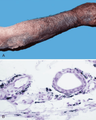
Minocycline-Induced Dermal Pigmentation
Major Organ Patterns of Drug-Induced Pathology
- Bone Marrow and Hematopoietic Elements: Granulocytopenia, thrombocytopenia, and pure red cell aplasia. Anti-neoplastic agents, chloramphenicol, and gold salts can precipitate irreversible aplastic anemia (marrow failure with pancytopenia), whereas penicillins and methyldopa induce secondary autoimmune hemolytic anemia.
- Cutaneous Eruptions: Encompasses urticaria, pruritic maculopapular morbilliform rashes, fixed drug eruptions, erythema multiforme, and life-threatening Stevens-Johnson Syndrome (SJS) and Toxic Epidermal Necrolysis (TEN) featuring extensive mucosal detachments.
- Renal Parenchymal Injury: Aminoglycosides and amphotericin B induce direct acute tubular injury; non-steroidal anti-inflammatory drugs (NSAIDs) trigger acute interstitial nephritis and papillary necrosis; penicillamine provokes membranous nephropathy.
- Pulmonary Fibrosis: Bleomycin and busulfan generate free radicals that trigger chronic alveolar pneumonitis and fatal subpleural interstitial fibrosis; nitrofurantoin induces acute eosinophilic or chronic pneumonitis.
- Cardiac Toxicity: Doxorubicin (Adriamycin) generates intracellular hydroxyl radicals, inducing myofibrillar drop-out, vacuolization, and dilated cardiomyopathy with congestive heart failure.
Pathology of Exogenous Hormonal Therapies
Hormone Replacement Therapy (HRT) and Oral Contraceptives (OCPs)
Menopausal Hormone Therapy (MHT):
- Endometrial Carcinoma: In women with an intact uterus, unopposed systemic estrogen administration continuously stimulates endometrial epithelial proliferation, markedly elevating the risk of endometrial hyperplasia and adenocarcinoma. Co-administration of a progestin counteracts this proliferative drive. Following surgical hysterectomy, progestin addition is unneeded.
- Breast Carcinoma: Combined estrogen-progestin regimens slightly increase the incidence of invasive breast adenocarcinoma over baseline.
- Thromboembolism & Stroke: Oral estrogens increase hepatic synthesis of coagulation factors (II, VII, IX, X) and suppress antithrombin III, increasing deep vein thrombosis (DVT), pulmonary embolism (PE), and ischemic stroke.
Oral Contraceptive Pills (OCPs) and Malignancy Risks:
- Endometrial and Ovarian Cancers: OCPs confer a robust, lasting protective reduction in both endometrial and epithelial ovarian cancers by suppressing ovulation and inducing cyclic progestational endometrial differentiation.
- Cervical Carcinoma: Prolonged use (>5 years) slightly elevates the incidence of cervical cancer in women with persistent oncogenic Human Papillomavirus (HPV) infection.
- Hepatic Adenoma: Rare benign vascular tumor of hepatocytes associated with prolonged high-dose oral contraceptive use, carrying a risk of spontaneous rupture and fatal intra-abdominal hemorrhage.
Specific Analgesic Toxicity: Acetaminophen and Aspirin
Acetaminophen (Paracetamol) Hepatotoxicity
At therapeutic doses, 95% of acetaminophen is safely conjugated in the liver with glucuronide (60%) or sulfate (35%) and excreted in urine. Roughly 5% is metabolized by CYP2E1 into the electrophilic toxic metabolite N-acetyl-p-benzoquinone imine (NAPQI), which is immediately neutralized by conjugation with intracellular glutathione (GSH).
Toxicity Mechanism: In acute overdose, glucuronidation and sulfation pathways are saturated. Increased flux through CYP2E1 rapidly depletes hepatocellular glutathione pools. Once GSH drops below 30% of normal, free NAPQI binds covalently to mitochondrial and plasma membrane proteins, precipitating mitochondrial permeability transition, calcium release, ATP exhaustion, and catastrophic centrilobular (Zone 3) hepatic necrosis.
Antidotal Therapy: N-acetylcysteine (NAC) provides cysteine as an essential substrate to replenish hepatic glutathione and directly conjugates unbound NAPQI if administered promptly within 8–12 hours of ingestion.
Aspirin (Acetylsalicylic Acid) Toxicity
Therapeutic Mechanisms & GI Complications: Aspirin irreversibly acetylates and inactivates cyclooxygenase (COX-1 and COX-2), blocking synthesis of thromboxane A2 (suppressing platelet aggregation) and protective mucosal prostaglandins (PGE2, PGI2). Loss of mucosal prostaglandins eliminates bicarbonate secretion and reduces microvascular perfusion, leading to petechiae, erosive gastritis, peptic ulceration, and gastrointestinal bleeding.
Salicylism (Chronic Overdose): Features tinnitus, high-frequency hearing loss, dizziness, nausea, and disorientation.
Acute Salicylate Overdose Acid-Base Dynamics:
- Primary Respiratory Alkalosis: Salicylates directly stimulate the medullary respiratory center, inducing severe tachypnea and respiratory alkalosis (decreased PaCO2).
- Primary High-Anion-Gap Metabolic Acidosis: Salicylates uncouple oxidative phosphorylation, driving anaerobic glycolysis, accumulation of lactic acid, and accumulation of pyruvic acid and ketoacids.
- The classic diagnostic presentation in adult toxicity is a mixed respiratory alkalosis and high-anion-gap metabolic acidosis.
12. Pathology of Non-Therapeutic Substances (Drugs of Abuse)
Non-therapeutic recreational substances exert severe tissue damage through receptor hyperstimulation, direct cardiotoxicity, adulterants, toxic overdoses, and secondary complications related to injection practices.
| Pharmacological Class | Specific Molecular Target | Representative Agents | Dominant Clinical Pathological Toxicity |
|---|---|---|---|
| Opioid Narcotics | μ-opioid receptor (agonist) | Heroin, morphine, fentanyl, oxycodone, hydromorphone | Profound medullary respiratory depression, non-cardiogenic pulmonary edema, pinpoint miosis, fatal anoxic brain injury |
| Sedative-Hypnotics | GABAA receptor (allosteric agonist) | Barbiturates, ethanol, benzodiazepines, methaqualone | Synergistic CNS depression, loss of protective airway reflexes, respiratory arrest, hypothermia |
| Psychomotor Stimulants | Dopamine, norepinephrine, and serotonin reuptake transporters (inhibitor/antagonist) | Cocaine, methamphetamine, amphetamine, MDMA ("Ecstasy") | Severe coronary vasospasm, myocardial infarction, hyperthermia, malignant ventricular arrhythmias, intracranial hemorrhage |
| Dissociative Anesthetics | NMDA glutamate receptor channel (antagonist) | Phencyclidine (PCP), ketamine | Rotatory nystagmus, extreme agitation, analgesia leading to traumatic injury, rhabdomyolysis |
| Cannabinoids | CB1 and CB2 cannabinoid receptors (agonist) | Marijuana, hashish, Δ9-tetrahydrocannabinol (THC) | Acute tachycardia, impaired sensorimotor coordination, cognitive fragmentation, airway irritation |
| Hallucinogens | 5-HT2A serotonin receptors (agonist) | Lysergic acid diethylamide (LSD), psilocybin, mescaline | Visual illusions, acute panic reactions ("bad trips"), sympathomimetic stimulation |
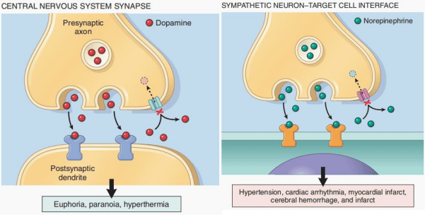
Cellular Mechanism of Cocaine Toxicity at Central and Peripheral Synapses
Detailed Pathology of Cocaine
Cocaine is a water-soluble alkaloid extracted from the leaves of Erythroxylon coca. It blocks the presynaptic reuptake of dopamine, serotonin, and norepinephrine:
- Cardiovascular Myocardial Infarction: Cocaine precipitates acute myocardial infarction in young patients with entirely normal coronary arteries through three simultaneous mechanisms:
- Intense Coronary Vasospasm: Excessive α1-adrenergic receptor activation on coronary smooth muscle constriction.
- Massive Increase in Myocardial Oxygen Demand: Driven by β1-mediated tachycardia and enhanced cardiac contractility.
- Platelet Aggregation and Intravascular Thrombosis: Promotes platelet adherence and localized release of thromboxane A2.
- Cardiac Arrhythmias and Sudden Death: Cocaine blocks cardiac voltage-gated myocyte sodium (Na+) channels (exerting a local anesthetic/quinidine-like effect), prolonging QRS complexes and precipitating fatal ventricular tachycardia or fibrillation.
- Central Nervous System: High fever (hyperthermia due to continuous muscle activity and impaired heat dissipation), intracranial berry aneurysm rupture, ischemic strokes, seizures, and agitation-induced rhabdomyolysis.
- Local Ischemic Perforation: Chronic intranasal snorting induces sustained mucosal and periosteal ischemia, culminating in ischemic necrosis and complete perforation of the cartilaginous nasal septum.
Detailed Pathology of Heroin and Opiates
Heroin (diacetylmorphine) is a semi-synthetic opioid metabolized rapidly into 6-monoacetylmorphine and morphine. It acts upon central and peripheral μ-opioid receptors to produce profound analgesia, euphoria, and drowsiness:
- Lethal Overdose Triad: Manifests with:
- Profound Central Respiratory Depression: Blunts the responsiveness of medullary respiratory centers to arterial carbon dioxide tension (PaCO2).
- Comatose Stupor / Depressed Consciousness.
- Pinpoint Pupils (Symmetrical Miosis): Driven by stimulation of the parasympathetic Edinger-Westphal nucleus via the oculomotor nerve.
Pharmacological Reversal: Prompt administration of Naloxone (Narcan), an antagonist at the μ-opioid receptor, reverses respiratory arrest.
- Non-Cardiogenic Pulmonary Edema: Capillary leakage produces lungs heavy with protein-rich fluid, alveolar hemorrhage, and pink froth pouring from tracheobronchial airways.
- Secondary Complications of Intravenous Drug Use:
- Infective Endocarditis: High frequency of right-sided valvular involvement (tricuspid valve) classically caused by Staphylococcus aureus, seeded by skin flora entering veins during unsterile injection. Septic emboli seed the pulmonary circulation, causing wedge-shaped septic pulmonary infarcts and lung abscesses.
- Foreign Body Granulomas: Insoluble cutting agents and adulterants (talc, cellulose, cornstarch) lodge within pulmonary microvessels, eliciting foreign-body giant cell granulomas visible under polarized light microscopy.
- Cutaneous & Vascular Scars: Repeated needle punctures produce fibrous "track marks", venous thrombosis, thrombophlebitis, and cutaneous cellulitis.
- Renal Amyloidosis & Glomerulopathy: Chronic cutaneous skin popping with recurrent abscesses drives systemic inflammation and serum amyloid A (SAA) synthesis, precipitating secondary AA amyloidosis and focal segmental glomerulosclerosis (FSGS) with nephrotic syndrome.
13. Mechanical Trauma and Forensic Pathology of Wounds
Mechanical trauma arises when physical forces applied to the body overcome tissue tensile strength, elasticity, and anatomical resilience. Patterns are classified based on the nature of the striking implement and tissue response:
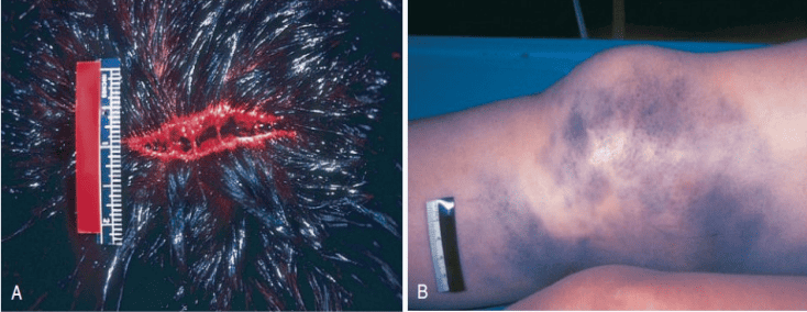
Gross Morphology of Laceration versus Contusion
Detailed Morphological Classification of Mechanical Wounds
| Wound Classification | Primary Physical Mechanism | Defining Gross Morphological Characteristics |
|---|---|---|
| Abrasion | Friction and tangential shearing forces scraping off the superficial epithelial layers | Epithelial denudation confined to epidermis or superficial dermal papillae; leaves a serous crust without true scarring; preserves dermal architecture |
| Contusion (Bruise) | Blunt mechanical impact compressing and rupturing underlying dermal and subcutaneous microvessels without disrupting epidermal continuity | Extravasation of red blood cells into interstitial soft tissues under intact skin. Macrophages phagocytose damaged erythrocytes, metabolizing red-blue hemoglobin → green biliverdin → yellow-brown hemosiderin.
Forensic Caveat: Discoloration alone cannot determine the precise age of a contusion. |
| Laceration | Blunt force impact (crushing, stretching, or shearing) tearing and splitting skin and underlying tissues over bone | Ragged, irregular, crushed margins with abraded edges; presence of intact tissue bridges (strands of untorn fibrous tissue, nerves, and vessels bridging the base of the wound gap) |
| Incised Wound (Cut) | Sharp-edged cutting implement (scalpel, broken glass, knife) drawn across tissues | Clean, sharply demarcated, linear margins without crushing or surrounding abrasion; absolute absence of tissue bridging; surface length exceeds wound depth |
| Puncture / Stab Wound | Narrow, pointed object thrust deeply into anatomical tissue | Small, deceptively benign external skin opening; depth of penetration significantly exceeds surface length; high risk of visceral organ perforation and tetanus entry |
| Gunshot Wound (GSW) | High-velocity kinetic energy transfer from ballistic projectiles | Contact wounds exhibit soot deposition, carbon monoxide-hemoglobin cherry discoloration, and star-shaped (stellate) tearing over bone. Distant entrance wounds show circular defects with a peripheral marginal abrasion ring; exit wounds are irregular, larger, and lack soot or abrasion collars |
14. Thermal Injuries: Burns, Hyperthermia, and Hypothermia
Thermal pathology encompasses destructive changes produced when excess heat or cold directly coagulates living tissues or overwhelms central homeostatic thermoregulation.
Thermal Burns: Classification of Depth and Tissue Impact
Clinical prognosis depends on depth of injury, total body surface area (TBSA) calculated by the Rule of Nines, presence of inhalation injury, and host age:
| Burn Depth Classification | Anatomical Layers Destroyed | Gross Appearance & Sensation | Regenerative Healing Capacity |
|---|---|---|---|
| Superficial (1st Degree) | Confined exclusively to the epidermis | Erythematous, dry, blanching, acutely painful; no blisters | Heals spontaneously within days without residual scarring via basal keratinocyte proliferation |
| Superficial Partial-Thickness (2nd Degree) | Epidermis and upper papillary dermis | Moist, pink-red, intact bullae/blisters, intense pain sensation | Regenerates completely within 2–3 weeks from surviving epidermal cells lining hair follicles and sweat ducts; minimal scarring |
| Deep Partial-Thickness (2nd Degree) | Epidermis and reticular dermis | Mottled white, waxy, dry, sluggish capillary blanching, reduced pain sensation | Slow healing over months; high risk of hypertrophic scar formation; often requires surgical debridement and grafting |
| Full-Thickness (3rd Degree) | Complete destruction of epidermis and entire dermal depth | Dry, leathery, charred black or porcelain white; centrally painless and insensate due to destruction of dermal nerve endings | Zero spontaneous re-epithelialization from within wound bed; strictly requires surgical excision and split-thickness skin grafting |
| Deeper Soft Tissue (4th Degree) | Extends through subcutaneous fat into fascia, muscle, and bone | Extensive charring, carbonized muscle tissue, exposed skeletal elements | Severe functional disability; requires extensive flap reconstruction or limb amputation |
Systemic Complications of Major Thermal Burns
- Hypovolemic Burn Shock: Massive systemic inflammatory release of histamine, kinins, and leukotrienes triggers generalized microvascular endothelial permeability. Huge volumes of protein-rich intravascular fluid leak into interstitial tissues, precipitating severe hypovolemic shock within hours without vigorous fluid resuscitation.
- Inhalation Injury: Trapped fire victims inhale superheated gases, scorching the supraglottic airway and causing rapid laryngeal edema with airway obstruction. Deeper chemical smoke inhalation introduces water-insoluble combustion products, damaging alveolar epithelium and triggering Acute Respiratory Distress Syndrome (ARDS).
- Sepsis and Septic Shock: Loss of the protective epithelial barrier combined with avascular, proteinaceous necrotic burn eschar creates an ideal medium for microbial colonization. Classical opportunistic pathogens include Pseudomonas aeruginosa, Staphylococcus aureus, and opportunistic fungi (Candida, Mucor).
- Stress-Induced Acute Gastric Ulceration: Splanchnic vasoconstriction during burn shock induces ischemic injury to gastric mucosa, presenting as acute erosive gastroduodenal ulcers known as Curling ulcers.
Hyperthermic Syndromes
Hyperthermia denotes elevation in core body temperature due to heat production or environmental gain exceeding physical heat dissipation capacity. It is not mediated by a pyrogen-induced change in the hypothalamic set point (which defines clinical fever):
- Heat Cramps: Painful, sustained skeletal muscle spasms occurring during vigorous physical exertion in high ambient heat, driven by massive perspiration-induced water and sodium/electrolyte loss. Core body temperature remains normal.
- Heat Exhaustion: Acute cardiovascular collapse caused by failure of compensatory mechanisms. Profuse sweating reduces intravascular volume, while cutaneous vasodilation pools blood peripherally, resulting in lightheadedness, nausea, weakness, tachycardia, and syncope. Core body temperature is elevated but remains <40 °C (<104 °F), and mental status remains preserved.
- Heat Stroke: A life-threatening medical emergency where thermoregulatory homeostatic mechanisms fail entirely. Core body temperature rises above 40 °C (104 °F). Patients develop central nervous system dysfunction (delirium, convulsions, coma). Cutaneous vasodilation causes circulatory shock, while direct thermal injury induces diffuse vascular endothelial death, triggering Disseminated Intravascular Coagulation (DIC), rhabdomyolysis, and multi-organ failure. Sweating is frequently absent (classical anhidrosis), but can persist in exertional heat stroke.
- Malignant Hyperthermia: An inherited autosomal dominant pharmacogenetic channelopathy involving mutations in the skeletal muscle sarcoplasmic reticulum calcium release channel (RYR1 gene). Administration of volatile halogenated anesthetics (e.g., halothane) or succinylcholine triggers uncontrolled, sustained efflux of Ca2+ into the myoplasm, producing continuous muscle rigidity, hypermetabolism, severe lactic acidosis, and rapid hyperthermia.
Hypothermic and Cold Injuries
- Systemic Hypothermia: Defined as a drop in core body temperature below 35 °C (95 °F). As core temperatures drop, early shivering and peripheral vasoconstriction give way to slurred speech, ataxia, confusion, and bradycardia. Below 30 °C, consciousness is lost, reflexes disappear, and fatal cardiac arrhythmias (ventricular fibrillation or asystole) develop.
Alcohol Interaction: Ethanol induces peripheral vasodilation (accelerating heat loss to cold ambient environments) and impairs judgment, predisposing intoxicated individuals to fatal hypothermia. - Frostbite (Freezing Cold Injury): Direct freezing of tissue water into extracellular and intracellular ice crystals, drawing water from cells and concentrating electrolytes. Concurrent severe cold-induced vasoconstriction and microvascular endothelial damage lead to platelet thrombosis and ischemic necrosis, producing mummified dry or wet gangrene.
- Trench Foot (Non-Freezing Cold Injury): Prolonged exposure of feet to cold, wet, non-freezing temperatures (0–15 °C) over days to weeks. Neurovascular dampening, continuous vasoconstriction, and endothelial injury produce severe ischemic damage, edema, and secondary necrosis without ice crystal formation.
15. Pathology of Electrical and Lightning Injury
Electrical trauma occurs when an external electric current passes through bodily tissues, producing cellular electrocution (disrupting membrane resting potentials), Joule heating (thermal burns), and secondary blunt mechanical trauma.
Physical Determinants of Electrical Injury
- Amperage (Current Strength): The volume of electrons flowing per second dictates physiological injury. Currents above 20 mA cause tetanic contraction of respiratory muscles; currents of 75–100 mA crossing the myocardium provoke fatal ventricular fibrillation.
- Tissue Resistance: Tissues resist electrical flow differently. Dry, intact keratinized skin offers high resistance, generating surface heat and thermal burns. Wet, macerated skin has lower resistance, permitting current to penetrate deep viscera, producing fatal arrhythmias without significant surface skin burns.
- Current Type: Alternating Current (AC) vs. Direct Current (DC):
- Alternating Current (AC): Standard 50–60 Hz household electricity induces sustained tetanic muscular contraction. When an individual grasps a live conductor, flexor muscles of the hand lock, preventing release ("no-let-go" phenomenon) and lengthening contact time.
- Direct Current (DC): High-voltage DC (e.g., lightning strikes, industrial direct lines) typically causes a single massive muscular spasm that throws the victim away from the source, causing secondary blunt injuries.
- Lightning Injury: Delivers high-voltage DC current over milliseconds. It disrupts the cardiac cycle (causing asystole) and paralyzes medullary respiratory centers. Survivors display pathognomonic cutaneous fern-like, arborizing erythematous patterns known as Lichtenberg figures (keraunopathologic branching patterns caused by dermal capillary injury along current pathways).
16. Injury Produced by Ionizing Radiation
Ionizing radiation comprises electromagnetic waves (X-rays, gamma rays) and high-energy subatomic particles (alpha particles, beta particles, neutrons) possessing sufficient energy to strip orbital electrons from atoms, creating biologically destructive ions and free radicals.
Dosimetry Units in Radiation Biology
| Radiation Metric | Standard SI Unit | Physical Quantity & Biological Relevance |
|---|---|---|
| Radioactivity | Becquerel (Bq) | Measures the rate of nuclear decay; 1 Bq = 1 nuclear disintegration per second |
| Absorbed Dose | Gray (Gy) | Measures physical energy deposited per unit mass of absorbing tissue; 1 Gy = 1 Joule per kilogram |
| Equivalent & Effective Dose | Sievert (Sv) | Incorporates radiation weighting factors (RBE) and tissue-specific radiosensitivities to evaluate biological damage and human cancer risk |
Molecular Mechanisms of Radiation Damage: Direct vs. Indirect Action
- Direct Action: High-energy radiation directly ionizes molecular bonds within nuclear DNA, producing base chemical modifications, single-strand breaks, and particularly double-strand DNA breaks (DSBs). If misrepaired by non-homologous end joining, DSBs produce chromosomal deletions, translocations, and cell death.
- Indirect Action (Radiolysis of Water): The dominant mechanism in low-LET electromagnetic radiation (X-rays, γ-rays). Radiation strikes cellular water (H2O), generating hydroxyl free radicals (•OH) that diffuse to adjacent DNA, oxidatively cleaving phosphodiester backbones.
Oxygen Enhancement Effect: Molecular oxygen (O2) reacts with radiation-induced DNA free radicals to form stable, chemically permanent organic peroxides ("fixing" the injury). Consequently, well-oxygenated normal tissues and tumor zones are radiosensitive, whereas hypoxic central tumor masses exhibit marked radioresistance.
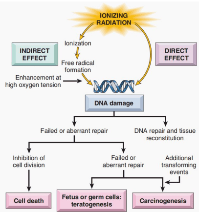
Direct and Indirect Molecular Consequences of Ionizing Radiation
Determinants of Tissue Radiosensitivity
According to the classical Law of Bergonié and Tribondeau, cellular radiosensitivity is directly proportional to reproductive metabolic rate and inversely proportional to cell differentiation:
- Extremely Radiosensitive: Hematopoietic stem cells in bone marrow, intestinal crypt stem cells, germ cells (spermatogonia, ovarian granulosa cells), and embryonic tissues.
Critical Pathological Exception: Mature circulating lymphocytes are non-proliferating cells that are hypersensitive to radiation, undergoing rapid interphase apoptosis within hours of low-dose exposure via p53-dependent mechanisms. - Intermediate Sensitivity: Vascular endothelial cells, fibroblasts, cutaneous basal keratinocytes.
- Radio-Resistant: Terminally differentiated, non-dividing mature skeletal myocytes, cardiac myocytes, and neurons.
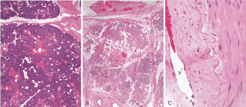
Microscopic Radiation-Induced Vascular Obliteration and Fibrosis
Microvascular Obliteration and Radiation Atypia
Vascular Injury & Progressive Fibrosis: Subacute and late radiation pathology is driven by endothelial damage. Irradiation triggers endothelial apoptosis, subendothelial edema, and microvascular thrombosis. Chronic wound healing drives fibrointimal proliferation and hyaline thickening of arteriolar walls, narrowing lumens. Downstream tissue develops chronic ischemia, leading to progressive atrophy and dense interstitial fibrosis.
Radiation Cytomegaly (Pseudomalignancy): Surviving irradiated cells exhibit marked nuclear and cytoplasmic enlargement (pleomorphism), multinucleation, and hyperchromasia. Pathologists must distinguish this benign, treatment-induced radiation atypia from recurrent malignancy.
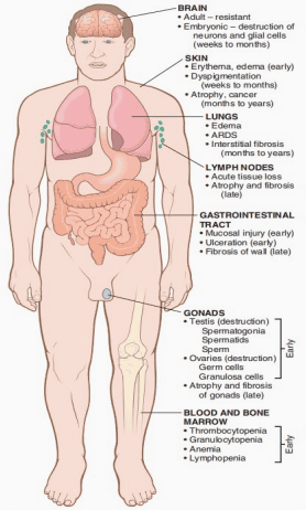
Temporal Organ Pathology Following Whole-Body Radiation Exposure
Acute Radiation Syndromes (ARS)
Following accidental high-dose, penetrating whole-body irradiation, patients develop predictable, dose-dependent clinical syndromes:
| Syndrome Name | Approximate Dose Range | Primary Anatomical Target | Clinical Manifestations & Prognosis |
|---|---|---|---|
| Hematopoietic Syndrome | 1 – 10 Gy | Hematopoietic stem cells within bone marrow stroma | Rapid lymphopenia within 24 hours, followed over 2–4 weeks by severe neutropenia, thrombocytopenia, and anemia. Death occurs secondary to systemic bacteremia/sepsis or massive hemorrhage unless supported with marrow stimulants or transplantation |
| Gastrointestinal Syndrome | 10 – 20 Gy | Mitotic stem cells within small intestinal crypts of Lieberkühn | Complete loss of mucosal epithelium, intractable secretory bloody diarrhea, massive electrolyte depletion, and intestinal bacterial translocation into the portal bloodstream. Death occurs within 1–2 weeks from shock and sepsis |
| Cerebrovascular / CNS Syndrome | > 20 – 50 Gy | Cerebral microvasculature and neurovascular membranes | Universal capillary endothelial breakdown with generalized cerebral edema, intractable convulsions, ataxia, coma, and cardiorespiratory arrest within 24–72 hours; universally fatal |
| Clinical Biological Effect | Critical Organ / Structure | Threshold Dose (Sieverts) |
|---|---|---|
| Temporary Sterility | Testes (damage to dividing spermatogonia) | 0.15 Sv |
| Depression of Hematopoiesis | Bone marrow (depletion of stem/progenitor cells) | 0.50 Sv |
| Reversible Skin Erythema | Cutaneous microvasculature and dermis | 1.0 – 2.0 Sv |
| Permanent Female Sterility | Ovaries (destruction of primary oocytes) | 2.5 – 6.0 Sv |
| Temporary Epilation (Hair Loss) | Hair follicle matrix keratinocytes | 3.0 – 5.0 Sv |
| Permanent Male Sterility | Testes (complete ablation of germinal epithelium) | 3.5 Sv |
| Cataract Formation | Lens epithelium (aberrant fiber differentiation) | 5.0 Sv |
Deterministic versus Stochastic Radiation Effects
- Deterministic Effects (Tissue Reactions): Characterized by a clear, predictable threshold dose below which the pathology does not manifest. Above the threshold, the clinical severity of tissue destruction increases proportionally with dose (e.g., radiation-induced marrow failure, cataract formation, erythema, and radiation necrosis).
- Stochastic Effects (Mutagenic/Carcinogenic): Non-threshold all-or-nothing biological phenomena governed by statistical probability. Increased radiation dose increases the probability that a malignancy will arise, but the clinical aggressiveness or histological severity of the resulting cancer is completely independent of the initiating dose. Classic radiation-induced neoplasms include acute leukemias (short latency of 5–7 years) and solid carcinomas of the thyroid, breast, and lung (latent intervals of decades).
Nutritional Disorders and Pathology
Nutritional Homeostasis
Nutritional disease develops whenever the bioavailable supply, cellular uptake, or metabolic utilization of essential nutrients fails to satisfy physiological requirements. Pathology encompasses the spectrum of undernutrition (protein-energy deprivation), isolated micronutrient deficiencies or toxic excesses, and overnutrition (obesity and metabolic syndrome). A patient presenting with morbid obesity may simultaneously harbor severe micronutrient deficiencies.
1. Biological Foundations of a Complete Diet
A nutritionally sufficient diet must deliver continuous substrates across three functional biochemical categories:
- Sufficient Energy Substrates: Digestible carbohydrates, neutral triglycerides, and proteins supply caloric energy to maintain resting basal metabolic functions, physical motor activity, thermogenesis, linear growth, and continuous tissue maintenance and repair. When caloric intake falls below daily metabolic expenditure, endogenous tissue reserves are consumed, culminating in sequential loss of adipose stores and skeletal muscle mass.
- Macromolecular Building Blocks: Ingested proteins supply essential amino acids required for the synthesis of structural cytoskeleton proteins, metabolic enzymes, peptide hormones, and transport immunoglobulins. Dietary fats provide essential polyunsaturated fatty acids (such as linoleic and α-linolenic acids) that cannot be synthesized de novo, serving as precursors for cell membrane phospholipid bilayers and eicosanoid signaling cascades.
- Vitamins and Trace Minerals: Organic coenzymes and inorganic elemental cofactors essential for metabolic enzymatic reactions, macromolecular synthesis, and physiological stability:
- Iron (Fe): Central catalytic constituent of hemoglobin and myoglobin for oxygen delivery, and mitochondrial cytochromes for oxidative phosphorylation.
- Calcium and Phosphate: Structural mineral phase of the crystalline hydroxyapatite skeleton; second messenger signaling and muscle contraction.
- Iodine (I): Substrate required by thyroid follicular cells for thyroxine (T4) and triiodothyronine (T3) synthesis.
- Vitamins: Complex organic cofactors supporting visual pigment cycles, epithelial integrity, genomic transcription, coagulation cascades, and free radical neutralization.
2. Classification: Primary versus Secondary Malnutrition
Malnutrition is broadly divided based on the underlying physiological mechanism:
- Primary Malnutrition: Results directly from inadequate dietary intake where the exogenous food supply lacks adequate energy, bioavailable protein, or essential micronutrients. Etiologies include economic poverty, regional food insecurity, famine, armed conflict, child neglect, and severely restricted elimination diets.
- Secondary (Conditional) Malnutrition: Occurs when nutrient supply is theoretically adequate, but an underlying illness or physiological condition impairs nutrient absorption, transport, storage, or utilization, or markedly increases metabolic demand or physical nutrient losses.
| Pathophysiological Mechanism | Underlying Clinical Disturbance | Representative Clinical Etiologies |
|---|---|---|
| Impaired Intestinal Absorption | Nutrients in the gastrointestinal tract fail to traverse the mucosal barrier into the circulation | Celiac disease, Crohn disease, cystic fibrosis with exocrine pancreatic insufficiency, chronic pancreatitis, and post-surgical short bowel syndrome |
| Impaired Utilization or Storage | Nutrients enter the vascular compartment but cannot be handled, converted, or stored by organs | Decompensated hepatic cirrhosis, inborn errors of metabolism, and metabolic enzyme deficiencies |
| Excessive Nutrient Losses | Essential nutrients are lost from bodily compartments at rates exceeding intake | Protein-losing enteropathy, nephrotic syndrome (urinary albumin loss), chronic enterocutaneous fistulae, and severe prolonged secretory diarrhea |
| Increased Metabolic Requirements | Systemic metabolic rate and tissue breakdown surpass available nutritional delivery | Extensive third-degree thermal burns, severe politrauma, systemic sepsis, hyperthyroidism, and advanced malignant cachexia |
Coexistence of Malnutrition Pathways
In clinical practice, primary and secondary mechanisms frequently operate simultaneously. An elderly patient with chronic heart failure or advanced chronic obstructive pulmonary disease (COPD) may suffer from chronic systemic inflammation-induced anorexia (secondary poor intake), hepatic congestion-induced malabsorption (secondary malabsorption), and poverty-related isolation that limits physical access to wholesome groceries (primary deficiency).
3. Comprehensive Etiological Drivers of Malnutrition
- Poverty, Socioeconomic Deprivation, and Food Insecurity: Severe global limitation of diverse foods results in concurrent caloric deficits and specific micronutrient deficiencies. Sustained starvation depletes glycogen within 24 hours, depletes adipose triglycerides over weeks, and leads to progressive skeletal muscle proteolysis, causing failure of cellular host defenses and secondary infection.
- Nutritional Ignorance and Inappropriate Infant Feeding: Failure to recognize shifting metabolic requirements across life stages (infancy, accelerated adolescent growth spurts, pregnancy, and lactation).
- Heavy dietary dependence on unfortified polished white rice precipitates thiamine deficiency (beriberi).
- Subsistence farming in iodine-deficient mountainous regions causes endemic goiter and congenital cretinism.
- Infant Nutrition Caveat: Commercially formulated infant formulas are designed to match human milk. They must not be equated with unmodified animal milk (e.g., cow or goat milk), which lacks appropriate iron and vitamin concentrations and places an excessive renal solute load on infant kidneys.
- Chronic Harmful Alcohol Use: Ethanol provides "empty" caloric energy (7 kcal/g) lacking amino acids, vitamins, and minerals, displacing nutritious food intake. Concomitant alcoholic gastritis, duodenitis, and pancreatic insufficiency impair intestinal nutrient absorption, while hepatic dysfunction disrupts storage of vitamins A, D, and B12.
- Acute and Chronic Consumptive Illness: Severe burns, systemic infections, and advanced cancers elevate systemic cytokine levels (TNF-α, IL-1, IL-6), promoting massive catabolic breakdown of skeletal muscle and fat stores that cannot be reversed by simple caloric supplementation alone.
- Self-Imposed Dietary Restriction: Severe psychological eating disorders, predominantly anorexia nervosa and bulimia nervosa.
4. Major Forms and Anthropometric Metrics of Childhood Undernutrition
Pediatric undernutrition presents in characteristic clinical configurations defined by standard anthropometric parameters:
| Anthropometric Entity | Objective Anthropometric Definition | Clinical & Pathological Interpretation |
|---|---|---|
| Wasting | Low weight-for-height (or length) below −2 standard deviations (−2 Z-scores) | Indicates acute, severe, recent nutritional deprivation or severe illness leading to rapid loss of somatic fat and muscle |
| Stunting | Low height-for-age below −2 standard deviations (−2 Z-scores) | Indicates chronic, long-term, cumulative nutritional deprivation and recurrent infections, reflecting failure of linear skeletal growth |
| Underweight | Low weight-for-age below −2 standard deviations (−2 Z-scores) | Composite index that captures acute wasting, chronic stunting, or a combination of both; non-specific for timing |
| Micronutrient Deficiency | Subnormal circulating or tissue levels of specific vitamins or trace elements | Frequently termed "hidden hunger"; can occur in severely wasted children or in normal-weight/overweight individuals lacking specific cofactors |
Dissociation Between Stunting and Wasting
Body weight alone does not distinguish acute from chronic malnutrition. A child with chronic stunting may exhibit an age-inappropriate short stature but maintain an entirely normal weight-for-height ratio. Conversely, a child suffering acute wasting displays profound emaciation and skeletal muscle loss with marked deficits in weight-for-height.
General Pathological Consequences of Systemic Undernutrition
- Mobilization and Loss of Fat and Muscle: Adipocytes mobilize stored triacylglycerols via hormone-sensitive lipase, causing profound loss of buccal, subcutaneous, and retroperitoneal fat depots. Sustained starvation forces the catabolism of skeletal myofibrillar proteins into alanine and glutamine for hepatic gluconeogenesis, producing thin, emaciated extremities.
- Growth Arrest: In children, chondrocyte proliferation in epiphyseal growth plates halts, causing linear growth failure and delayed bone age.
- Immunodeficiency and the Vicious Cycle of Infection: Severe undernutrition induces profound thymic involution (nutritional thymectomy), lymphoid tissue atrophy in the spleen and lymph nodes, and blunted cell-mediated immunity. Secretory IgA production falls along mucosal surfaces, permitting microbial invasion. Subsequent diarrhea and respiratory infections increase catabolic demand and worsen malabsorption, accelerating nutritional decline.
- Impaired Wound Healing and Tissue Repair: Lack of amino acids, zinc, and vitamin cofactors arrests fibroblast proliferation, collagen synthesis, and re-epithelialization.
- Multiple Organ Hypoplasia: Reduced protein synthesis causes mucosal atrophy of the small intestine, normocytic normochromic or mixed anemia due to marrow hypoplasia, bradycardia, hypotension, and impaired thermoregulation.
Clinical Assessment of Nutritional Status
- Comprehensive Clinical History: Dietary history (caloric intake, breastfeeding duration, weaning practices), food insecurity, feeding difficulties, vomiting, persistent diarrhea, and chronic fever.
- Targeted Physical Examination: Inspection for loss of temporal and buccal fat pads, loss of gluteal muscle mass, dependent bilateral pitting edema, follicular hyperkeratosis, dermal desquamation, and alterations in hair texture and pigmentation.
- Anthropometry: Precise measurement of nude body weight, recumbent length (infants <2 years) or standing height, calculation of age-adjusted Z-scores, and measurement of the Mid-Upper Arm Circumference (MUAC) using a standardized insertion tape.
- Laboratory Investigations: Complete blood count (anemia, lymphopenia), serum electrolytes (potassium, magnesium, phosphate), blood glucose (fasting hypoglycemia), urinalysis, stool microscopy, and serum total protein and albumin levels.
5. Severe Acute Malnutrition (SAM): Marasmus and Kwashiorkor
Severe Acute Malnutrition (SAM) in Children Aged 6–59 Months
A diagnosis of SAM is established if the child fulfills any one of the following three independent clinical criteria:
- Severe Wasting: Weight-for-height (or length) < −3 standard deviations (−3 Z-scores) of WHO growth standards.
- Severely Depleted Muscle Mass: Mid-Upper Arm Circumference (MUAC) < 115 mm.
- Bilateral Pitting Nutritional Edema: Present over both feet and lower legs, independent of weight-for-height metrics (automatically categorizes the child as having SAM).
Two Functional Body Protein Compartments
To understand the clinicopathological divergence between Marasmus and Kwashiorkor, the body's total protein reserves are conceptualized into two distinct compartments:
| Protein Compartment | Primary Anatomical Representation | Disease Syndrome Primarily Depleting It |
|---|---|---|
| Somatic Protein Compartment | Proteins within skeletal muscle tissue | Marasmus: Somatic muscle is catabolized to supply amino acids for gluconeogenesis, while visceral protein production in the liver is preserved |
| Visceral Protein Compartment | Proteins within visceral organs (liver, kidneys) and circulating plasma proteins (albumin, transferrin) | Kwashiorkor: Hepatic protein synthesis is compromised, depleting circulating albumin and visceral stores, while somatic muscle is relatively spared |
1. Marasmus (Non-Edematous Severe Wasting)
Pathophysiological Mechanism: Arises from a profound, prolonged deficiency of total caloric (energy) intake affecting all macronutrients (carbohydrates, fats, and proteins equally). The physiological response mimics prolonged starvation:
- The body mobilizes subcutaneous adipose tissue via lipolysis and catabolizes skeletal muscle via the ubiquitin-proteasome pathway to generate energy substrates and gluconeogenic precursors.
- Amino acids derived from somatic breakdown allow the liver to maintain near-normal rates of visceral protein synthesis. Consequently, serum albumin concentrations are normal or only mildly depressed.
- Because intravascular oncotic pressure is maintained by circulating albumin, nutritional edema is characteristically ABSENT.
Clinical Features:
- Profound loss of subcutaneous fat; skin appears loose, thin, and wrinkled ("baggy pants" sign over the buttocks).
- Severe skeletal muscle wasting with broomstick extremities; ribs and facial bones become prominent.
- Loss of buccal fat pads creates a shrunken, elderly, pinched facial appearance ("monkey facies" / "old man appearance").
- The head appears disproportionately large relative to the emaciated torso and limbs.
- Severe linear growth retardation (stunting); child is alert, irritable, and typically ravenously hungry when food is presented.
2. Kwashiorkor (Edematous Malnutrition)
Pathophysiological Mechanism: Characterized classically by severe protein deprivation disproportionately greater than the reduction in total caloric intake. It occurs frequently in infants weaned prematurely from protein-rich breast milk onto an exclusive, high-carbohydrate, protein-poor starchy gruel (e.g., cassava, yams, polished rice gruels; the Ga language term translates to "the disease of the deposed child when the next baby is born").
Contemporary Clarification: Pathogenesis is now recognized as multifactorial. In addition to low dietary protein intake, systemic mucosal inflammation, severe oxidant stress, peroxisomal dysfunction, and dysbiosis of the gut microbiome contribute to failure of tissue fluid balance and cellular leakage beyond simple hypoalbuminemia alone.
Pathological Hallmarks:
- Generalized Nutritional Edema: The defining diagnostic feature. Begins as dependent, bilateral pitting edema over the dorsum of the feet and pretibial areas, progressing to involve the thighs, hands, periorbital tissues, and peritoneal cavity (ascites). Edema masks underlying somatic tissue wasting, giving a false impression of nourishment.
- Severe Hypoalbuminemia: Visceral protein synthesis in the liver fails, dropping serum albumin levels profoundly (often <2.0 g/dL), lowering intravascular plasma oncotic pressure and driving fluid transudation into the interstitium.
- Enlarged Fatty Liver (Hepatic Steatosis): The liver becomes enlarged, soft, and yellow due to massive macrovesicular steatosis. Fat accumulates because hepatocytes cannot synthesize apolipoprotein B-100, halting the assembly and export of very-low-density lipoproteins (VLDL).
- Dermatological Alterations ("Flaky-Paint" Dermatosis): Alternating zones of hyperpigmentation, irregular desquamation, cracking, and pale hypopigmentation, resembling peeling old paint.
- Hair Alterations ("Flag Sign"): Hair becomes dry, brittle, sparse, and depigmented, turning reddish-brown or sandy yellow. Alternating periods of adequate and poor nutrition produce alternating dark and light bands along individual hair shafts (the flag sign).
- Psychological Apathy: The child is lethargic, profoundly apathetic, irritable when handled, and displays anorexia.
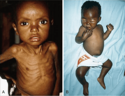
Clinical Comparison of Pediatric Marasmus and Kwashiorkor
Detailed Comparison: Marasmus versus Kwashiorkor
| Clinical & Pathological Feature | Marasmus (Caloric Deprivation) | Kwashiorkor (Protein Deprivation) |
|---|---|---|
| Primary Defining Appearance | Severe emaciation, wizened look, skeletal wasting | Puffy, edematous, "moon-face", abdominal ascites |
| Subcutaneous Adipose Tissue | Profoundly depleted; virtually absent | Relatively preserved; concealed beneath edema |
| Somatic Skeletal Muscle | Markedly atrophic and wasted (somatic depletion) | Variable wasting; masked by interstitial fluid |
| Bilateral Pitting Edema | Absent | Present (sine qua non diagnostic feature) |
| Serum Albumin Concentration | Normal or only slightly depressed | Markedly reduced (often < 2.0 g/dL) |
| Hepatic Morphology | Normal size or small; fatty change uncommon | Massive hepatomegaly with severe steatosis |
| Dermatological Manifestations | Dry, wrinkled skin; no flaky-paint pattern | "Flaky-paint" dermatosis with desquamation |
| Hair Texture & Pigmentation | Normal or minimal non-specific thinning | Sparse, brittle, reddish-blonde, flag sign |
| Behavior and Appetite | Alert, irritable, ravenous appetite maintained | Profound apathy, misery, severe anorexia |
Marasmic Kwashiorkor: An overlapping clinical state where a child with chronic severe caloric wasting (marasmus) sustains an acute reduction in dietary protein or an intercurrent infection, developing bilateral pitting edema, hepatomegaly, and hypoproteinemia on top of profound somatic emaciation.
Organ Pathology in Severe Acute Malnutrition
- Gastrointestinal Mucosa: Marked mitotic arrest within intestinal crypts of Lieberkühn leads to mucosal blunting, flattening of absorptive villi, and microvillar atrophy. Epithelial cells lose disaccharidase enzymes (particularly lactase). Ingestion of lactose results in osmotic fermentation, creating intractable diarrhea that compounds nutrient malabsorption.
- Bone Marrow: Hypoplasia of erythroid, myeloid, and megakaryocytic lineages. Anemia is universally present and multifactorial: iron deficiency, folate deficiency, and blunted erythropoietin release due to reduced metabolic demand.
- Lymphoid Involutive Changes: Severe hypoplasia and architectural atrophy of the thymus, Peyer patches, spleen, and peripheral lymph nodes.
- Central Nervous System: When SAM occurs during the first two years of life, impaired myelination, loss of dendritic branching, and reduced brain volume cause permanent neurocognitive and behavioral deficits.
Cancer Cachexia versus Uncomplicated Starvation
Cachexia is a hypercatabolic wasting syndrome seen in patients with advanced malignancies (gastric, pancreatic, pulmonary carcinomas) and chronic inflammatory states (AIDS, end-stage heart failure). While simple starvation (marasmus) is an adaptive response conserving energy by downregulating basal metabolic rate and preserving visceral protein, cachexia is an active, maladaptive pro-inflammatory catabolic state:
- Tumor cells and host macrophages release pro-inflammatory cytokines: TNF-α (cachectin), IL-1, IL-6, and Interferon-γ.
- Circulating tumor factors, such as Proteolysis-Inducing Factor (PIF), activate the ubiquitin-proteasome pathway in skeletal muscle, accelerating myofibrillar proteolysis.
- Lipid-Mobilizing Factor (LMF) increases adipocyte lipolysis and induces mitochondrial uncoupling protein (UCP-1) in adipose tissue, converting chemical energy into heat.
- Clinical Contrast: The resting energy expenditure in cachexia is elevated, and muscle wasting cannot be reversed by nutritional support or total parenteral nutrition without treating the underlying malignancy.
6. Pathology of Eating Disorders: Anorexia Nervosa and Bulimia Nervosa
Eating disorders represent severe psychiatric disturbances with serious physiological, endocrine, and morphological complications:
1. Anorexia Nervosa
Definition: Self-imposed severe restriction of energy intake relative to requirements, resulting in significantly low body weight (BMI < 18.5 kg/m2), an intense fear of gaining weight, and distorted body image perception. It is not a primary loss of appetite; patients actively suppress hunger.
Endocrine Alterations:
- Hypothalamic-Pituitary-Gonadal Suppression: Pulsatile release of Gonadotropin-Releasing Hormone (GnRH) is suppressed, causing low LH and FSH levels, culminating in hypoestrogenism and secondary amenorrhea.
- Euthyroid Sick Syndrome: Peripheral conversion of T4 to T3 is downregulated to conserve energy, resulting in low serum T3, normal T4, and normal TSH, presenting clinically with cold intolerance, hypothermia, constipation, and bradycardia.
- Hypothalamic-Pituitary-Adrenal Activation: Elevated circulating corticotropin-releasing hormone (CRH) and serum cortisol levels.
Systemic Morphological Changes:
- Skeletal Demineralization: Hypoestrogenism combined with elevated glucocorticoids accelerates osteoclastic resorption, causing early, severe osteopenia and osteoporosis with high pathological fracture risk.
- Cutaneous Changes: Dry, scaly, cracked skin covered by fine, soft downy hairs known as lanugo.
- Cardiovascular Collapse: Sinus bradycardia, hypotension, loss of left ventricular wall mass, mitral valve prolapse, and life-threatening prolongation of the QT interval predisposing to torsades de pointes and sudden death.
2. Bulimia Nervosa
Definition: Recurrent episodes of binge eating (consuming abnormally large amounts of food with a sense of loss of control) followed by inappropriate compensatory behaviors: self-induced vomiting, chronic laxative abuse, diuretic misuse, prolonged fasting, or compulsive exercise. Body weight typically remains within or slightly above the normal range (BMI 18.5–25.0 kg/m2), making the condition clinically occult.
Pathological Consequences of Chronic Purging:
- Acid-Base and Electrolyte Derangements:
- Self-Induced Vomiting: Massive loss of gastric hydrochloric acid (HCl) produces a hypochloremic, hypokalemic metabolic alkalosis. Aldosterone-mediated renal potassium wasting worsens hypokalemia.
- Chronic Laxative Abuse: Intestinal loss of alkaline secretions produces a normal-anion-gap hyperchloremic metabolic acidosis.
- Oral and Dental Pathology: Chronic regurgitation of gastric hydrochloric acid erodes the lingual surfaces of maxillary incisors and bicuspids (perimylolysis). Repeated reflex stimulation induces bilateral, painless hypertrophy of the salivary glands, particularly parotid sialadenosis.
- Gastrointestinal Mucosal Trauma: Severe chemical esophagitis, esophageal mucosal ulceration, and mechanical longitudinal tears at the gastroesophageal junction (Mallory-Weiss syndrome) or full-thickness rupture (Boerhaave syndrome).
- Cutaneous Clue (Russell's Sign): Abrasions, small lacerations, calluses, or hyperpigmentation over the dorsal aspect of the knuckles and metacarpophalangeal joints, caused by repeated friction against the incisor teeth during digital gag reflex induction.
4. Refeeding Syndrome
A life-threatening metabolic emergency occurring when aggressive enteral or parenteral nutritional rehabilitation is administered too rapidly to severely malnourished patients (anorexia nervosa, marasmus, chronic alcoholism):
- During prolonged starvation, cellular energy production relies on fatty acid oxidation and ketone bodies, depleting intracellular phosphate pools while serum phosphate remains deceptively normal.
- Sudden reintroduction of carbohydrates triggers an immediate surge of pancreatic insulin secretion.
- Insulin stimulates cellular uptake of glucose, driving rapid cellular co-uptake of phosphate, potassium, and magnesium to support glycolysis and ATP production.
- This causes catastrophic severe hypophosphatemia, accompanied by hypokalemia and hypomagnesemia.
- Depletion of ATP and 2,3-diphosphoglycerate (2,3-DPG) impairs myocyte contractility, resulting in acute biventricular congestive heart failure, pulmonary edema, diaphragmatic paralysis, rhabdomyolysis, seizures, and death.
7. Vitamin Deficiencies and Toxicities
Vitamin A: Physiological Roles and Deficiency States
Chemical Forms and Dietary Sources: Vitamin A designates a group of fat-soluble retinoids. Dietary sources comprise preformed vitamin A (retinyl esters) derived from animal tissues (liver, eggs, butter, oily fish) and provitamin A carotenoids (β-carotene) from yellow and dark-green leafy vegetables (carrots, sweet potatoes, spinach). Intestinal enterocytes cleave β-carotene into retinol, package retinyl esters into chylomicrons, and deliver them to the liver, where >90% of total body reserves are stored within hepatic stellate cells (Ito cells). Retinol is mobilized into the circulation bound to retinol-binding protein (RBP).
Three Core Physiological Functions:
- Phototransduction in the Retina: Retinol is converted to 11-cis-retinal, which complexes with the protein opsin in retinal rod cells to form rhodopsin. Light absorption isomerizes 11-cis-retinal to all-trans-retinal, triggering a neurochemical cascade that allows vision in low light.
- Regulation of Gene Expression and Epithelial Differentiation: All-trans-retinoic acid binds to nuclear receptors—the Retinoic Acid Receptors (RAR) and Retinoid X Receptors (RXR)—acting as transcription factors that regulate gene expression. This maintains specialized mucus-secreting columnar epithelia in the respiratory tract, gut, and ocular surface.
- Immune Defense and Host Resistance: Retinoids maintain epithelial basement membrane barriers and support antibody responses, natural killer cell activity, and T-helper cell differentiation.
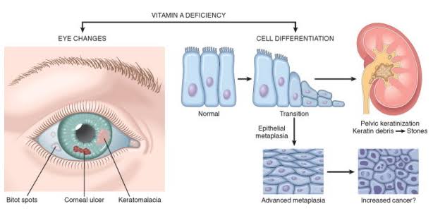
Ocular Pathology and Systemic Squamous Metaplasia in Vitamin A Deficiency
Clinical Spectrum of Vitamin A Deficiency
- Night Blindness (Nyctalopia): The earliest clinical symptom of deficiency, caused by failure to regenerate functional rhodopsin within rod photoreceptors.
- Xerophthalmia (Dry Eye Syndrome): Loss of mucus-secreting conjunctival goblet cells causes drying of the bulbar conjunctiva (conjunctival xerosis). Keratinized epithelial debris accumulates into triangular, foamy, opaque gray-white plaques termed Bitot spots.
- Keratomalacia: Untreated xerophthalmia causes corneal ulceration, liquefactive necrosis, and softening of the cornea (keratomalacia). The softened cornea perforates, leading to iris prolapse, endophthalmitis, and permanent blindness (the leading cause of preventable childhood blindness worldwide).
- Squamous Metaplasia of Respiratory and Urinary Tracts: Normal pseudo-stratified ciliated columnar epithelium of the respiratory tract undergoes squamous metaplasia with surface keratinization. Loss of the mucociliary escalator predisposes to secondary bronchopneumonia. In the renal pelvis and bladder, desquamated keratin plugs serve as niduses for urinary calculus formation.
- Follicular Hyperkeratosis (Phrynoderma): Occlusion of cutaneous hair follicles by hyperkeratotic plugs produces a rough, papular "toad-skin" dermatosis.
- Measles Complications: In vitamin A-deficient children, measles virus infection depletes remaining vitamin A stores, suppressing immunity and precipitating fatal giant cell pneumonia, severe enteritis, and rapid corneal perforation. Exogenous vitamin A supplementation reduces measles-associated pediatric mortality.
Hypervitaminosis A (Vitamin A Toxicity)
- Acute Toxicity: Follows massive single-dose ingestion (e.g., Arctic explorer ingestion of polar bear liver). Manifests as severe headache, nausea, vomiting, dizziness, blurred vision, and pseudotumor cerebri (idiopathic intracranial hypertension) with papilledema.
- Chronic Toxicity: Arises from prolonged ingestion of excessive preformed retinoids. Causes dry, scaling, fissured lips (cheilitis), alopecia, generalized bone pain, periosteal hyperostosis, and hepatomegaly with hepatic stellate cell lipid fibrosis.
- Teratogenic Risks: Systemic pharmaceutical retinoids (e.g., isotretinoin used for cystic acne) are potent teratogens, disrupting cranial neural crest cell migration during embryogenesis, leading to microtia, cleft palate, thymic hypoplasia, and conotruncal cardiac defects. Pregnancy must be excluded before retinoid therapy. Dietary β-carotene is non-teratogenic.
Vitamin D: Synthesis, Metabolism, and Bone Pathology
Cutaneous Synthesis and Activation Cascade:
- Cutaneous Photolysis: Solar ultraviolet B (UVB) radiation (290–315 nm) photolyzes epidermal 7-dehydrocholesterol into cholecalciferol (Vitamin D3). Dietary intake provides D3 from animal sources and ergocalciferol (D2) from fungal and plant sterols.
- Hepatic 25-Hydroxylation: Vitamin D binds vitamin D-binding protein in blood and is transported to the liver. Microsomal and mitochondrial 25-hydroxylases (CYP2R1) add a hydroxyl group to form 25-hydroxyvitamin D [25(OH)D; calcidiol], the primary circulating form and clinical marker of total body stores.
- Renal 1α-Hydroxylation: In the proximal renal tubular epithelium, the mitochondrial enzyme 1α-hydroxylase (CYP27B1) converts 25(OH)D into 1,25-dihydroxyvitamin D [1,25(OH)2D; calcitriol], the biologically active hormone. Renal 1α-hydroxylase is strongly stimulated by Parathyroid Hormone (PTH) and hypophosphatemia, and inhibited by high calcium, high calcitriol, and Fibroblast Growth Factor 23 (FGF23).
| Anatomical Site | Specific Enzymatic Transformation | Resulting Chemical Metabolite | Physiological Significance |
|---|---|---|---|
| Epidermis (Skin) | Photochemical conversion of 7-dehydrocholesterol by solar UVB | Cholecalciferol (Vitamin D3) | Inert precursor; dependent on sunlight, skin melanin density, and age |
| Hepatic Parenchyma | C-25 Hydroxylation by hepatic CYP2R1 enzyme | 25-hydroxyvitamin D [25(OH)D] | Major circulating storage form (half-life: 2–3 weeks); standard lab marker |
| Proximal Renal Tubule | C-1α Hydroxylation by renal CYP27B1 (stimulated by PTH) | 1,25(OH)2D (Calcitriol) | Active hormone; binds nuclear VDR to regulate calcium homeostasis |
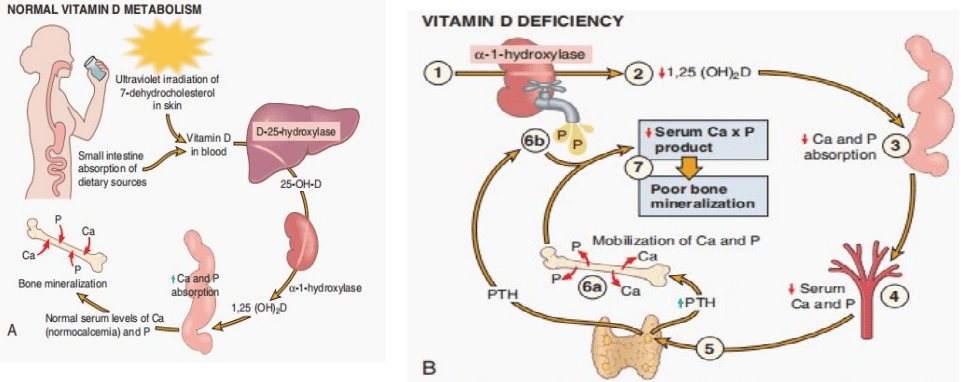
Pathophysiology of Vitamin D Deficiency and Secondary Hyperparathyroidism
Normal Functions and Pathophysiological Cascade of Deficiency
Active 1,25(OH)2D binds the nuclear Vitamin D Receptor (VDR) to increase intestinal absorption of calcium and phosphate, stimulate calcium reabsorption in distal renal tubules, and regulate osteoblast-mediated mineralization of newly synthesized organic osteoid matrix. The pathological sequence in deficiency comprises:
- Deficient Calcitriol Synthesis: Suboptimal 1,25(OH)2D impairs intestinal enterocyte absorption of both calcium and phosphorus.
- Secondary Hyperparathyroidism: A subtle drop in ionized serum calcium stimulates calcium-sensing receptors (CaSR) on parathyroid chief cells to hypersecrete Parathyroid Hormone (PTH).
- Bone Mineral Mobilization: PTH stimulates osteoblasts to express RANKL, activating osteoclasts to resorb bone calcium and phosphate, attempting to normalize circulating calcium.
- Renal Phosphate Wasting: High PTH levels downregulate sodium-phosphate co-transporters (NaPi-IIa) in the proximal renal tubule, causing marked phosphaturia and severe hypophosphatemia.
- Impaired Mineralization: Although PTH may maintain serum calcium near normal, the calcium-phosphate solubility product (Ca × P) drops below the critical threshold required for hydroxyapatite crystallization, leaving skeletal osteoid unmineralized.
Rickets (In Children) versus Osteomalacia (In Adults)
Rickets (Pediatric Skeleton with Open Epiphyses): Characterized by defective mineralization of newly formed organic osteoid alongside failure of provisional calcification and mineralization of cartilage at the epiphyseal growth plates. Chondrocytes continue proliferating in the growth plate, but because provisional mineralization fails, cartilage cells do not mature and undergo apoptosis. Irregular tongues of unmineralized, disorganized cartilage and excess soft osteoid accumulate, causing the growth plate to become widened, thick, cupped, and mechanically unstable.
Osteomalacia (Adult Skeleton with Closed Epiphyses): In adults, linear growth has ceased and epiphyseal plates are closed. Normal bone remodeling continues, with osteoclasts carving resorption pits that osteoblasts fill with organic matrix (osteoid). Due to mineral deficiency, this newly deposited osteoid fails to mineralize, forming abnormally thick, uncalcified osteoid seams coating trabecular and cortical surfaces. The bones lose mineral rigidity, becoming structurally weak, tender, and prone to insufficiency fractures.
| Characteristic Skeletal Deformity | Anatomical Location & Underlying Pathology | Clinical Manifestation |
|---|---|---|
| Craniotabes | Thinning and softening of parietal and occipital calvarial bones | The skull indents easily on palpation with a ping-pong ball sensation |
| Frontal Bossing | Excess unmineralized osteoid accumulates at cranial eminences | Prominent forehead, producing a characteristic square-shaped skull ("caput quadratum") |
| Rachitic Rosary | Nodular expansion of unmineralized osteoid and cartilage at costochondral junctions | Palpable, beaded prominences along both lateral borders of the anterior chest wall |
| Harrison Groove (Sulcus) | Lower ribcage softening subjected to inward diaphragmatic muscular traction | Horizontal, transverse depression along the lower thoracic margin |
| Pigeon Breast (Pectus Carinatum) | Weakened anterior costal cartilages yielding to respiratory forces | Anterior protrusion of the sternum |
| Widened Epiphyses | Overgrowth and lateral flaring of uncalcified growth-plate cartilage | Visible, bulbous enlargement of the wrists and ankles |
| Bowed Legs (Genu Varum / Genu Valgum) | Weight-bearing pressure bowing softened, poorly mineralized long bones | Exaggerated lateral curvature of the femora and tibiae upon walking |
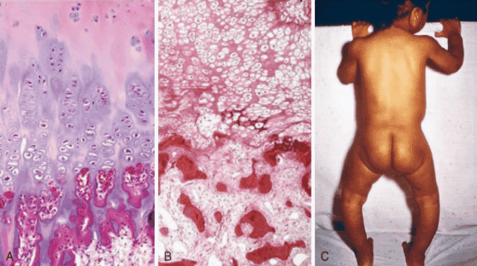
Morphological Disorganization of the Growth Plate and Bowed Legs in Rickets
Comparison: Osteomalacia versus Osteoporosis
| Diagnostic Parameter | Osteomalacia (Mineralization Defect) | Osteoporosis (Reduction in Bone Mass) |
|---|---|---|
| Primary Pathological Defect | Qualitative defect: Inadequate mineralization of newly formed organic bone matrix | Quantitative defect: Reduction in total bone mass with normal mineral-to-matrix ratio |
| Histological Osteoid State | Abnormally thick, widened unmineralized osteoid seams coating trabecular bone | Trabeculae are thin, attenuated, and sparse, but normally mineralized |
| Classic Radiological Sign | Pseudofractures (Looser zones / Milkman lines): symmetrical radiolucent bands perpendicular to cortex | Generalized radiolucency, vertebral compression fractures, femoral neck fractures |
| Serum Biochemistry Profile | Low 25(OH)D; low/normal Ca; low phosphate; high PTH; high Alkaline Phosphatase | Completely normal serum calcium, phosphate, PTH, and alkaline phosphatase |
Hypervitaminosis D: Caused exclusively by excessive oral pharmacological supplementation (sunlight cannot induce toxicity due to cutaneous degradation into inactive lumisterol and tachysterol). High 1,25(OH)2D produces severe hypercalcemia and hypercalciuria, manifesting clinically with polyuria, polydipsia, nephrolithiasis, and metastatic calcification of soft tissues (kidneys, lungs, stomach).
Vitamin C (Ascorbic Acid) Deficiency: Scurvy
Physiological Role & Mechanism of Collagen Defect: Humans cannot synthesize ascorbic acid endogenously due to the genetic evolutionary absence of the enzyme L-gulonolactone oxidase. Ascorbic acid functions as an essential electron donor and reducing cofactor for the enzymes prolyl hydroxylase and lysyl hydroxylase. These enzymes hydroxylate proline and lysine residues on newly translated procollagen polypeptide chains inside the rough endoplasmic reticulum:
- Hydroxyproline is essential for hydrogen bonding that stabilizes the triple-helix configuration of mature collagen.
- Hydroxylysine is required for intra- and intermolecular covalent cross-linking that confers mechanical tensile strength to connective tissues.
- In vitamin C deficiency, unhydroxylated procollagen polypeptides cannot cross-link, are degraded intracellularly, or form mechanically unstable collagen with low melting temperatures.
- This causes fragility of vascular walls, defective dentin and osteoid formation, and failure of wound healing.
Additional Functions: Vitamin C acts as an antioxidant, supports catecholamine biosynthesis (dopamine β-hydroxylase), and reduces ferric iron (Fe3+) to the ferrous state (Fe2+) in the stomach, promoting non-heme dietary iron absorption.
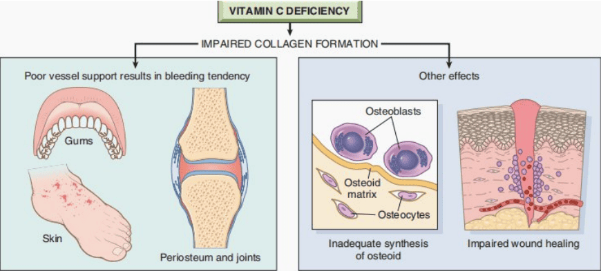
Pathological Consequences of Defective Collagen Hydroxylation in Scurvy
Clinical Manifestations of Scurvy
- Hemorrhagic Diathesis: Capillaries and venules lack supportive perivascular collagen, causing vessel rupture under minimal hydrostatic pressure. Manifests as:
- Perifollicular Hemorrhages: Petechial hemorrhages centered around hair follicles, accompanied by coiled, fragmented corkscrew hairs and follicular hyperkeratosis.
- Cutaneous Ecchymoses and Purpura: Extensive subcutaneous hematomas on dependent extremities.
- Gingival Pathology: The periodontal ligament consists of collagen with a rapid metabolic turnover. Deficiency causes marked swelling, sponginess, bleeding, and ulceration of the gingiva, with tooth loosening and secondary bacterial infection (confined to dentate individuals).
- Subperiosteal Hemorrhages and Skeletal Disease: In growing children, vessel fragility produces large, painful subperiosteal hematomas stripping the periosteum from long bones. Osteoblasts fail to synthesize organic osteoid matrix. Children adopt a motionless flexed posture ("frog-leg position") due to excruciating bone and joint pain.
- Impaired Wound Healing: Failure of collagen deposition prevents scars from acquiring tensile strength, leading to surgical wound dehiscence.
- Anemia: Multifactorial, resulting from chronic blood loss, decreased dietary iron reduction, and co-existent folate deficiency.
8. Other Fat-Soluble Vitamins: Vitamin E and Vitamin K
1. Vitamin E (α-Tocopherol)
Physiological Role: Acts as a lipid-soluble antioxidant, scavenging free radicals and terminating lipid peroxidation chain reactions in polyunsaturated membrane phospholipids. It protects red blood cell membranes and myelinated axons.
Clinical Deficiency Syndrome: Rare in dietary restriction; occurs secondary to fat malabsorption (cystic fibrosis, cholestasis, abetalipoproteinemia). Manifests as:
- Spinocerebellar Degeneration: Axonal degeneration of the posterior columns (loss of proprioception and vibratory sense) and lateral spinocerebellar tracts (ataxic gait).
- Peripheral Neuropathy: Absent deep tendon reflexes and limb muscle weakness.
- Hemolytic Anemia: Oxidative lysis of erythrocyte membranes, particularly in premature infants.
- Differential Clue: The neurological phenotype mimics Vitamin B12 subacute combined degeneration, but Vitamin E deficiency produces no megaloblastic anemia and serum methylmalonic acid levels are normal.
2. Vitamin K (Phylloquinone & Menaquinone)
Physiological Role: Serves as an essential cofactor for hepatic microsomal γ-glutamyl carboxylase, which post-translationally carboxylates glutamic acid residues on coagulation proteins to form γ-carboxyglutamic acid (Gla) domains. These Gla domains bind Ca2+ to negative platelet phospholipid membranes. Coagulation factors dependent on vitamin K are:
Pro-coagulant Factors II (Prothrombin), VII, IX, and X; and Anticoagulant Proteins C and S
Laboratory and Clinical Features:
- Prolonged Prothrombin Time (PT/INR): Factor VII has the shortest biological half-life (~6 hours) among coagulation proteins, making PT the earliest laboratory indicator of deficiency. In severe cases, partial thromboplastin time (aPTT) also becomes prolonged.
- Vitamin K Deficiency Bleeding (VKDB) of the Newborn: Neonates are born with low hepatic reserves, have a sterile gut lacking vitamin K-producing bacterial microflora, and breast milk contains low vitamin K concentrations. Without prophylactic intramuscular Vitamin K administration at birth, infants can develop intracranial hemorrhage, umbilical cord bleeding, or retroperitoneal hematomas.
- Adult Deficiency: Occurs secondary to broad-spectrum antibiotic therapy (depleting gut microflora), severe obstructive cholestasis (blocking biliary micelle formation required for absorption), or pharmacotherapy with the competitive antagonist Warfarin.
9. Water-Soluble B-Complex Vitamins
| Vitamin Name | Active Biochemical Coenzyme | Key Enzymatic Reaction | Primary Clinical Deficiency Syndrome |
|---|---|---|---|
| Thiamine (Vitamin B1) | Thiamine Pyrophosphate (TPP) | Pyruvate dehydrogenase, α-ketoglutarate dehydrogenase, and transketolase (HMP shunt) | Dry Beriberi (peripheral neuropathy, muscle wasting) Wet Beriberi (dilated cardiomyopathy, biventricular failure, high-output edema) Wernicke-Korsakoff Syndrome (ophthalmoplegia, ataxia, confusion, amnesia) |
| Riboflavin (Vitamin B2) | FAD and FMN | Redox electron transfer in the respiratory chain and mitochondrial succinate dehydrogenase | Ariboflavinosis: Angular cheilitis (fissuring at corners of mouth), glossitis (magenta tongue), stomatitis, and corneal neovascularization |
| Niacin (Vitamin B3) | NAD+ and NADP+ | Hydride transfer coenzymes for dehydrogenases in glycolysis, Krebs cycle, and fatty acid oxidation | Pellagra ("The 4 Ds"): Symmetrical bilateral photosensitive Dermatitis (Casal necklace), intractable secretory Diarrhea, encephalopathic Dementia, and ultimately Death |
| Pyridoxine (Vitamin B6) | Pyridoxal Phosphate (PLP) | Transamination of amino acids, synthesis of neurotransmitters (GABA, serotonin), and δ-ALA synthase | Peripheral sensorimotor neuropathy, microcytic sideroblastic anemia, seizures, and glossitis. Can be provoked by isoniazid (INH) therapy |
| Cobalamin (Vitamin B12) | Methylcobalamin & Adenosylcobalamin | Homocysteine methyltransferase (methionine synthase) and methylmalonyl-CoA mutase | Megaloblastic Anemia (hypersegmented neutrophils) accompanied by Subacute Combined Degeneration of spinal cord (demyelination of posterior and lateral columns) |
| Folate (Vitamin B9) | Tetrahydrofolate (THF) | One-carbon transfer reactions in purine and thymidylate (dTMP) synthesis for DNA replication | Megaloblastic Anemia indistinguishable from B12, but WITHOUT neurological cord degeneration; maternal deficiency causes Neural Tube Defects (spina bifida, anencephaly) |
| Pantothenic Acid (Vitamin B5) | Coenzyme A (CoA) | Acyl carrier protein for fatty acid metabolism and citric acid cycle entry | Extremely rare isolated deficiency; non-specific malaise, paresthesias ("burning feet syndrome") |
| Biotin (Vitamin B7) | Biotin-Enzyme Complex | Cofactor for carboxylase enzymes (pyruvate carboxylase, acetyl-CoA carboxylase) | Dermatitis, alopecia, and neurological paresthesias; can be induced by ingestion of raw egg whites (contains avidin) |
Differential Diagnosis: Vitamin B12 versus Folate Deficiency
Both deficiencies impair DNA synthesis, causing nuclear-cytoplasmic asynchrony, megaloblastic transformation in bone marrow, macro-ovalocytic red cells, and hypersegmented neutrophils (≥5 lobes). However, their pathological profiles and laboratory markers differ critically:
| Diagnostic Parameter | Vitamin B12 (Cobalamin) Deficiency | Folate (Vitamin B9) Deficiency |
|---|---|---|
| Neurological Cord Degeneration | Present: Subacute Combined Degeneration (SCD). Demyelination of posterior columns (loss of position/vibration) and lateral corticospinal tracts (spastic paraparesis). Can occur in the absence of anemia | Absent: Neurological tracts are structurally preserved; no subacute combined degeneration |
| Serum Methylmalonic Acid (MMA) | Elevated: Conversion of methylmalonyl-CoA to succinyl-CoA requires B12. Accumulation of toxic methylmalonic acid damages myelin | Completely Normal: Methylmalonyl-CoA mutase does not require folate |
| Serum Homocysteine | Elevated: Methylation of homocysteine to methionine requires both B12 and THF | Elevated: Homocysteine remethylation pathway is blocked |
| Clinical Danger of Misdiagnosis | Treating B12 deficiency with folate resolves the megaloblastic anemia ("folate trap" bypass) but allows irreversible neurological spinal degeneration to progress | Isolated folate therapy corrects hematological abnormalities without adverse masking |
10. Selected Essential Trace Elements
Trace elements are inorganic transition metals and minerals required in minute daily quantities (milligrams or micrograms) serving as catalytic active sites for structural and metabolic enzymes.
| Trace Element | Metabolic Enzymes / Functions | Underlying Basis of Deficiency | Clinical Pathological Features |
|---|---|---|---|
| Zinc (Zn) | Essential component of >300 metalloenzymes (carbonic anhydrase, alkaline phosphatase, RNA polymerase) and "zinc-finger" transcription factors | Inadequate dietary supply, total parenteral nutrition lacking zinc, inborn defect of absorption (Acrodermatitis enteropathica) | Pustular, erythematous periorificial and acral dermatitis (around eyes, mouth, anus, hands/feet), intractable diarrhea, complete alopecia, impaired wound healing, hypogonadism, and immune deficiency |
| Iron (Fe) | Heme prosthetic group in hemoglobin, myoglobin, and catalase; iron-sulfur mitochondrial complexes | Chronic occult blood loss (gastrointestinal neoplasms, menorrhagia), dietary deficiency, increased pregnancy demand, intestinal celiac sprue | Microcytic, hypochromic anemia, fatigue, angular cheilosis, glossitis, and spoon-shaped concave nails (koilonychia) |
| Iodine (I) | Substrate for thyroid follicular biosynthesis of thyroid hormones T3 and T4 | Deficiency in soil and crops in mountainous inland geography without dietary iodized salt fortification | Compensatory hypersecretion of TSH producing endemic colloid goiter, clinical hypothyroidism, and infant cretinism (profound intellectual disability, short stature, deaf-mutism) |
| Copper (Cu) | Cofactor for cytochrome c oxidase (ATP synthesis), lysyl oxidase (collagen/elastin cross-linking), and ferroxidase ceruloplasmin | Total parenteral nutrition without trace minerals, excessive zinc supplementation (competes for intestinal metallothionein), gastric bypass | Microcytic anemia resistant to iron, neutropenia, defective vascular and bone matrix cross-linking, and sensory ataxia mimicking subacute combined degeneration |
| Fluoride (F) | Incorporates into dental enamel crystal lattice, converting hydroxyapatite into acid-resistant fluorapatite | Low concentration in local geographic water supplies lacking municipal fluoridation | Increased susceptibility to dental bacterial caries.
Toxicity: Excess consumption causes chalky, mottled, brown-stained dental enamel (dental fluorosis) and skeletal fluorosis. |
| Selenium (Se) | Essential constituent of glutathione peroxidase, protecting cells from peroxide-mediated oxidative damage; iodothyronine deiodinase | Soil depletion of selenium in specific agricultural basins (e.g., Keshan province, China) | Myopathy and an endemic juvenile congestive cardiomyopathy designated as Keshan disease |
11. Executive Pathological Revision Summary
| Deficient Factor | Primary Pathognomonic Clinical Association |
|---|---|
| Vitamin A | Night blindness (nyctalopia), Bitot spots, keratomalacia, and widespread squamous metaplasia |
| Vitamin D | Rickets in children (growth plate widening, bowed legs); Osteomalacia in adults (unmineralized osteoid seams) |
| Vitamin C | Scurvy: perifollicular hemorrhages, corkscrew hairs, bleeding spongy gums, and subperiosteal hematomas |
| Vitamin E | Spinocerebellar ataxia, posterior column sensory loss, and hemolytic anemia without megaloblastosis |
| Vitamin K | Prolonged prothrombin time (PT/INR) and bleeding diathesis (VKDB of the newborn) |
| Thiamine (B1) | Dry beriberi (neuropathy), Wet beriberi (high-output heart failure), and Wernicke-Korsakoff encephalopathy |
| Riboflavin (B2) | Angular cheilitis, glossitis, stomatitis, and corneal neovascularization |
| Niacin (B3) | Pellagra: photosensitive dermatitis, intractable diarrhea, dementia, and death |
| Pyridoxine (B6) | Peripheral neuropathy, sideroblastic anemia, and cheilosis (provoked by isoniazid) |
| Cobalamin (B12) | Megaloblastic anemia with elevated methylmalonic acid and subacute combined degeneration of the spinal cord |
| Folate (B9) | Megaloblastic anemia with normal methylmalonic acid; periconceptional maternal deficiency causes neural tube defects |
| Zinc | Acrodermatitis enteropathica: periorificial/acral rash, diarrhea, alopecia, and impaired wound healing |
| Iron | Microcytic, hypochromic anemia and koilonychia |
| Iodine | Endemic multinodular goiter, adult hypothyroidism, and infantile cretinism |
| Copper | Microcytic anemia, neutropenia, and posterior column spinal cord degeneration |
| Selenium | Skeletal myopathy and endemic dilated cardiomyopathy (Keshan disease) |