INTRODUCTION & SUBDIVISION OF THE NERVOUS SYSTEM
MENINGES, VENTRICLES & CSF
Divisions of the Nervous System
| Primary Division | Subdivision | Structural & Functional Characterization |
|---|---|---|
| Central NS (CNS) (The body's master control unit) |
Spinal Cord | A column of nerves between the brain and peripheral nervous system. |
| Central NS (CNS) | Brain stem | Connects the brain to the spinal cord. |
| Central NS (CNS) | Brain | Divided into three major parts: the hindbrain (lower part), the midbrain, and the forebrain. |
| Peripheral NS (PNS) (The body's link to the outside world) |
The Autonomic NS | Regulates involuntary bodily processes, including heart rate, respiration, digestion and pupil contraction; operates automatically without conscious direction. |
| Peripheral NS (PNS) | Sympathetic NS | Prepares the body for action and stress. This is called "fight or flight". |
| Peripheral NS (PNS) | Parasympathetic NS | Calms the body and helps the body to conserve energy. |
| Peripheral NS (PNS) | The Somatic NS | Carries sensory information from sensory organs to the CNS and relays motor (movement) commands to muscles; controls voluntary movements. |
Autonomic Functional Divergence
Sympathetic ("E Division"): Exercise, Excitement, Emergency, Embarrassment (Thoracolumbar outflow, short preganglionic fibers, catabolic expenditure).
Parasympathetic ("D Division"): Digestion, Defecation, Diuresis (Craniosacral outflow, long preganglionic fibers, anabolic conservation).
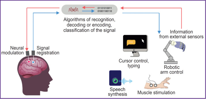
Intracortical recording interface and action potential profiling during fine motor tasks.
General Plan of the Nervous System
| Anatomical Level | Component Structures | Functional & Protective Orientation |
|---|---|---|
| Cranial Cavity | Skull & Brain | Houses cerebral hemispheres, diencephalon, cerebellum, brainstem, and cranial nerves. |
| Cranial Nerves | Two cranial nerves & Cranial nerve X | Peripheral nerves originating from brain/brainstem; CN X (vagus) forms a major parasympathetic component of the autonomic system. |
| Vertebral Canal | Spinal cord & Spinal nerves | Extends within elements of vertebral column; gives rise to segmental cervical, thoracic, lumbar, and sacral nerve pairs. |
| Encephalic Subdivisions | Telencephalon, Diencephalon, Brainstem, Cerebellum | Telencephalon (cerebral hemisphere), Diencephalon, Midbrain, Pons, Medulla oblongata, and Cerebellum continuous inferiorly with spinal cord. |
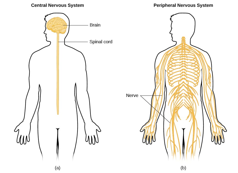
General plan of the nervous system.
Neuron
| Neuronal Domain | Histological Component | Cytological Specialization & Functional Role |
|---|---|---|
| Receptive Zone | Dendrites & Perikaryon | Extensive branching covered with synapses; soma contains the nucleus, prominent nucleolus, and abundant Nissl bodies (rough endoplasmic reticulum). |
| Trigger Zone | Axon hillock & Initial segment | Tapered region devoid of Nissl substance; high density of voltage-gated sodium channels where action potentials are initiated. |
| Transmission Zone | Axon & Collateral branches | Cylindrical process propagating impulses; insulated by myelin sheaths interrupted at Nodes of Ranvier for saltatory conduction. |
| Glial Insulation (CNS vs. PNS) | Oligodendrocyte vs. Schwann cell | Demarcation line separates central myelination (Oligodendrocytes myelinating multiple axons) from peripheral myelination (Schwann cells myelinating single internodes). |
| Terminal Zone | Telodendria & Motor end-plates | Terminal arborization releasing neurotransmitters at synaptic junctions or neuromuscular junction motor end-plates. |
Myelination Pathology: CNS vs. PNS
Multiple Sclerosis (MS): Autoimmune demyelination selectively targeting oligodendrocytes within the central nervous system (optic nerve, periventricular white matter, spinal cord).
Guillain-Barré Syndrome (GBS): Acute inflammatory demyelinating polyneuropathy selectively destroying peripheral Schwann cells, sparing central myelin tracts.
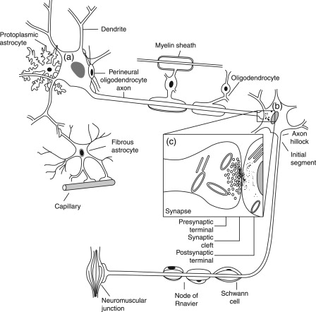
Structural architecture of a typical neuron and the transition between central and peripheral myelination.
Neurons (Functional Classifications & Synaptic Sequences)
| Morphological Pattern | Structural Zones | Neuronal Class & Synaptic Connectivity |
|---|---|---|
| Type 1 (Multipolar) | Dendritic receptive zone → Long transmission axon → Terminal arborization | Classic alpha motor neuron projecting from spinal ventral horn to peripheral somatic motor end-plates. |
| Type 2 (Bipolar) | Single dendrite → Central cell body → Terminal zone | Specialized sensory neurons (retinal bipolar cells, olfactory epithelium, vestibular/cochlear ganglia). |
| Type 3 (Pseudounipolar) | Peripheral receptive arbor → T-junction axon bypassing soma → Central terminal | Primary sensory neurons located in dorsal root ganglia (DRG) and cranial sensory ganglia conveying somatic sensation. |
| Autonomic Disynaptic Chain | Preganglionic cell → Intervening autonomic ganglion synapse → Postganglionic cell | Two-neuron motor pathway: CNS preganglionic myelinated axon synapses on unmyelinated postganglionic cells targeting smooth muscle, cardiac muscle, or glands. |
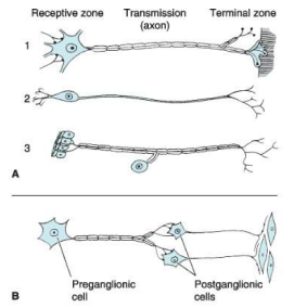
Morphological zones of primary neurons and autonomic synaptic architecture.
The Central Nervous System (CNS)
- The Central Nervous System (CNS) has two major structures: the brain and the spinal cord.
- The brain is protected by the cranial cavity and the spinal cord is surrounded by the spinal cavity in the vertebral column.
- They are both covered by protective membranes called meninges.
| Spinal Segment Level | Primary Motor & Sensory Territory | Critical Autonomic & Functional Milestone |
|---|---|---|
| C1 – C4 | Breathing and head/neck movement | Phrenic nerve (C3–C5): "C3, 4, 5 keeps the diaphragm alive". Controls respiration. |
| C4 – C6 | Heart rate and shoulder movement (C5) | Deltoid abduction and diaphragm contribution. |
| C6 – C7 | Wrist and elbow movement | Biceps/brachioradialis flexion (C6) and triceps extension (C7). |
| C7 – T1 | Hand and finger movement | Intrinsic hand musculature, fine motor manipulation, finger flexion/abduction. |
| T1 – T12 | Sympathetic tone (including temperature regulation) and trunk stability | Intercostal muscle innervation, abdominal wall musculature, and complete thoracolumbar sympathetic outflow. |
| T11 – L2 | Ejaculation and hip motion (L2) | Sympathetic control of genital organs (emission phase) and iliopsoas hip flexion. |
| L3 | Knee extension | Quadriceps femoris innervation via the femoral nerve; patellar reflex arc. |
| L4 – S1 | Foot motion and knee flexion (L5) | Tibialis anterior dorsiflexion (L4), extensor hallucis longus (L5), hamstring knee flexion (L5), gastrocnemius plantarflexion (S1); Achilles reflex arc. |
| S2 – S4 | Penile erection and bowel and bladder activity (S2–S3) | Parasympathetic outflow: Pelvic splanchnic nerves controlling micturition, defecation, and erectile tumescence. |
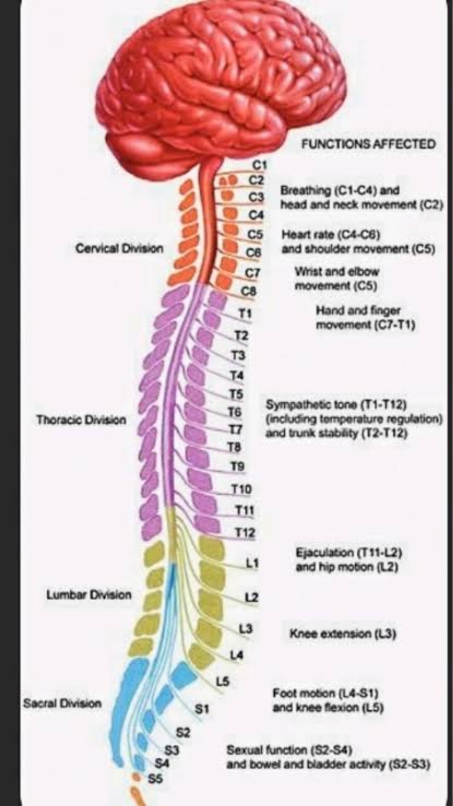
Segmental functional map of spinal nerves and descending CNS controls.
The Brain
- Is semisolid in consistency and conforms to the shape of its container.
- Weighs approximately 1400 g in an adult.
- Male brain is on average slightly heavier than the female brain.
- Protected from the external environment by three barriers:
- Skull
- Meninges
- Cerebrospinal fluid
Meninges
The brain and spinal cord are enclosed within three distinct membranous envelopes:
- Dura mater: Outermost, thickest, pachymeninx.
- Arachnoid mater: Intermediate avascular web-like leptomeninx.
- Pia mater: Delicate, highly vascular, adherent inner leptomeninx.
| Dural Specialization / Reflection | Anatomical Course | Contained Venous Sinuses & Functional Significance |
|---|---|---|
| Falx cerebri | Sickle-shaped vertical dural partition descending vertically into the longitudinal cerebral fissure between cerebral hemispheres | Contains the superior sagittal sinus along its upper attached convex border and the inferior sagittal sinus in its lower free margin. |
| Tentorium cerebelli | Crescentic horizontal dural roof over the posterior cranial fossa; separates occipital lobes from cerebellum | Contains transverse sinuses laterally and straight sinus along its junction with the falx cerebri; features the tentorial notch (incisura) accommodating the midbrain. |
| Falx cerebelli | Small sickle-shaped vertical reflection below tentorium cerebelli | Separates the cerebellar hemispheres within the posterior cranial fossa; contains the occipital sinus. |
| Diaphragma sellae | Small horizontal dural sheet roofing the sella turcica | Covers the pituitary gland (hypophysis cerebri); pierced centrally by the infundibulum (pituitary stalk). |
Tentorial Herniation (Uncal Herniation)
Supratentorial mass lesions (epidural hematoma, tumors) raise intracranial pressure, forcing the medial temporal lobe (uncus) inferiorly through the tentorial notch. This compresses:
- Oculomotor nerve (CN III): Ipsilateral pupillary dilation and "down-and-out" eye deviation.
- Cerebral peduncle: Contralateral hemiparesis (or ipsilateral hemiparesis via Kernohan's notch phenomenon).
- Posterior cerebral artery (PCA): Ipsilateral occipital infarction and contralateral homonymous hemianopia.
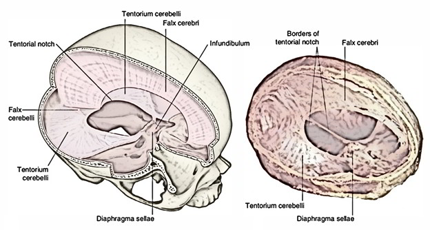
Dural partitions: Falx cerebri, tentorium cerebelli, tentorial notch, and diaphragma sellae.
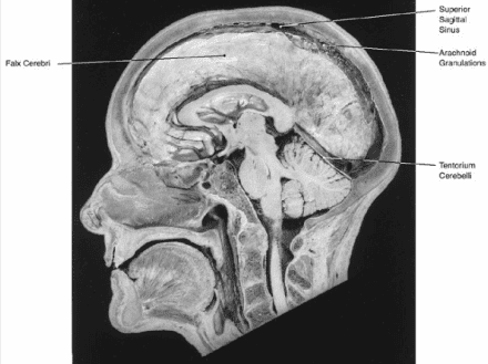
The brain showing the arachnoid meningeal layer and arachnoid granulations in situ.
SPACES BETWEEN MENINGES
- Epidural space
- Subdural space
- Subarachnoid space
| Meningeal Space | Anatomical Boundary & Nature | Contained Structures & Clinical Pathology |
|---|---|---|
| Epidural (Extradural) space | Potential space in the cranium between the bone (internal table of skull) and periosteal dura; true space in spinal canal | Contains fat and internal vertebral venous plexus in spinal cord. In cranium, skull fractures (pterion) lacerate the middle meningeal artery → Epidural Hematoma (lens-shaped/biconvex hyperdensity on CT, limited by suture lines). |
| Subdural space | Potential space between the inner meningeal dura mater and arachnoid mater | Traversed by delicate bridging veins draining into superior sagittal sinus. Acceleration/deceleration shearing leads to Subdural Hematoma (crescent-shaped, crosses suture lines, common in elderly/alcoholics). |
| Subarachnoid space | True space between the arachnoid mater and the pia mater | Filled with Cerebrospinal Fluid (CSF) and contains the major cerebral blood vessels. Rupture of saccular (berry) aneurysms in the circle of Willis causes Subarachnoid Hemorrhage (SAH) ("worst headache of life", bloody LP). |
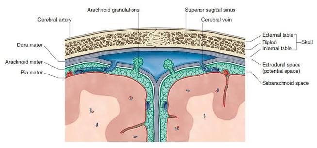
Arrangement of the cranial meninges, meningeal spaces, and superior sagittal sinus with arachnoid granulations.
CEREBRAL DURAL VENOUS SINUSES
Endothelium-lined venous channels situated between the endosteal (periosteal) and meningeal layers of the dura mater, collecting blood from the brain and CSF from the subarachnoid space.
| Dural Venous Sinus | Anatomical Location | Drainage Trajectory & Connections |
|---|---|---|
| Superior sagittal sinus | Upper fixed border of falx cerebri, from crista galli to confluence | Receives superior cerebral veins and CSF via arachnoid granulations; drains into the confluence of sinuses (usually right transverse). |
| Inferior sagittal sinus | Lower free concave margin of falx cerebri | Unites with the great cerebral vein (of Galen) at the anterior edge of tentorium cerebelli to form the straight sinus. |
| Straight sinus | Line of junction between falx cerebri and tentorium cerebelli | Runs downward and backward to empty into the confluence of sinuses (usually left transverse). |
| Confluence of sinuses (Torcular Herophili) | Internal occipital protuberance | Connecting confluence uniting superior sagittal, straight, and occipital sinuses with the bilateral transverse sinuses. |
| Transverse sinuses (Right & Left) | Attached posterolateral border of tentorium cerebelli | Courses horizontally along internal occipital groove, turning inferiorly at the petrous temporal bone to become the sigmoid sinus. |
| Sigmoid sinus | S-shaped groove in mastoid temporal and occipital bones | Direct continuation of transverse sinus; exits through the jugular foramen to become the internal jugular vein. |
| Cavernous sinus | Bilateral body of sphenoid bone in middle cranial fossa | Receives superior/inferior ophthalmic veins and sphenoparietal sinuses; connected by intercavernous sinuses; drains via superior and inferior petrosal sinuses into transverse sinus and internal jugular vein. |
| Basilar sinus | Clivus of the skull posterior to dorsum sellae | Interconnects bilateral inferior petrosal sinuses and communicates with internal vertebral venous plexuses. |
Cavernous Sinus Thrombosis (CST)
Facial infections in the "danger area" (nasolabial triangle) spread retrograde via valveless ophthalmic veins into the cavernous sinus. Structures at risk include:
- Internal carotid artery and Abducens nerve (CN VI) — traverse the center of the sinus; CN VI palsy (loss of lateral gaze) is the earliest sign.
- CN III, CN IV, CN V1 (ophthalmic), CN V2 (maxillary) — embedded in the lateral dural wall.
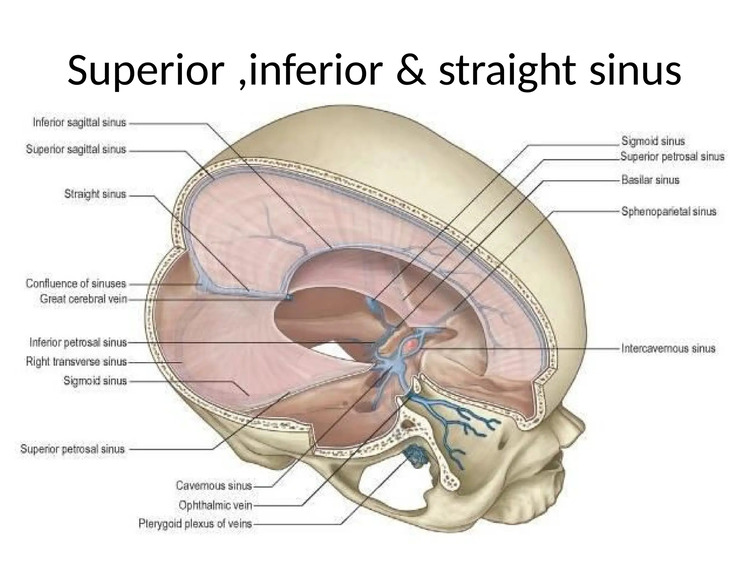
Lateral (A) and medial (B) surfaces of brain showing sites of dural venous sinuses.
Spinal Cord Meninges
| Spinal Meningeal Structure | Anatomical Architecture | Functional & Protective Relationship |
|---|---|---|
| Spinal Dura mater | Single-layered tube (lacks periosteal layer of cranial dura); separated from vertebrae by true epidural space | Extends inferiorly to level of S2 vertebra, where it tapers and invests the filum terminale to anchor on the coccyx. |
| Spinal Arachnoid mater | Avascular membrane lining dural sac | Maintains the expansive lumbar cistern containing CSF, cauda equina, and filum terminale below L1/L2. |
| Spinal Pia mater | Vascular membrane closely adhering to spinal cord parenchymal surface | Forms the denticulate ligaments (21 pairs of lateral tooth-like anchors pinning the cord to dura) and the filum terminale. |
| Vascular Supply & Innervation | Anterior and Posterior spinal arteries; Recurrent meningeal nerves | Anterior spinal artery runs in anterior median fissure (supplies anterior 2/3 of cord); two posterior spinal arteries supply posterior 1/3; recurrent meningeal nerves innervate dura and periosteum. |
Lumbar Puncture (Spinal Tap) Layers
Performed to safely sample CSF from the lumbar cistern without injuring the spinal cord (which terminates at L1–L2 in adults). The needle is typically inserted at L3/L4 or L4/L5 (supracristal plane), penetrating the following layers in sequence:
1. Skin → 2. Subcutaneous tissue → 3. Supraspinous ligament → 4. Interspinous ligament → 5. Ligamentum flavum ("pop" felt) → 6. Epidural space → 7. Dura mater ("pop" felt) → 8. Arachnoid mater → Subarachnoid space (lumbar cistern).
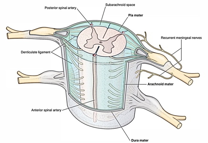
Spinal cord meninges, denticulate ligaments, subarachnoid space, and nerve roots.
Ventricular system
- Ventricles are developmentally derived from the cavity of the neural tube.
- Lined throughout with ependyma and are filled with cerebrospinal fluid.
- They are:
- Lateral ventricles (paired within cerebral hemispheres)
- Third ventricle (midline cleft within diencephalon)
- Fourth ventricle (cavity posterior to pons/medulla, anterior to cerebellum)
Lateral ventricles
- Two lateral ventricles and one is present in each cerebral hemisphere.
- Each is a roughly C-shaped cavity and includes a body, which occupies the parietal lobe.
- Anterior, posterior and inferior horns extend from the body into the frontal, occipital, and temporal lobes respectively.
- Communicate with the cavity of the 3rd ventricle through the interventricular foramen (of Monro), which lies in the medial wall of each ventricle.
- The choroid plexus of the lateral ventricles projects into each ventricle on its medial aspect.
| Ventricular Component | Lobar Location | Key Boundaries & Anatomical Relations |
|---|---|---|
| Anterior (Frontal) horn | Frontal lobe | Medially by septum pellucidum; roof and anterior wall by corpus callosum; floor and lateral wall by the bulbous head of the caudate nucleus. Lacks choroid plexus. |
| Body (Central part) | Parietal lobe | Extends from interventricular foramen to splenium of corpus callosum. Roofed by corpus callosum; floor formed by fornix, choroid plexus, thalamus, and stria terminalis. |
| Posterior (Occipital) horn | Occipital lobe | Tapers backward into white matter; medial wall marked by bulb of posterior horn (forceps major) and calcar avis (calcarine sulcus). Lacks choroid plexus. |
| Inferior (Temporal) horn | Temporal lobe | Curving anteroinferiorly around posterior thalamus. Floor contains the prominent hippocampus, fimbria, and collateral eminence; roof formed by tail of caudate nucleus and stria terminalis. |
| Atrium (Trigone) | Confluence area | Junction of body, posterior horn, and inferior horn; contains the largest clump of choroid plexus (glomus choroideum, frequently calcified on adult CT). |
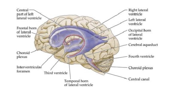
Three-dimensional anatomical configuration and components of the cerebral ventricles.
A cast of the ventricular cavities
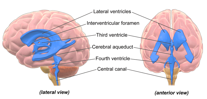
Casts of the ventricular cavities in lateral, coronal, and schematic views.
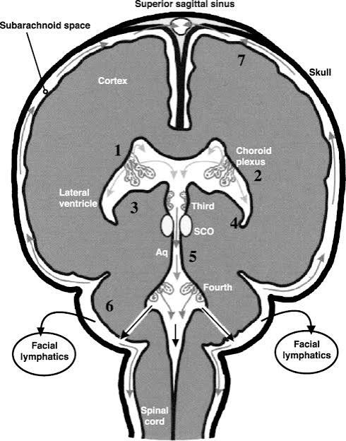
Coronal sections showing choroid plexus attachments in the lateral and third ventricles.
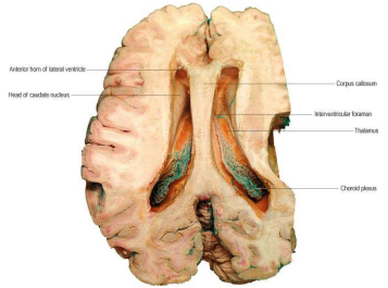
Horizontal brain slice demonstrating lateral ventricular anatomy, head of caudate, and choroid plexus.
Third Ventricle
- A slit-like cleft between the two thalami.
- Communicates anteriorly with the lateral ventricles through the interventricular foramina (of Monro) and posteriorly with the 4th ventricle through cerebral aqueduct (of Sylvius).
- Blood supply of the choroid plexuses of the 3rd and lateral ventricles is derived from the choroidal branches of the internal carotid and basilar arteries.
- Venous blood drains into the internal cerebral veins.
- Cerebral aqueduct of Sylvius is a narrow channel about 1.8 cm long which connects the 3rd with the 4th ventricle.
- It is lined with ependyma and is surrounded by a layer of gray matter called the central gray (periaqueductal gray).
| Boundary Wall | Constituent Anatomical Structures |
|---|---|
| Lateral Walls | Medial surfaces of the thalamus superiorly and hypothalamus inferiorly, separated by the hypothalamic sulcus; bridged across midline in ~70% of individuals by the interthalamic adhesion (massa intermedia). |
| Anterior Wall | Lamina terminalis and anterior commissure. |
| Posterior Wall | Pineal gland, habenular commissure, and posterior commissure; opens inferiorly into the cerebral aqueduct. |
| Roof | Delicate ependymal layer covered by the tela choroidea (double layer of pia mater harboring internal cerebral veins) below the fornix. |
| Floor | Formed by hypothalamic structures: optic chiasm, infundibulum, tuber cinereum, and mammillary bodies, plus the ventral tegmentum of the midbrain. |
Fourth Ventricle
- A cavity situated anterior to the cerebellum and posterior to the pons and the superior half of the medulla oblongata.
- Continuous above with the cerebral aqueduct and below with central canal of the spinal cord.
- Possesses lateral boundaries, a roof and a rhomboid-shaped floor.
- Lateral boundaries are formed by the superior and inferior cerebellar peduncles.
Fourth Ventricle (Roof & Apertures)
- The tent-shaped roof projects into the cerebellum.
- Formed by the superior medullary velum above and the inferior medullary velum below.
- Inferior medullary velum is pierced in the midline by a large opening, the median aperture, the foramen of Magendie.
Ventricular Exit Foramina
Foramen of Magendie (x1): Magendie = Median aperture (midline exit to cisterna magna).
Foramina of Luschka (x2): Luschka = Lateral apertures (lateral recesses opening to cerebellopontine angle).
Fourth Ventricle (Floor / Rhomboid Fossa)
- Floor is formed by the posterior surface of the pons and the cranial half of the medulla oblongata.
- In the midline is the median sulcus.
- On each side of this sulcus is the median eminence, which is bounded laterally by sulcus limitans.
- Lateral to sulcus limitans, there is the vestibular area, beneath which lie the vestibular nuclei.
- The facial colliculus, produced by the fibres of the facial nerve looping over the abducens nucleus, lies at the inferior end of the medial eminence.
Fourth Ventricle (Internal Features & Vascularization)
- Strands of nerve fibers, the stria medullaris, emerge from the median sulcus and pass laterally to enter the inferior cerebellar peduncle.
- Inferior to the stria, lies the hypoglossal triangle formed by the underlying hypoglossal nucleus and lateral to this lies the vagal triangle produced by the underlying dorsal motor nucleus of the vagus.
- The choroidal plexus of the 4th ventricle is suspended from the interior half of the roof of the ventricle.
- The blood supply to the plexus is from the inferior cerebellar arteries (PICA and AICA).
| Rhomboid Fossa Landmark | Underlying Neural Substrate | Clinical & Topographical Significance |
|---|---|---|
| Facial Colliculus | Abducens (CN VI) nucleus encircled by internal genu of Facial (CN VII) nerve | Lesions produce combined ipsilateral facial paralysis and lateral rectus palsy (internal strabismus). |
| Sulcus Limitans | Embryological boundary groove | Separates medial motor derivatives (basal plate) from lateral sensory derivatives (alar plate). |
| Substantia Ferruginea | Locus coeruleus | Bluish melanin-rich region at rostral floor; primary noradrenergic projection center of CNS. |
| Hypoglossal Triangle | Hypoglossal (CN XII) nucleus | Upper medullary floor; motor control of ipsilateral tongue musculature. |
| Vagal Triangle (Trigone) | Dorsal motor nucleus of Vagus (CN X) | Parasympathetic outflow to thoracic and abdominal viscera. |
| Area Postrema | Chemoreceptor trigger zone (CTZ) | Caudal floor border near obex; circumventricular organ lacking BBB, regulates emesis. |
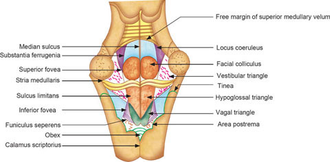
Floor of the fourth ventricle (rhomboid fossa) and underlying cranial nerve nuclei topography.
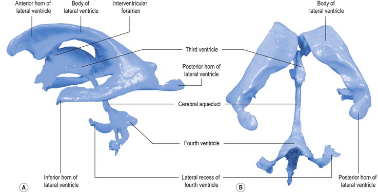
Morphological cast of cerebral ventricles demonstrating foramina, horns, and recesses.
Embryonic Brain Subdivisions, Adult Derivatives & Ventricles
| Primary Vesicle (Embryonic) | Secondary Vesicle | Adult Brain Derivatives | Associated Ventricular Space |
|---|---|---|---|
| Prosencephalon (Forebrain) |
Telencephalon | Cerebral cortex, Basal ganglia, Hippocampus, Olfactory bulb, Basal forebrain | Lateral ventricles |
| Diencephalon | Dorsal thalamus, Hypothalamus, Epithalamus (pineal), Subthalamus | Third ventricle | |
| Mesencephalon (Midbrain) |
Mesencephalon | Midbrain (superior and inferior colliculi, cerebral peduncles) | Cerebral aqueduct |
| Rhombencephalon (Hindbrain) |
Metencephalon | Cerebellum, Pons | Fourth ventricle |
| Myelencephalon | Medulla oblongata | Fourth ventricle | |
| Spinal cord | Spinal cord | Spinal cord | Central canal |
Central Canal
- Central Canal of the spinal cord and medulla oblongata opens superiorly into the 4th ventricle.
- Inferiorly, it extends through the inferior half of the medulla oblongata and through the entire length of the spinal cord.
- In the conus medullaris of the spinal cord, it expands to form the terminal ventricle.
- The central canal is closed at its lower end.
- It is filled with CSF, and is lined with ependyma.
- The central canal is surrounded by gray matter, the gray commissure.
- There is no choroid plexus in the central canal.
Subarachnoid Space
- Is the interval between the arachnoid mater and pia mater and envelopes the brain and spinal cord.
- Filled with CSF and contains the large blood vessels of the brain.
- Inferiorly, extends beyond the lower end of the spinal cord and invests the cauda equina.
- Ends below at the level of the interval between the second and third sacral vertebrae.
Subarachnoid Cisterns
- In certain locations around the base of the brain, the subarachnoid does not closely follow the surface of the brain so that the subarachnoid space expands to form cisterns.
- The cerebellomedullary cistern (cisterna magna) lies between the cerebellum and the medulla oblongata.
- The pontine cistern lies on the anterior surface of the pons.
- The Interpeduncular cistern lies on the anterior surface of the midbrain between the crura cerebri.
| Subarachnoid Cistern | Anatomical Boundary & Location | Traversing Structures & Diagnostic Use |
|---|---|---|
| Cerebellomedullary Cistern (Cisterna Magna) | Between inferior surface of cerebellum and posterior surface of medulla oblongata | Largest cranial cistern; receives CSF from fourth ventricle via foramen of Magendie; site of cisternal puncture when lumbar puncture is contraindicated. |
| Pontine Cistern | Ventral surface of pons | Contains the basilar artery and origins of abducens (CN VI) nerves. |
| Interpeduncular Cistern | Between cerebral peduncles (crura cerebri) of midbrain | Contains the circle of Willis (cerebral arterial circle) and oculomotor (CN III) nerves. |
| Superior Cistern (Quadrigeminal Cistern) | Posterior to midbrain tectum, between splenium of corpus callosum and superior cerebellar surface | Contains the great cerebral vein (of Galen) and pineal gland. |
| Lumbar (Spinal) Cistern | From L2 (termination of conus medullaris) down to S2 vertebra | Contains CSF, cauda equina nerve roots, and filum terminale; standard safe site for diagnostic lumbar puncture. |
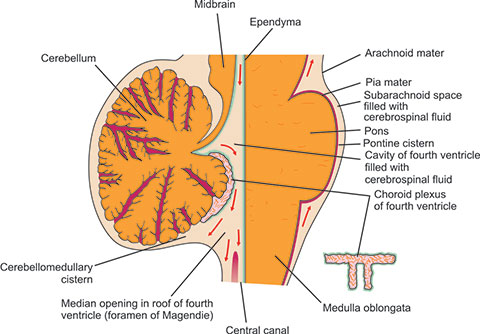
Fourth ventricle roof, foramen of Magendie, and adjacent cerebellomedullary and pontine cisterns.
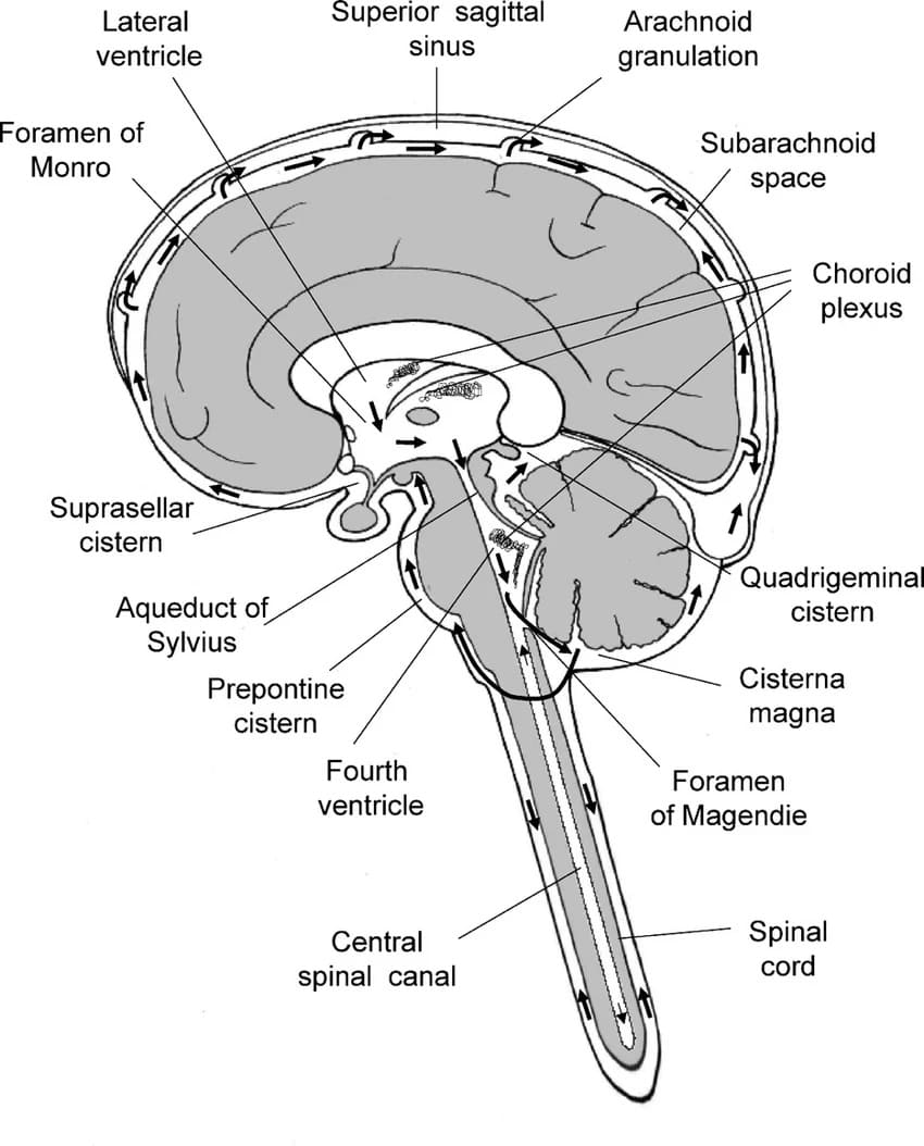
Comprehensive view of basal subarachnoid cisterns, ventricular connections, and spinal cistern.
Extensions of the subarachnoid space
- Around the optic nerve to the back of the eyeball where the arachnoid mater and pia mater fuse with the sclera.
- The central artery and vein of the retina cross this extension of the subarachnoid space to enter with the optic nerve and they may be compressed in patients with raised CSF pressure.
- Around the other cranial and spinal nerves.
- Around the arteries and veins of the brain and spinal cord at points where they penetrate the nervous tissue (forming perivascular Virchow-Robin spaces).
Pathophysiology of Papilledema
Because the subarachnoid space directly surrounds the optic nerve (CN II) within its dural sheath up to the sclera:
- Elevated intracranial pressure (ICP) is transmitted directly along the nerve sheath.
- Elevated CSF pressure compresses the thin-walled central retinal vein crossing the subarachnoid space.
- Venous outflow obstruction leads to axoplasmic stasis, retinal vein engorgement, blurring of optic disc margins, and papilledema observed on fundoscopy.
Cerebrospinal Fluid
Cerebrospinal fluid
- A clear, colorless liquid that fills the ventricles of the brain and bathes the external surface of the brain and spinal cord.
- Formed from the choroid plexus within the ventricles and circulates through the three openings in the roof of the fourth ventricle to reach the subarachnoid space.
- Produced continuously at a rate of about 0.5 ml per minute and with a total volume of about 30 ml (infant volume; adult total volume is approximately 140–150 ml): this corresponds to a turnover time of about 5 hours.
Formation of CSF
- Formed in the choroid plexuses of the lateral, third, and fourth ventricles; some originates as tissue fluid in the brain substance.
- The choroid plexuses have a folded surface and consist of a core of vascular connective tissue covered with cuboidal epithelium of the ependyma.
- The blood of the capillaries are separated from the ventricular lumen by fenestrated endothelium, a basement membrane and the surface epithelium. The ependymal cells of the choroid plexuses actively secrete the CSF.
Composition
- CSF has a specific gravity of about 1.007.
- Possesses in solution, inorganic salts similar to those in the blood plasma.
- Glucose content is about half that of blood and there is only a trace of protein.
- A few cells are present and these are lymphocytes.
- Normal lymphocyte count is 0 to 3 cells per cu mm.
- In the lateral recumbent position, CSF pressure, as measured by lumbar puncture, is about 60 to 150 mm of water.
- This pressure may be easily raised by straining, coughing or compressing the internal jugular veins in the neck (Queckenstedt test).
Composition Summary
- Contains not more than 5 lymphocytes/μl and is usually sterile.
| CSF Laboratory Parameter | Normal Reference Value | Clinical Significance |
|---|---|---|
| pH | 7.35 | Maintains precise central hydrogen ion concentration for medullary chemoreceptors. |
| Specific gravity | 1.007 | Matches neural tissue specific gravity to maintain neutral mechanical buoyancy. |
| Glucose | 66% of plasma glucose (~45–80 mg/dL) | Decreases profoundly (<40% of blood glucose) in bacterial and fungal meningitis due to microbial consumption. |
| Total protein | <45 mg/dl in the lumbar cistern | Elevated in infections, Guillain-Barré syndrome (albuminocytologic dissociation), and MS (oligoclonal bands). |
| Opening Pressure | 60 to 150 mm H₂O | Elevated (>200–250 mm H₂O) in intracranial tumors, hydrocephalus, and meningitis. |
Functions of the CSF
- Serves as a protective cushion between the central nervous system and the surrounding bones.
- Because the density of the brain is only slightly greater than that of the CSF, it provides mechanical buoyancy and support for the brain (reducing effective net weight of brain from ~1400 g to ~50 g).
- The close relationship of the fluid to the nervous tissue and the blood enables it to serve as a reservoir and assist in the regulation of the contents of the skull (Monro-Kellie doctrine).
- Is an ideal physiological substrate and probably plays an active part in the nourishment of the nervous tissue;
- Assists in the removal of products of neuronal metabolism.
- The secretions of the pineal gland possibly influence the activities of the pituitary gland by circulating through the CSF in the third ventricle.
Circulation of CSF
- Passes from the lateral ventricles into the 3rd ventricle through the interventricular foramina (of Monro).
- Then passes into the 4th ventricle through the cerebral aqueduct.
- The circulation is aided by the arterial pulsations of the choroid plexuses and the cilia on the ependymal cells lining the ventricles.
- From the 4th ventricle, the fluid passes through the median aperture (foramen of Magendie) and the lateral foramina of Luschka of the lateral recesses of the 4th ventricle and enters the subarachnoid space.
- The fluid then flows superiorly through the interval in the tentorium cerebelli to reach the inferior surface of the cerebrum.
- It now moves superiorly over the lateral aspect of each cerebral hemisphere.
- Some of the CSF moves inferiorly in the subarachnoid space around the spinal cord and cauda equina.
- The pulsations of the cerebral and spinal arteries and the movements of the vertebral column facilitate this flow of fluid.
Complete Pathway Flow of CSF
Choroid Plexuses (Lateral Ventricles) → Interventricular Foramina of Monro → Third Ventricle → Cerebral Aqueduct of Sylvius → Fourth Ventricle → Foramen of Magendie & Foramina of Luschka → Subarachnoid Space (Basal Cisterns & Spinal Canal) → Arachnoid Granulations → Superior Sagittal Sinus (Venous System).
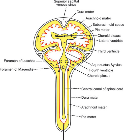
Complete circuit of CSF formation, ventricular circulation, cisternal flow, and dural venous reabsorption.
Detailed outflow architecture of fourth ventricle apertures into surrounding subarachnoid cisterns.
Absorption of CSF
- CSF is absorbed into the arachnoid villi that project into the dural venous sinuses, especially the superior sagittal sinus.
- The arachnoid villi are grouped together to form arachnoid granulations (Pacchionian bodies). Each arachnoid villus is a diverticulum of the subarachnoid space that pierces the dura mater.
- Absorption of CSF into the venous sinuses occurs when the CSF pressure exceeds that in the sinus (acts as a one-way pressure-dependent valve).
- Studies of the arachnoid villi indicate that fine tubules lined with endothelium permit a direct flow of fluid from the subarachnoid space into the lumen of the venous sinuses.
Blood – Brain Barrier
| Barrier Component | Ultrastructural Element | Physiological Barrier Mechanism |
|---|---|---|
| 1. Endothelial Cells | Endothelial cells in the wall of the capillary joined by continuous tight junctions (zonula occludens) | Primary physical barrier; non-fenestrated in brain parenchyma, abolishes paracellular diffusion. |
| 2. Basement Membrane | Continuous basement membrane surrounding the capillary outside the endothelial cells | Continuous extracellular matrix sheet providing structural support and charge selectivity. |
| 3. Astrocyte Foot Processes | Foot processes of astrocytes (podocytes / end-feet) that adhere to the outer surface of the capillary wall | Induce and maintain tight junction formation; regulate local microvascular transport and metabolism. |
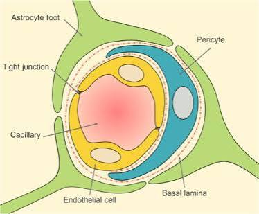
Structure: Capillary endothelial cells, continuous basement membrane, and astrocyte end-feet.
Blood – Brain Barrier Absence (Circumventricular Organs)
In select specialized neural midline regions known as Circumventricular Organs (CVOs), the blood-brain barrier is absent because the capillary endothelium possesses fenestrations, allowing direct neuroendocrine sampling or hormone secretion into the bloodstream.
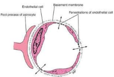
Blood-Brain Barrier Absence: Capillaries showing fenestrations in specialized neuroendocrine zones.
Circumventricular Organs ("Areas Without a Barrier")
Key structures lacking a blood-brain barrier include:
- Area Postrema: Chemoreceptor trigger zone in fourth ventricle floor; detects toxins to trigger vomiting.
- Posterior Pituitary (Neurohypophysis): Secretes vasopressin (ADH) and oxytocin directly into circulation.
- Median Eminence of Hypothalamus: Releases hypothalamic releasing/inhibiting factors into hypophyseal portal system.
- Pineal Gland: Secretes melatonin into blood.
- Subfornical Organ (SFO) & OVLT: Regulate osmoregulation and response to angiotensin II.
Blood – Brain Barrier (Permeability Characteristics)
- Gases and water pass readily through the barrier (rapid lipid diffusion of O₂, CO₂, and aquaporin channels).
- Glucose and electrolytes pass more slowly (require specialized facilitative carrier-mediated transporters like GLUT-1 and ion channels).
- The barrier is almost impermeable to plasma proteins and other large organic molecules.
- Compounds with molecular weights of about 60,000 and higher remain within the blood circulatory system.
Clinical Notes
| Clinical Condition / Investigation | Primary Neuropathophysiology | Diagnostic Manifestations & Clinical Management |
|---|---|---|
| The Optic Nerve, Raised Cerebrospinal Fluid Pressure, and Papilledema | Subarachnoid space surrounds optic nerve inside dural sheath to eyeball; raised ICP compresses central retinal vein. | Venous stasis causes bilateral edema and blurring of optic disc margins (papilledema), optic cup loss; heralds life-threatening intracranial mass lesions. |
| Hydrocephalus (Communicating & Noncommunicating) | Abnormal accumulation of CSF causing ventricular enlargement.
• Non-communicating (Obstructive): Structural block within ventricles (e.g., aqueductal stenosis, Dandy-Walker, colloid cyst). • Communicating: Free passage between ventricles, but impaired reabsorption at arachnoid granulations (post-meningitis, subarachnoid hemorrhage). |
Infants: Macrocephaly, bulging anterior fontanelle, "setting-sun" eye sign. Adults: Headache, vomiting, papilledema. Treated by ventriculoperitoneal (VP) shunting or endoscopic third ventriculostomy. |
| Clinical Investigation of the Cerebral Ventricles (CT & MRI) | Cross-sectional neuroimaging evaluating size, symmetry, and displacement of ventricular cavities. | Detects midline shift, mass effect, ventricular effacement, hydrocephalic dilatation, and intraventricular hemorrhages. |
| Cerebrospinal Fluid Pressure and Composition in Disease | Pathological alteration of CSF fluid dynamics and biochemistries during infection, hemorrhage, or demyelination. | Bacterial meningitis presents with cloudy CSF, polymorphonuclear pleocytosis, high protein, and low glucose. Viral presents with clear CSF, lymphocytic pleocytosis, and normal glucose. |
| Tumors of the Fourth Ventricle | Neoplasms (e.g., Ependymoma, Medulloblastoma in children) arising in roof or floor of fourth ventricle. | Causes rapid acute obstructive hydrocephalus via aqueduct/aperture occlusion; manifests with cerebellar ataxia, truncal instability, and persistent emesis. |
| Blood-Brain Barrier in the Fetus and Newborn | Immature development of tight junctions and astrocyte coverage in neonatal microvasculature. | Permits substances like unconjugated bilirubin to cross easily into basal ganglia, leading to Kernicterus (toxic encephalopathy with choreoathetosis and sensorineural deafness). |
| Brain Trauma and the Blood-Brain Barrier | Mechanical disruption of capillary tight junctions and glial architecture following contusion or concussive injury. | Rapid extravasation of plasma proteins and fluid into the extracellular space produces severe vasogenic cerebral edema and acute intracranial hypertension. |
| Tumors and the Blood-Brain Barrier | High-grade malignant primary brain neoplasms (e.g., Glioblastoma Multiforme) and metastases stimulate abnormal, disorganized angiogenesis. | Tumor vessels completely lack functional tight junctions, creating prominent leakage of contrast agents on MRI/CT and massive surrounding vasogenic edema. |
| Meningitis | Inflammation of leptomeninges (pia-arachnoid and subarachnoid CSF space) typically by bacterial (e.g., N. meningitidis, S. pneumoniae) or viral pathogens. | Triad of nuchal rigidity, fever, and altered mental status. Positive Kernig and Brudzinski signs; marked disruption of blood-brain barrier permits antibiotic penetration. |