Anatomy of The Brain and spinal cord
Topography
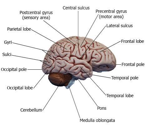
Introduction
I. The Brain
- is the part of the central nervous system (CNS)
- It lies within the cranial vault, the encephalon.
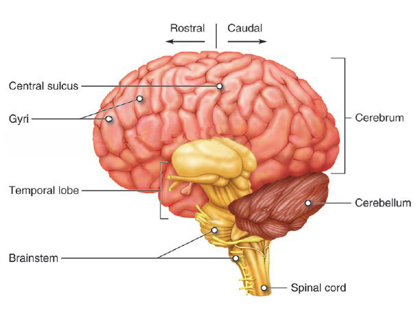
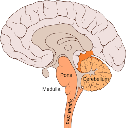
- consists of:
- the cerebrum
- cerebral hemispheres and diencephalon
- the brainstem
- (midbrain, pons, and medulla)
- the cerebellum.
- the cerebrum
- weighs 350 g in the newborn and 1400 g in the adult.
- is covered by three connective tissue membranes:
- the meninges.
- is surrounded by cerebrospinal fluid (CSF):
- which supports it and protects it from trauma.
Meningeal Coverings & CSF Buoyancy
The brain is enveloped by three connective tissue membranes: dura mater (outer tough pachymeninx), arachnoid mater, and pia mater (inner delicate leptomeninges). The cerebrospinal fluid (CSF) in the subarachnoid space provides hydraulic cushioning, reducing the effective weight of the 1400 g adult brain to approximately 50 g, preventing mechanical compression against the bony skull base.
Divisions of the Brain
The brain is classified into six postembryonic divisions:
- Telencephalon
- Diencephalon
- Mesencephalon
- Pons
- medulla oblongata
- cerebellum.
Telencephalon
Telencephalon consists of:
- the cerebral hemispheres
- (which comprise both cerebral cortex and white matter)
- the basal ganglia.
The cerebral hemispheres contain the lateral ventricles.
| Primary Brain Vesicle | Secondary Postembryonic Division | Adult Neural Structures & Cavities |
|---|---|---|
| Prosencephalon (Forebrain) | Telencephalon | Cerebral cortex, subcortical white matter, basal ganglia; Cavity: Lateral ventricles |
| Diencephalon | Thalamus, hypothalamus, epithalamus, subthalamus; Cavity: Third ventricle | |
| Mesencephalon (Midbrain) | Mesencephalon | Tectum (colliculi), tegmentum, cerebral peduncles; Cavity: Cerebral aqueduct (of Sylvius) |
| Rhombencephalon (Hindbrain) | Metencephalon | Pons and Cerebellum; Cavity: Superior fourth ventricle |
| Myelencephalon | Medulla oblongata; Cavity: Inferior fourth ventricle & central canal |
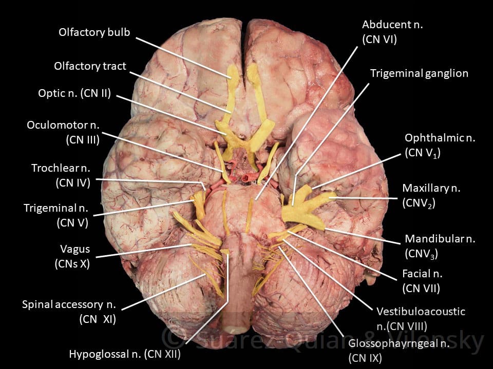
Ventral view of the brain displaying Olfactory tract, various cranial nerves and Internal carotid artery
The cerebral hemisphere
- is derived from the embryological telencephalon.
- It is the largest part of the forebrain and it reaches the greatest degree of development in the human brain.
- Superficially, the cerebral hemisphere consists of:
- a layer of grey matter called the cerebral cortex,
- It is highly convoluted to form a complex pattern of ridges (gyri) and furrows (sulci).
- This serves to maximise the surface area of the cerebral cortex:
- about 70% of which is hidden within the depths of sulci
- Beneath the surface:
- axons running to and from the cells of the cortex form an extensive mass of white matter.
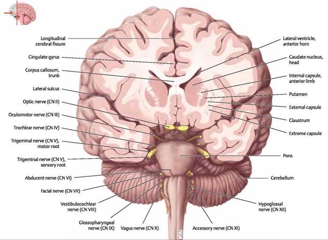
Coronal sections showing Head of caudate nucleus, Anterior limb of internal capsule, Corpus callosum, and Anterior horn of lateral ventricle
Cerebral hemispheres
- are separated by the longitudinal cerebral fissure and the falx cerebri.
- are interconnected by the corpus callosum.
- consist of four anatomical lobes and other functional lobes and the olfactory structures:
- a. Frontal lobe
- extends from the central sulcus to the frontal pole.
- lies above the lateral sulcus and anterior to the central sulcus.
- contains the following gyri:
- a. Frontal lobe
Frontal lobe
- (1) Precentral gyrus
- consists of the motor area (area 4).
- (2) Superior frontal gyrus
- contains the supplementary motor cortex on the medial surface (area 6).
- (3) Middle frontal gyrus
- contains the frontal eye field (area 8).
- (4) Inferior frontal gyrus
- contains the Broca speech area in the dominant hemisphere (areas 44 and 45).
- (5) Gyrus rectus and orbital gyri
- are separated by the olfactory sulcus.
- (6) Anterior paracentral lobule
- is found on the medial surface between the superior frontal gyrus (paracentral sulcus) and the central sulcus.
- represents a continuation of the precentral gyrus on the medial hemispheric surface.
Frontal Lobe Lesion Syndromes
Broca Area (Brodmann 44, 45): Lesion in the dominant hemisphere (inferior frontal gyrus) results in expressive (non-fluent) motor aphasia; comprehension remains intact, but fluency and speech production are impaired.
Frontal Eye Field (Brodmann 8): Destructive lesion of middle frontal gyrus causes conjugate gaze deviation toward the side of the lesion (ipsilesional preference) due to unopposed action of the contralateral frontal eye field.
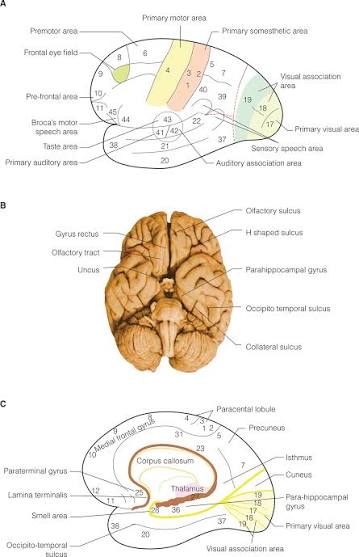
Topographical maps showing functional cortical areas and cerebral surface landmarks
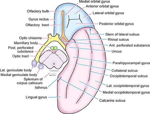
Fig. 8.7. Structures to be seen on the inferior aspect of the cerebrum. The midbrain has been cut across.
Parietal lobe
- extends from the central sulcus to the occipital lobe and lies superior to the temporal lobe.
- contains the following lobules and gyri:
- (1) Postcentral gyrus
- is the primary somatosensory area of the cerebral cortex (areas 3, 1, and 2).
- (2) Superior parietal lobule
- comprises association areas involved in somatosensory functions (areas 5 and 7).
- (3) Inferior parietal lobule
- (a) Supramarginal gyrus
- interrelates somatosensory, auditory, and visual input (area 40).
- (b) Angular gyrus (area 39)
- receives impulses from primary visual cortex.
- (a) Supramarginal gyrus
- (4) Precuneus
- is located between the paracentral lobule and the cuneus.
- (5) Posterior paracentral lobule
- is located on the medial surface between the central sulcus and the precuneus.
- represents a continuation of the postcentral gyrus on the medial hemispheric surface.
- (1) Postcentral gyrus
Gerstmann Syndrome (Angular Gyrus Lesion)
Lesions of the dominant angular gyrus (Brodmann 39) produce Gerstmann Syndrome, characterized by the classic tetrad:
- Agraphia (inability to write)
- Acalculia (inability to perform mathematical operations)
- Finger agnosia (inability to recognize own or examiner's fingers)
- Right-left disorientation
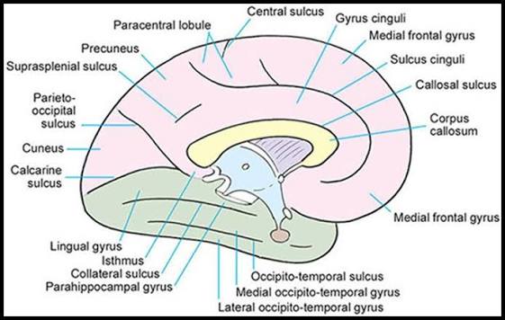
Medial cerebral landmarks: central sulcus, paracentral lobule, precuneus, parieto-occipital sulcus, cuneus, and calcarine sulcus
Temporal lobe
- extends from the temporal pole to the occipital lobe, lying below the lateral sulcus.
- extends from the lateral sulcus to the collateral sulcus.
- contains the following gyri:
- (1) Transverse temporal gyri of Heschl
- lie buried within the lateral sulcus.
- extend from the superior temporal gyrus toward the medial geniculate body
- are the primary auditory areas of the cerebral cortex (areas 41 and 42).
- (2) Superior temporal gyrus
- is associated with auditory functions.
- contains the Wernicke speech area in the dominant hemisphere (area 22).
- contains the planum temporale on its superior hidden surface.
- (3) Middle temporal gyrus
- (4) Inferior temporal gyrus
- (5) Lateral occipitotemporal gyrus (fusiform gyrus)
- lies between the inferior temporal sulcus and the collateral sulcus.
- (1) Transverse temporal gyri of Heschl
Language Cortex Differentiation
Wernicke Area (Area 22, Superior Temporal Gyrus): Receptive language center. Damage causes sensory (fluent) aphasia, characterized by word salad, neologisms, and impaired comprehension with preserved fluency.
Fusiform (Occipitotemporal) Gyrus: Houses the fusiform face area (FFA). Bilateral damage results in prosopagnosia (inability to recognize familiar faces).
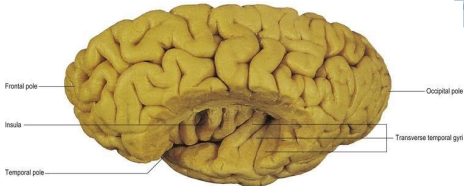
Lateral cerebral dissection exposing the Insula and Transverse temporal gyri
Occipital lobe
lies posterior to a line connecting the parieto-occipital sulcus and the preoccipital notch.
contains two structures:
- (1) Cuneus
- lies between the parieto-occipital sulcus and the calcarine sulcus.
- contains the visual cortex (areas 17, 18, and 19).
- (2) Lingual gyrus (medial occipitotemporal gyrus)
- lies below the calcarine sulcus.
- contains the visual cortex (areas 17, 18, and 19).
Calcarine Sulcus Retinotopic Mapping
Cuneus (Cranial/Upper bank): Represents the inferior contralateral visual quadrant ("Pie on the floor").
Lingual Gyrus (Lower bank): Represents the superior contralateral visual quadrant ("Pie in the sky").
Insular lobe (insula)
- lies buried within the lateral sulcus.
- has short and long gyri.
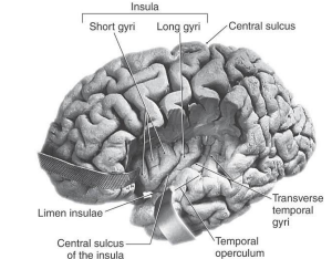
Topography of the Insular lobe showing Short gyri, Long gyri, and Limen insulae
Limbic lobe
- is a C-shaped structure of the medial hemispheric surface that encircles the corpus callosum and the lateral aspect of the midbrain.
- includes the following structures:
- (1) Paraterminal gyrus and subcallosal area
- are located anterior to the lamina terminalis and ventral to the rostrum of the corpus callosum.
- (2) Cingulate gyrus
- lies directly above the corpus callosum.
- merges with the parahippocampal gyrus at the isthmus.
- (3) Parahippocampal gyrus
- lies between the hippocampal and collateral sulci and terminates in the uncus.
- (4) Hippocampal formation
- lies between the choroidal and hippocampal fissures.
- is jelly-rolled into the parahippocampal gyrus.
- is connected to the hypothalamus and septal area via the fornix.
- includes the following three structures:
- (a) Dentate gyrus
- (b) Hippocampus
- (c) Subiculum
- (1) Paraterminal gyrus and subcallosal area
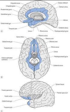
Limbic lobe boundaries along the medial and basal cerebral hemispheres
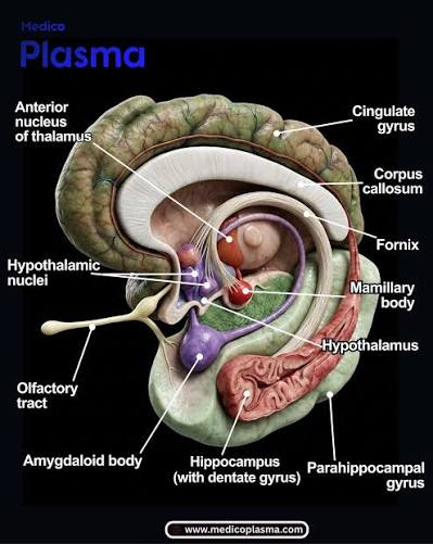
Limbic system anatomy
Uncal Herniation (Transtentorial Herniation)
Expanding supratentorial mass lesions (e.g., epidural hematoma) displace the medial temporal lobe uncus inferomedially across the tentorium cerebelli. This compresses:
- Oculomotor nerve (CN III): Ipsilateral fixed, dilated pupil ("blown pupil") and ptosis.
- Cerebral peduncle (Crus cerebri): Contralateral hemiparesis (or ipsilateral hemiparesis via Kernohan notch phenomenon).
- Posterior cerebral artery (PCA): Contralateral homonymous hemianopia with macular sparing.
Olfactory structures
are found on the orbital surface of the brain and include:
| Structure | Embryology / Origin | Functional Anatomy & Connections |
|---|---|---|
| (1) Olfactory bulb and tract | Telencephalic outgrowth | Are an outpouching of the telencephalon |
| (2) Olfactory bulb | Peripheral sensory relay | Receives the olfactory nerve (cranial nerve [CN] I) through cribriform plate |
| (3) Olfactory trigone and striae | Axonal divergence | Bifurcates into medial and lateral olfactory striae |
| (4) Anterior perforated substance | Vascular entry zone | Is created by penetrating striate arteries (lenticulostriate branches of MCA) |
| (5) Diagonal band of Broca | Telencephalic white fiber bundle | Interconnects the amygdaloid nucleus and the septal area |
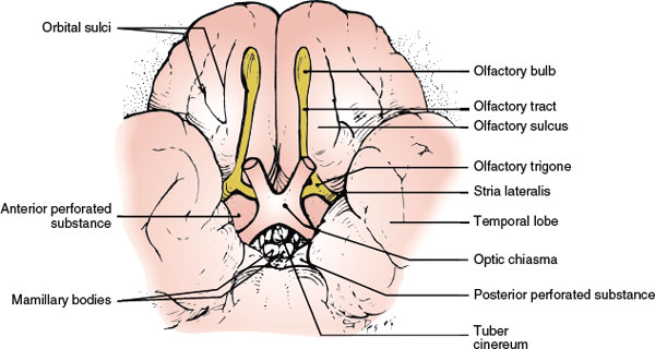
Ventral orbital surface showing olfactory tracts, olfactory trigone, and anterior perforated substance
Basal ganglia
are the subcortical nuclei of the telencephalon.
include the following structures:
- a. Caudate nucleus
- is part of the striatum, together with the putamen.
- b. Putamen
- is part of the striatum, together with the caudate nucleus.
- is part of the lentiform nucleus, together with the globus pallidus.
- c. Globus pallidus
- part of the lentiform nucleus, together with the putamen.
- d. Amygdaloid nuclear complex
- (amygdala)
Basal Ganglia Terminology Matrix
- Corpus Striatum = Caudate nucleus + Putamen + Globus pallidus
- Striatum (Neostriatum) = Caudate nucleus + Putamen
- Lentiform Nucleus = Putamen + Globus pallidus (external and internal segments)
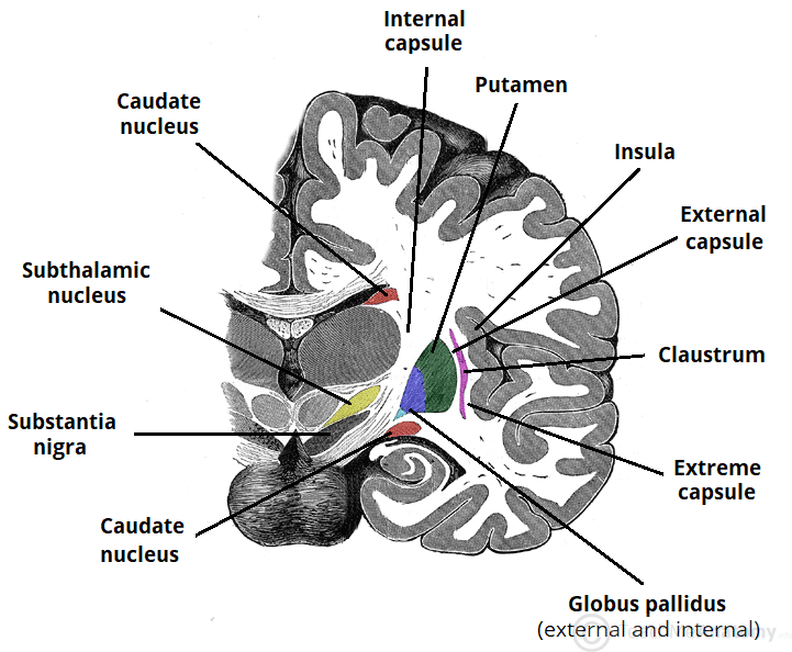
Coronal section displaying Caudate nucleus, Putamen, Globus pallidus, Internal capsule, and Substantia nigra
Lateral ventricles
- are ependyma-lined cavities of the cerebral hemispheres.
- contain CSF and choroid plexus.
- communicate with the third ventricle via the two interventricular foramina of Monro.
- are separated from each other by the septa pellucida.
Other features of the cerebrum
Cerebral cortex
- consists of a thin layer or mantle of gray substance.
- covers the surface of each cerebral hemisphere.
- is folded into gyri that are separated by sulci.
White matter
- includes the cerebral commissures and the internal capsule.
a. Cerebral commissures
interconnect the cerebral hemispheres and include:
- (1) Corpus callosum
- is the largest commissure of the brain.
- interconnects the two hemispheres.
- has four parts:
- (a) Rostrum
- (b) Genu
- (c) Body
- (d) Splenium
- (2) Anterior commissure
- is located in the midsagittal section between the lamina terminalis and the column of the fornix.
- interconnects the olfactory bulbs and the middle and inferior temporal lobes.
- (3) Hippocampal commissure
- (commissure of the fornix)
- is located between the fornices and ventral to the splenium of the corpus callosum.
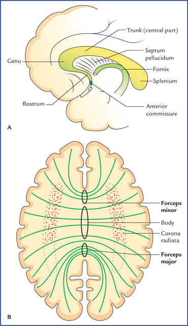
Morphology and fiber projections of the Corpus callosum: Forceps minor, Body, Corona radiata, and Forceps major
Diencephalon
- is located between the telencephalon and mesencephalon and between the interventricular foramen and the posterior commissure.
- receives the optic nerve (CN II).
- consists of:
- the epithalamus
- Thalamus
- Hypothalamus
- subthalamus,
- and the third ventricle and associated structures.
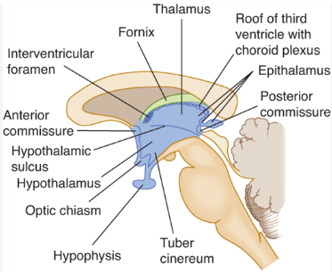
Diencephalic divisions and third ventricular boundaries
Mesencephalon (midbrain)
- is located between the diencephalon and the pons.
- extends from the posterior commissure to the frenulum of the superior medullary velum.
- contains the cerebral aqueduct, which interconnects the third and fourth ventricles.
- 1. Ventral surface
- a. Cerebral peduncle
- b. Interpeduncular fossa
- (1) Oculomotor nerve (CN III)
- (2) Posterior perforated substance: is created by penetrating branches of the posterior cerebral and posterior communicating arteries.
- 2. Dorsal surface
- a. Superior colliculus (visual system)
- b. Brachium of the superior colliculus
- c. Inferior colliculus (auditory system)
- d. Brachium of the inferior colliculus
- e. Trochlear nerve (CN IV): is the only cranial nerve to exit the brainstem from the dorsal aspect.
Tectal Colliculi Functional Relay
Superior Colliculus: Mediates visual reflexes and eye tracking movements (Superior = Sight).
Inferior Colliculus: Mediates auditory reflexes and acoustic processing (Inferior = Information heard).
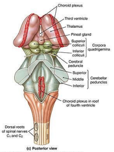
Posterior view of the brainstem showing Corpora quadrigemina and Cerebellar peduncles
Pons
- is located between the midbrain and the medulla.
- extends from the inferior pontine sulcus to the superior pontine sulcus.
- 1. Ventral surface
- a. Base of the pons
- b. Cranial nerves:
- (1) Trigeminal nerve (CN V)
- (2) Abducent nerve (CN VI)
- (3) Facial nerve (CN VII)
- (4) Vestibulocochlear nerve (CN VIII)
- 2. Dorsal surface (rhomboid fossa)
- a. Locus ceruleus
- contains the largest collection of norepinephrinergic neurons in the CNS.
- b. Facial colliculus
- contains the abducent nucleus and internal genu of the facial nerve.
- c. Sulcus limitans
- separates the alar plate from the basal plate.
- d. Striae medullares of the rhomboid fossa
- divide the rhomboid fossa into the superior pontine portion and the inferior medullary portion.
- a. Locus ceruleus
Cranial Nerve Pontine Junctions
- Anterolateral mid-pons: Exit site of the large sensory and small motor root of Trigeminal nerve (CN V).
- Pontomedullary junction: From medial to lateral emerges Abducent (CN VI) at the sulcus above the pyramid, followed by Facial (CN VII) and Vestibulocochlear (CN VIII) at the cerebellopontine angle.
Medulla oblongata
- is located between the pons and the spinal cord.
- extends from the first cervical nerve (C1) to the inferior pontine sulcus (also called the pontobulbar sulcus).
- 1. Ventral surface
- a. Pyramid: contains the corticospinal tract.
- b. Olive: contains the inferior olivary nucleus.
- c. Cranial nerves:
- (1) Glossopharyngeal nerve (CN IX)
- (2) Vagal nerve (CN X)
- (3) Accessory nerve (CN XI)
- (4) Hypoglossal nerve (CN XII)
- 2. Dorsal surface
- a. Gracile tubercle
- b. Cuneate tubercle
- c. Rhomboid fossa:
- (1) Striae medullares of the rhomboid fossa
- (2) Vagal trigone
- (3) Hypoglossal trigone
- (4) Sulcus limitans
- (5) Area postrema (vomiting center)
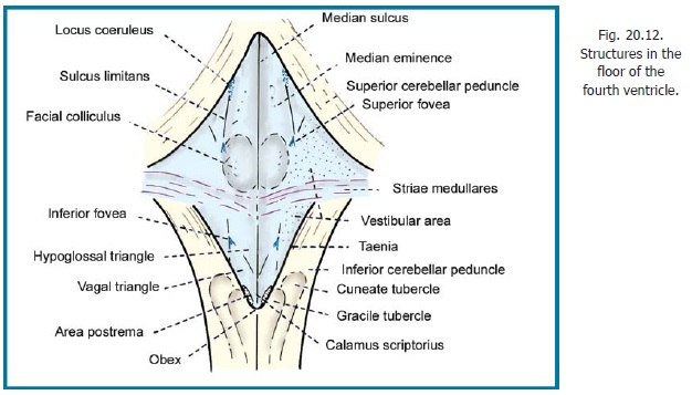
Topographical anatomy of the Rhomboid fossa (Floor of the Fourth Ventricle)
Area Postrema (Chemoreceptor Trigger Zone)
Located on the dorsal medulla immediately rostral to the obex, the area postrema is a circumventricular organ lacking a blood-brain barrier. It detects circulating toxins, chemotherapeutic agents, and emetic stimuli in blood and CSF to trigger the vomiting reflex.
Cerebellum
- is located in the posterior cranial fossa.
- is attached to the brainstem by three cerebellar peduncles.
- forms the roof of the fourth ventricle.
- is separated from the occipital and temporal lobes by the tentorium cerebelli.
- consists of folia and fissures on its surface.
- contains the following surface structures:
- 1. Hemispheres: are made up of two lateral lobes.
- 2. Vermis: is a midline structure.
- 3. Flocculus and vermal nodulus: form the flocculonodular lobule.
- 4. Tonsil:
- is a rounded lobule on the inferior surface of each cerebellar hemisphere.
- may herniate, with increased intracranial pressure, through the foramen magnum.
- 5. Superior cerebellar peduncle: connects the cerebellum to the pons and midbrain.
- 6. Middle cerebellar peduncle: connects the cerebellum to the pons.
- 7. Inferior cerebellar peduncle: connects the cerebellum to the medulla.
- 8. Anterior lobe: lies anterior to the primary fissure.
- 9. Posterior lobe: is located between the primary and posterolateral fissures.
- 10. Flocculonodular lobe: lies posterior to the posterolateral fissure.
| Peduncle | Primary Connection | Dominant Pathway |
|---|---|---|
| Superior cerebellar peduncle (Brachium conjunctivum) | Pons and Midbrain | Mainly efferent (dentatorubrothalamic tract to red nucleus & VL thalamus) |
| Middle cerebellar peduncle (Brachium pontis) | Pons | Entirely afferent (pontocerebellar fibers from contralateral basilar pons) |
| Inferior cerebellar peduncle (Corpus restiforme) | Medulla oblongata | Mainly afferent (dorsal spinocerebellar, olivocerebellar, cuneocerebellar, and vestibulocerebellar fibers) |
Tonsillar Herniation (Chiari Malformations)
Mass effect within the posterior cranial fossa forces the cerebellar tonsils downward through the foramen magnum. This causes life-threatening compression of the ventrolateral medulla, targeting vital cardiorespiratory centers and leading to respiratory arrest.
Spinal Cord
I. Introduction:
Spinal Cord
- is derived from the caudal part of the neural tube.
- maintains segmental organization throughout development.
- is surrounded by three membranes, the meninges.
- weighs about 30 g, comprising 2% of the weight of the adult brain.
External Morphology
Location:
- extends, in adults, from the foramen magnum to the lower border of the first lumbar vertebra (L1);
- in newborns, it extends to the third lumbar vertebra (L3).
- is continuous with the medulla oblongata at the spinomedullary junction:
- a plane defined by three structures:
- the foramen magnum,
- the pyramidal decussation
- and the emergence of the first cervical nerve ventral rootlets.
- a plane defined by three structures:
- lies within the subarachnoid space:
- which extends caudally to the level of the second sacral vertebra (S2).
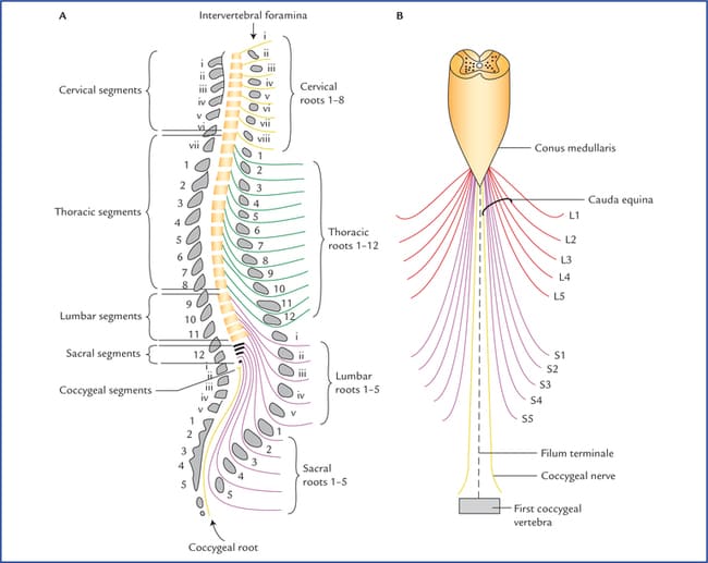
Alignment of spinal cord segments with vertebrae, conus medullaris, and dural termination
Attachments
- suspend and anchor the spinal cord within the dural sac.
- arise from the vascular pia mater, which closely invests the spinal cord.
1. Denticulate ligaments
- are two flattened bands of pial tissue that attach to the spinal dura with about 21 attachments.
2. Filum terminale
- is a pial filament extending from the conus medullaris to the end of the dural sac, with which it fuses.
3. Spinal nerve roots
- provide the strongest anchorage and fixation of the spinal cord to the vertebral canal.
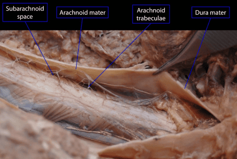
Spinal cord & meninges, posterolateral view
Lumbar Puncture (Spinal Tap) Landmarks
Because the adult spinal cord terminates at the lower border of L1 (conus medullaris) while the subarachnoid space (lumbar cistern) extends to S2, a lumbar puncture is safely performed at the L3/L4 or L4/L5 intervertebral space (aligned with the supracristal plane of the iliac crests), accessing CSF without risking needle injury to the spinal cord.
Shape of the spinal cord
- is an elongated
- nearly cylindrical structure
- flattened dorsoventrally
- and is approximately 1 cm in diameter.
- has cervical (C5–T1) and lumbar (L1–S2) enlargements:
- for the nerve supply of the upper and lower extremities (the brachial and lumbosacral plexuses).
- terminates caudally as the conus medullaris.
- averages, in length, 45 cm in males and 42 cm in females.
- consist of 31 pairs of nerves that emerge from the spinal cord:
- 8 cervical,
- 12 thoracic
- 5 lumbar
- 5 sacral
- and 1 coccygeal.
Other features
- contain both motor and sensory fibers.
1. Special considerations
- a. The first cervical nerve and the coccygeal nerve:
- usually have no dorsal (sensory) roots and no corresponding dermatomes.
- b. The first cervical nerve:
- passes between the atlas and the skull.
- c. The second cervical nerve:
- passes between the atlas and the axis.
- d. With the exception of C1:
- spinal nerves exit the vertebral canal via intervertebral or sacral foramina.
Functional components of spinal nerve fibers
| Functional Fiber Type | Origin / Pathway | Peripheral Distribution & Function |
|---|---|---|
| a. General somatic afferent (GSA) fibers | Pseudounipolar cells in dorsal root ganglia | Convey sensory input from skin, muscle, bone, and joints to the central nervous system (CNS). |
| b. General visceral afferent (GVA) fibers | Pseudounipolar neurons with visceral projections | Convey sensory input from visceral organs to the CNS. |
| c. General somatic efferent (GSE) fibers | Ventral horn alpha & gamma motor neurons | Convey motor output to skeletal muscle. |
| d. General visceral efferent (GVE) fibers | Intermediolateral cell column neurons & sacral parasympathetic nucleus | Convey sympathetic motor output via paravertebral/prevertebral ganglia to glands, smooth muscle, and visceral organs; convey parasympathetic motor output from sacral nucleus to pelvic viscera via intramural ganglia. |
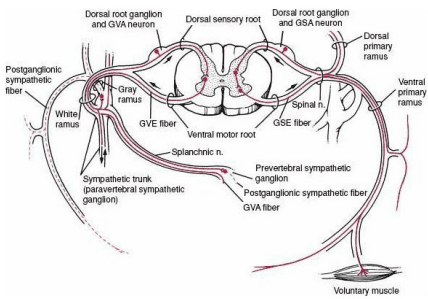
Neural pathways and functional fiber components (GSA, GVA, GSE, GVE) of a spinal nerve
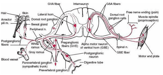
Somatic and autonomic reflex circuitry: GSA, GVA, GSE, and GVE functional trajectories
Spinal Nerve Fiber Components Summary
- GSA (General Somatic Afferent): Body wall sensations (pain, temperature, touch, proprioception from skin and joint capsules).
- GSE (General Somatic Efferent): Voluntary skeletal muscle movement.
- GVA (General Visceral Afferent): Distension and ischemia sensations from internal visceral organs.
- GVE (General Visceral Efferent): Autonomic motor control (sympathetic from T1–L2 lateral horn; parasympathetic from S2–S4 sacral segments).
