CEREBRUM & FUNCTIONAL AREAS
Brain Functions
- Motor Cortex
- Sensory Cortex
- Vision
- Taste
- Cognition
- Emotion
- Speech
- Language
- Hearing
- Autonomic Functions
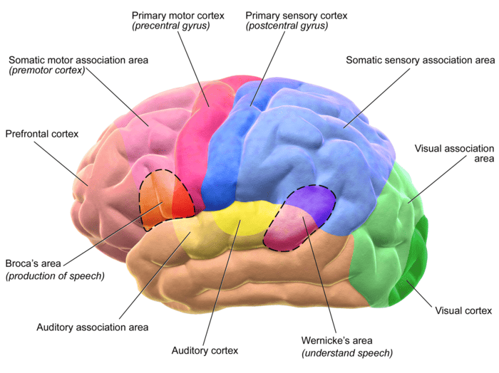
Functional areas of the cerebral cortex
Cortical Topography & Somatotopic Organization
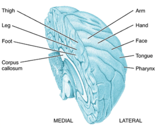
Medial vs. Lateral representation across the cortical mantle
Motor Homunculus
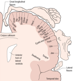
Motor Homunculus
Sensory Homunculus
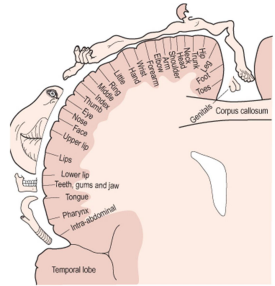
Sensory Homunculus
Vascular Homunculus Rule ("Legs in the River, Face on the Street")
Anterior Cerebral Artery (ACA): Supplies the medial surface of the motor and sensory strips → controls the foot, leg, and perineum/genitals ("feet dangling in the midline river"). Occlusion leads to contralateral lower extremity motor and sensory loss.
Middle Cerebral Artery (MCA): Supplies the lateral convexity → controls the trunk, arm, hand, face, and speech areas ("face and arms out on the lateral street"). Occlusion causes contralateral upper extremity and facial deficits plus aphasia.
Brodmann areas
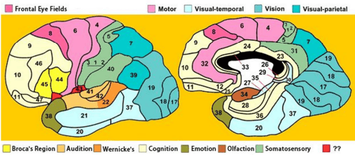
Brodmann areas
Frontal Lobe
- Primary motor cortex - Area 4
- Premotor and supplementary motor cortex - Area 6
- Frontal eye fields - Area 8
- Dorsolateral prefrontal cortex - Area 9
- Prefrontal cortex - Areas 9, 10
- Anterior prefrontal cortex - Area 10
- Broca's area, involved in motor speech - Areas 44, 45
Parietal Lobe
- Primary somatosensory cortex - Areas 1, 2, 3
- Somatosensory association cortex - Areas 5, 7
- Supramarginal gyrus, involved in language perception - Area 40
- Angular gyrus, important for language, math, and cognition - Area 39
Temporal Lobe
- Primary auditory cortex - Areas 41, 42
- Part of the superior temporal gyrus, included in Wernicke's area - Area 22
- Middle temporal gyrus - Area 21
- Inferior temporal gyrus - Area 20
Occipital Lobe
- Primary visual cortex - Area 17
- Secondary visual cortex (V2) - Area 18
- Associative visual cortex (V3) - Area 19
| Cerebral Lobe | Brodmann Area | Anatomical / Functional Designation |
|---|---|---|
| Frontal | Area 4 | Primary motor cortex (Precentral gyrus) |
| Frontal | Area 6 | Premotor cortex and supplementary motor area (SMA) |
| Frontal | Area 8 | Frontal eye fields (conjugate voluntary saccades) |
| Frontal | Areas 9, 10 | Dorsolateral and anterior prefrontal cortex (executive cognition) |
| Frontal | Areas 44, 45 | Broca's area (pars opercularis & pars triangularis; motor speech) |
| Parietal | Areas 3, 1, 2 | Primary somatosensory cortex (Postcentral gyrus) |
| Parietal | Areas 5, 7 | Somatosensory association cortex (Superior parietal lobule) |
| Parietal | Area 39 | Angular gyrus (reading, math, cross-modal integration) |
| Parietal | Area 40 | Supramarginal gyrus (phonological processing & language perception) |
| Temporal | Areas 41, 42 | Primary auditory cortex (Heschl's transverse gyri) |
| Temporal | Area 22 | Wernicke's area / Superior temporal gyrus (receptive language) |
| Temporal | Areas 21, 20 | Middle and inferior temporal gyri (semantic and visual object recognition) |
| Occipital | Area 17 | Primary visual cortex / Striate cortex (V1; Calcarine sulcus) |
| Occipital | Areas 18, 19 | Secondary (V2) and associative (V3) visual cortex (extrastriate) |
Functional areas (regions) of cerebral neocortex
- Motor area: somatotopically organized around the cruciate sulcus.
- drives voluntary movement and it
- is the primary source of pyramidal tract fibers to cranial nerve nuclei and spinal cord (corticospinal tracts).
- Primary sensory areas: receive specific afferents of a given modality from the thalamus or geniculate bodies.
- Somesthetic (somatosensory) area: receives specific tactile input as well as information related to pain, temperature and pressure sensation.
- Visual area: receives visual input. The area is retinotopically organized in the occipital lobe around the marginal sulcus.
- Auditory area: receives auditory input. The area is cochleotopically organized around the apex of the pseudosylvian fissure.
- Vestibular area: receives vestibular apparatus input. It is rostral to the auditory area.
- Note: Taste is represented in the somesthetic area near the tongue region. Olfaction is conscious detected at the piriform lobe (paleocortex).
Association areas:
- These are cortical areas that receive their specific input from other cortical areas, and so they are not involved directly with processing sensory and motor information.
- Principally involved integrating and interpreting information derived from primary sensory areas.
Motor Cortex
The light red area is the premotor cortex, which is responsible for repetitive motions of learned motor skills. The dark red area is the primary motor area, and is responsible for control of skeletal muscles.
Different areas of the brain are associated with different parts of the body.
Injury to the motor cortex can result in motor disturbance in the associated body part.
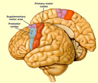
Primary motor area and premotor cortex
Cognition
The prefrontal cortex is involved with intellect, complex learning, and personality.
Injuries to the front lobe can cause mental and personality changes.
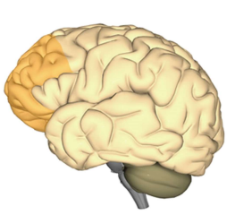
Prefrontal cortex involved with cognition and personality
Frontal Lobe Syndrome (Phineas Gage)
Lesions to the orbitofrontal and dorsolateral prefrontal cortex cause loss of executive function, disinhibition, emotional lability, poor judgment, lack of foresight, and profound changes in social behavior and personality while preserving baseline intelligence.
Speech
Broca’s area is where we formulate speech and the area of the brain that sends motor instructions to the motor cortex.
Injury to Broca’s area can cause difficulty in speaking. The individual may know what words he or she wishes to speak, but will be unable to do so.
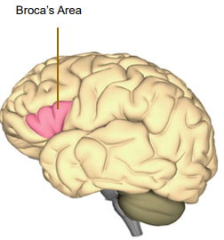
Broca’s Area
Broca's (Expressive / Non-Fluent) Aphasia
Presentation: Speech is effortful, halting, and agrammatic ("broken speech"), but comprehension remains intact. Patients possess insight into their deficit, resulting in significant frustration. Caused typically by left superior division MCA infarction.
Sensory Cortex
The sensory portion of the cerebrum is illustrated here.
Injury to the sensory cortex can result in sensory disturbance in the associated body part.
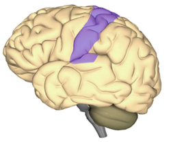
Sensory cortex (postcentral gyrus)
Taste
The gustatory complex (green circle) is the part of the sensory cortex (purple area) that is responsible for taste.
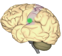
Gustatory complex on the sensory cortex
Hearing
There are two auditory areas of the brain:
- The primary auditory area (brown circle) is what detects sounds that are transmitted from the ear. It is located in the sensory cortex.
- The auditory association area (purple circle) is the part of the brain that is used to recognize the sounds as speech, music, or noise.
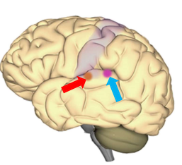
Primary auditory area and auditory association area
Language
Wernicke’s area is a specialized portion of the parietal lobe that recognizes and understands written and spoken language.
Wernicke’s area surrounds the auditory association area.
Damage to this part of the brain can result in someone hearing speech, but not understanding it.
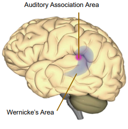
Wernicke’s Area & Auditory Association Area
Wernicke's (Receptive / Fluent) Aphasia
Presentation: Speech is fluent, rapid, and effortless, but entirely nonsensical and devoid of meaning ("word salad" with paraphasic errors). Auditory comprehension and repetition are severely impaired. Patients lack insight (anosognosia). Typically caused by left inferior division MCA infarction.
Vision
The visual cortex resides in the occipital lobe of the brain.
Sensory impulses travel from the eyes via the optic nerve to the visual cortex.
Damage to the visual cortex can result in blindness.
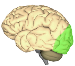
Visual cortex residing in the occipital lobe
Emotion
Emotions are an extremely complex brain function.
The emotional core of the brain is the limbic system. This is where senses and awareness are first processed in the brain.
Mood and personality are mediated through the prefrontal cortex. This part of the brain is the center of higher cognitive and emotional functions.
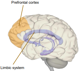
Prefrontal cortex and Limbic system
Limbic System Functions ("The 5 F's")
The functional spectrum of the limbic system encompasses: Feeding (satiety/hunger), Fleeing (fear response/amygdala), Fighting (aggression), Feeling (emotion), and Sex (reproduction/mating drive), anchored by Forming memories (hippocampus).
Injury Mechanisms
The brain is a complex and delicate organ, and one that is vulnerable to injury from a variety of different traumas. These include:
- Frontal Lobe Injury
- Occipital Lobe Injury
- Temporal Lobe Injury
- Side Impact Injury
- Coup/Contre-coup Injury
- Diffuse Axonal Injury
- Epidural Hematoma
- Subdural Hematoma
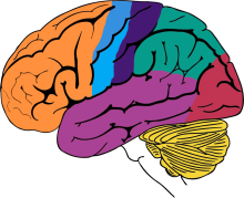
Cerebral topography and traumatic vulnerability
Frontal Lobe Injury
The frontal lobe of the brain can be injured from direct impact on the front of the head.
During impact, the brain tissue is accelerated forward into the bony skull. This can cause bruising of the brain tissue and tearing of blood vessels.
Frontal lobe injuries can cause changes in personality, as well as many different kinds of disturbances in cognition and memory.
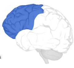
Frontal lobe injury location
Occipital Lobe Injury
Occipital lobe injuries occur from blows to the back of the head.
This can cause bruising of the brain tissue and tearing of blood vessels.
These injuries can result in vision problems or even blindness.
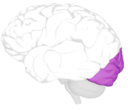
Occipital lobe injury location
Temporal Lobe Injury
The temporal lobe of the brain is vulnerable to injury from impacts of the front of the head.
The temporal lobe lies upon the bony ridges of the inside of the skull, and rapid acceleration can cause the brain tissue to smash into the bone, causing tissue damage or bleeding.
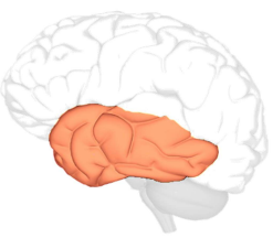
Temporal lobe injury location
Injury to Specific Gyri
- Occipitotemporal (fusiform) gyrus: a visual agnosia for objects, prosopagnosia, and pure alexia (the inability to read).
- Angular gyrus: alexia and agraphia.
- Orbital gyrus: impaired processing of olfactory and gustatory inputs and integration of emotions and memories associated with sensory experiences.
- ETC
Gerstmann Syndrome (Dominant Angular Gyrus Lesion)
Damage to the dominant angular gyrus (Area 39) produces the classic tetrad:
- Agraphia (inability to write)
- Acalculia (inability to perform mathematical operations)
- Finger agnosia (inability to distinguish fingers)
- Right-left disorientation
Lesions to the fusiform gyrus specifically cause prosopagnosia (inability to recognize familiar faces).
Side Impact Injury
Injuries to the right or left side of the brain can occur from injuries to the side of the head.
Injuries to this part of the brain can result in language or speech difficulties, and sensory or motor problems.
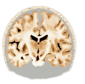
Coronal cross-section vulnerable to lateral impacts
Coup/Contre-coup Injury
A French phrase that describes bruises that occur at two sites in the brain.
When the head is struck, the impact causes the brain to bump the opposite side of the skull. Damage occurs at the area of impact and on the opposite side of the brain.
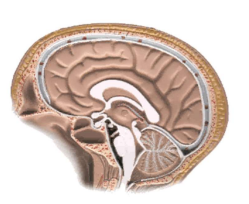
Coup / Contre-coup injury mechanism
Diffuse Axonal Injury
Brain injury does not require a direct head impact. During rapid acceleration of the head, some parts of the brain can move separately from other parts. This type of motion creates shear forces that can destroy axons necessary for brain functioning.
These shear forces can stretch the nerve bundles of the brain.
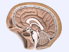
Shear force injury to deep cerebral nerve bundles
The brain is a complex network of interconnections. Critical nerve tracts can be sheared and stressed during an acceleration-type of injury.
Diffuse axonal injury is a very serious injury, as it directly impacts the major pathways of the brain.
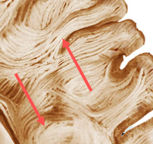
Shearing and destruction of critical white matter pathways in diffuse axonal injury
Intracranial Hematomas Differential
| Feature | Epidural Hematoma (EDH) | Subdural Hematoma (SDH) |
|---|---|---|
| Vessel Ruptured | Middle meningeal artery (pterion fracture) | Bridging cortical veins (acceleration/shear) |
| CT Morphology | Biconvex / Lenticular; does NOT cross suture lines | Crescent-shaped; crosses suture lines |
| Clinical Course | Lucid interval followed by rapid herniation | Slowly progressive headache, confusion in elderly/alcoholics |
CYTOARCHITECTURE: GRAY MATTER
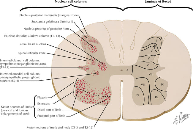
Ventral Horn Motor Neuron Topography
Medial-to-Lateral: Proximal to distal musculature → Medial neurons supply the axial/trunk muscles, while lateral neurons supply the extremities/hands.
Anterior-to-Posterior: Action → Anterior (ventral) neurons supply extensors, while posterior (dorsal) neurons supply flexors.
CYTOARCHITECTURE: WHITE MATTER
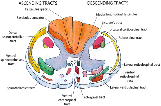
Ascending and descending tracts and somatotopic organization of the dorsal columns
Dorsal Column Somatotopy ("Gracilis = Ground")
Fasciculus Gracilis (Medial): Carries sensory fibers from the lower body (Sacral & Lumbar) — "Gracilis touches the grass/ground".
Fasciculus Cuneatus (Lateral): Carries sensory fibers from the upper body (Thoracic T6 and above & Cervical) — "Cuneatus carries the Cervical arms".
CLINICALLY IMPORTANT SPINAL CORD STRUCTURES
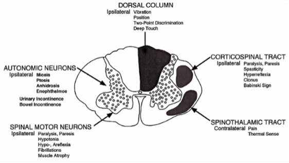
Schematic diagram of clinically important spinal cord structures and effects of lesions in each
| Spinal Structure | Functional Modality | Clinical Manifestations of Lesion |
|---|---|---|
| DORSAL COLUMN | Epicritic sensation: Vibration, conscious proprioception (position), two-point discrimination, deep touch | Ipsilateral: Loss of vibration sense, loss of position sense, impaired two-point discrimination, sensory ataxia (positive Romberg test) |
| CORTICOSPINAL TRACT | Upper Motor Neuron (UMN) voluntary skeletal motor control | Ipsilateral: Paralysis, paresis, spasticity, hyperreflexia, clonus, extensor plantar response (Babinski sign) |
| SPINOTHALAMIC TRACT | Protopathic sensation: Pain, temperature, and crude touch | Contralateral: Loss of pain and thermal sense starting 1–2 segments below the lesion level |
| SPINAL MOTOR NEURONS (Anterior Horn) | Lower Motor Neuron (LMN) execution to muscle motor units | Ipsilateral: Flaccid paralysis, paresis, hypotonia, hypo-/areflexia, fibrillations, fasciculations, severe muscle atrophy |
| AUTONOMIC NEURONS (Intermediolateral Column T1–L2 / S2–S4) | Sympathetic & parasympathetic visceromotor regulation | Ipsilateral: Horner's syndrome (miosis, ptosis, anhidrosis, enophthalmos); neurogenic bladder (urinary incontinence), bowel incontinence |
SPINAL CORD SYNDROMES
In clinical practice, the following clinicopathologic syndromes are encountered:
- Segmental Lower Motor Neuron Syndrome (seen in poiliomyelitis)
- Hemisection (Brown-Sequard Syndrome)
- Anterior Horn and Lateral Corticospinal Tract Syndrome (Motor Neuron Disease)
- Lesions Around Central Canal (Syringomyelia)
- Combined System Degeneration Syndrome (seen in Pernicious anaemia patients)
- Anterior Spinal Artery Syndrome
- Transection
- Conus Medullaris Syndrome
- Cauda Equina Syndrome
- Autonomic Syndromes
| Syndrome | Primary Pathology & Affected Structures | Hallmark Clinical Features |
|---|---|---|
| Segmental LMN Syndrome | Infection/destruction of anterior horn motor neurons (e.g., poliomyelitis, enterovirus) | Pure LMN signs: Asymmetric flaccid paralysis, fasciculations, muscle atrophy, absent deep tendon reflexes; no sensory loss |
| Brown-Séquard Syndrome | Hemicord transection/compression (e.g., stab wound, tumor) | Ipsilateral: UMN spastic paralysis below lesion, loss of vibration/proprioception. Contralateral: Loss of pain/temperature 1–2 levels below |
| Motor Neuron Disease | Degeneration of anterior horn cells (LMN) + lateral corticospinal tracts (UMN) (e.g., ALS) | Combined UMN and LMN deficits: Spasticity and hyperreflexia alongside muscle wasting and fasciculations; completely intact sensation |
| Syringomyelia | Fluid-filled cavitation (syrinx) expanding from the central canal (cervical spine; Chiari malformation) | Compression of crossing anterior white commissure fibers → bilateral "cape-like" loss of pain and temperature over arms and shoulders; dorsal columns spared |
| Subacute Combined Degeneration | Vitamin B12 deficiency (seen in pernicious anaemia); demyelination of dorsal columns & lateral corticospinal tracts | Bilateral loss of vibration/proprioception, sensory ataxia, spastic paresis, positive Babinski sign, paresthesias in stocking-glove pattern |
| Anterior Spinal Artery Syndrome | Ischemia/infarction of anterior 2/3 of spinal cord (anterior spinal artery thrombosis or aortic cross-clamping) | Bilateral paraplegia (corticospinal), bilateral loss of pain/temperature (spinothalamic), autonomic bowel/bladder dysfunction; dorsal columns spared |
| Complete Spinal Transection | Complete traumatic severance or severe transverse myelitis across full spinal cord diameter | Initial spinal shock (flaccidity, areflexia), followed by permanent bilateral spastic paralysis, complete sensory anesthesia below level, loss of all sphincter control |
| Conus Medullaris Syndrome | Lesion at vertebral levels L1–L2 terminating the spinal cord (sacral cord segments S3–S5) | Early and severe symmetric perianal numbness ("saddle anesthesia"), bilateral early urinary and fecal incontinence, symmetric hyperreflexic/spastic lower limb signs |
| Cauda Equina Syndrome | Compression of lumbosacral nerve roots below L2 (massive midline disc herniation, trauma, tumor) | Markedly asymmetric radicular pain, asymmetric flaccid leg weakness (LMN), hyporeflexia/areflexia, late bowel/bladder overflow incontinence; neurosurgical emergency |
| Autonomic Syndromes | Disruption of sympathetic outflow / descending hypothalamospinal tracts (e.g., high thoracic cord lesions) | Autonomic dysreflexia (life-threatening hypertension, reflex bradycardia triggered by visceral distension), neurogenic shock, orthostatic hypotension |
Conus Medullaris vs. Cauda Equina Syndrome
Conus Medullaris: Involves the distal spinal cord itself → Symmetric onset, early prominent urinary/bowel incontinence, mixed UMN/LMN signs, bilateral symmetrical saddle numbness.
Cauda Equina: Involves individual peripheral nerve roots → Asymmetric onset, severe unilateral radicular pain, pure LMN signs (flaccid paralysis, absent knee/ankle jerks), delayed urinary incontinence.