Tissue Healing, Cellular Regeneration, and Wound Repair Pathology
Key Definitions in Tissue Restoration
- Biological Adaptation to Insult: Multicellular organisms must execute organized physiological countermeasures to withstand chemical, mechanical, and inflammatory microenvironmental trauma.
- Tissue Repair / Healing: The coordinated biological cascade mobilized by the host following traumatic or pathological disruption, structured to reinstate anatomical architecture and physiological competency.
- Fundamental Pathways: Following the eradication of inciting injurious agents and the clearance of acute inflammatory exudates, tissue reconstitution proceeds via parenchymal regeneration, fibrous connective tissue repair, or a synchronous combination of both modalities.
1. Fundamental Mechanisms of Tissue Reconstitution
Host structural recovery bifurcates into two distinct cellular pathways that determine whether original architecture is perfectly re-established or replaced by structural collagen:
| Restorative Process | Primary Cellular Mediators | Structural Outcome | Mandatory Prerequisite |
|---|---|---|---|
| Parenchymal Regeneration | Mitotically active, uninjured native parenchymal cells and resident stem cell populations. | Comprehensive restoration of original histology and specialized physiological function without scarring. | Preservation of the underlying extracellular matrix (ECM framework and basement membrane). |
| Connective Tissue Repair (Fibrosis) | Recruited fibroblasts, transdifferentiated myofibroblasts, and proliferating capillary endothelial cells. | Deposition of fibrous collagenous stroma resulting in permanent fibrous scar formation (cicatrization). | Occurs when extracellular stromal scaffolding is destroyed or native tissue lacks proliferative potential. |
In extensive traumatic injuries, parenchymal restoration and fibrovascular scar synthesis frequently operate simultaneously in adjacent micro-domains.
2. Biology of Cellular Regeneration
For parenchymal restoration to achieve complete anatomical integrity, two core criteria must be fulfilled simultaneously:
- The damaged tissue compartment must contain cells endowed with mitotic replication competence or harbor an uninjured resident stem cell pool capable of asymmetrical division and terminal lineage differentiation.
- The underlying connective tissue stroma and basement membranes must remain structurally stable to supply the mechanical polarity and architectural guidance necessary for organized cellular layering.
High regenerative capacity is prominent within cutaneous epithelia, gastrointestinal mucosal linings, and hepatic parenchyma. Mitogenic stimulus is provided through paracrine signaling cascades orchestrated by macrophage-derived polypeptide growth factors. Conversely, tissues populated by permanent post-mitotic cells—such as ventricular cardiomyocytes and central nervous system neurons—cannot mount regenerative hyperplasia and inevitably heal via permanent fibrotic scar tissue.
3. Cell Cycle Dynamics and Proliferative Classifications
Parenchymal turnover varies significantly across tissue compartments: while some cell populations turn over rapidly within days, others remain arrested in basal metabolic states throughout the lifetime of the host. Tissue stability requires strict governance via the eukaryotic cell cycle.
Phases of the Eukaryotic Cell Cycle
- Mitosis Phase (M): Active karyokinesis (prophase, metaphase, anaphase, telophase) and cytokinesis, generating two diploid daughter cells.
- First Gap Phase (G1): Post-mitotic interval characterized by intense organelle biogenesis, metabolic preparation, and cellular growth prior to genome duplication.
- Synthesis Phase (S): Critical interval where coordinated semiconservative replication of nuclear genomic DNA occurs.
- Second Gap Phase (G2): Pre-mitotic checkpoint phase focusing on protein synthesis, DNA mismatch error scanning, and structural preparation for spindle assembly.
- Quiescence Phase (G0): Non-proliferative, metabolically viable resting interval into which cells exit following mitosis.
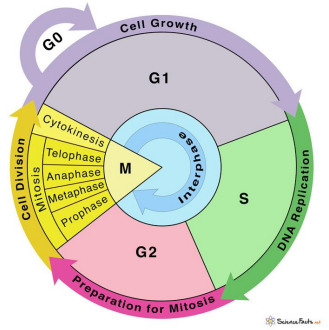
The Regulatory Stages of the Eukaryotic Cell Cycle
The principal kinetic variable dictating the rate of tissue turnover is the chronological duration of the G1 phase. Rapidly renewing cellular systems complete cycle progression every 16 to 24 hours, whereas slow-cycling populations remain elongated in G1 or reversibly arrested within G0.
| Cell Classification | Proliferative Capacity | Typical Tissue Distribution | Repair Capacity |
|---|---|---|---|
| Labile (Continuously Dividing) Cells | Undergo continuous baseline mitotic division throughout the life of the individual, deriving from local stem cells. | Stratified squamous epidermis; linings of the gastrointestinal, respiratory, and urinary tracts; vaginal and cervical mucosa; uterine endometrium; hematopoietic marrow; splenic and lymph node lymphoid cells. | Exceptional regenerative capacity; returns to normal histology if stromal architecture is maintained. |
| Stable (Quiescent) Cells | Display low basal mitotic turnover in adulthood (arrested in G0), but retain the competence to rapidly re-enter G1 upon chemical or mechanical injury. | Visceral parenchymal organs (hepatocytes, renal tubular cells, pancreatic acini, adrenal cortex, thyroid); mesenchymal lineages (vascular endothelial cells, fibroblasts, smooth muscle cells, osteoblasts, chondrocytes). | Substantial compensatory hyperplasia and regeneration following injury (e.g., partial hepatectomy). |
| Permanent (Non-dividing) Cells | Irreversibly exited the cell cycle around perinatal development; completely lack replicative machinery in postnatal life. | Central nervous system neurons, skeletal myocytes, and left/right ventricular cardiac myocytes. | Zero parenchymal replication; necrotic lesions are repaired exclusively by connective tissue scarring and gliosis. |
4. Morphological Stages of Fibrovascular Repair
When substantial tissue damage disrupts both parenchymal populations and the supporting extracellular stromal scaffold, structural reconstitution can occur only via connective tissue substitution. This fibrovascular repair process comprises two synchronized biological actions:
- Granulation Tissue Synthesis: A three-phase reaction encompassing an acute inflammatory response, enzymatic debris clearance, and vascular/fibroblastic ingrowth.
- Wound Contraction: Physical reduction of the wound defect mediated by phenotypically altered stromal cells.
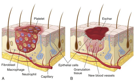
Evolution of Acute Inflammation into Organizing Granulation Tissue
The Triphasic Formation of Granulation Tissue
- Phase 1: Acute Exudative Inflammation (0–24 Hours): Physical trauma causes microvascular hemorrhage, activating the extrinsic and intrinsic clotting cascades. A stable fibrin-platelet meshwork fills the wound void to limit hemorrhage and prevent desiccation. Chemotactic signals draw an acute cellular infiltrate composed of plasma proteins, high concentrations of polymorphonuclear neutrophils, and early circulating monocytes.
- Phase 2: Enzymatic Clearance and Phagocytosis (Days 1–3): Coordinated debridement of necrotic devitalized cells, spent erythrocytes, and matrix debris. Neutrophil degranulation releases neutral proteases, complemented by endogenous autolytic lysosomal enzymes. By day 3, recruited tissue macrophages dominate the field, orchestrating active phagocytosis and releasing cytokines essential for fibroblastic induction.
- Phase 3: Angiogenic and Fibrogenic Ingrowth (Days 3–7+): Endothelial and mesenchymal cells aggressively invade the provisional fibrin lattice. The term granulation tissue derives from its granular, hyperemic surface, where each minute pink granule corresponds histologically to newly sprouted, loop-forming capillary arcades suspended in edematous ground substance rich in fibroblasts and young collagen.
5. Molecular Drivers of Angiogenesis (Neovascularization)
Angiogenesis represents the formation of new vascular channels from pre-existing blood vessels. Within healing wounds, this occurs predominantly through endothelial sprouting:
- Local tissue hypoxia and protease activation trigger proteolytic degradation of the parent basement membrane via matrix metalloproteinases (MMPs).
- Endothelial pericytes detach under the influence of angiopoietin signaling, allowing vascular dilation mediated by nitric oxide and Vascular Endothelial Growth Factor (VEGF).
- A specialized endothelial "tip cell" extends filopodia and migrates toward chemotactic growth factor gradients.
- Trailing stalk endothelial cells proliferate aggressively to construct a solid cord, which subsequently undergoes vacuolization and canalization to form a functional vascular lumen.
- Perivascular adventitial cells and pericytes are recruited via Platelet-Derived Growth Factor (PDGF) to stabilize the newly developed vascular wall.
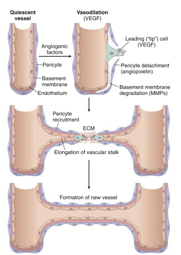
Signaling Events and Molecular Steps of Angiogenic Sprouting
Microvascular Hyperpermeability in Early Repair
Nascent capillary sprouts exhibit loose, immature inter-endothelial junctions and incomplete basement membrane coverage. Elevated local concentrations of VEGF dramatically amplify vascular permeability. This structural leakiness causes marked plasma fluid and protein extravasation, explaining the characteristically edematous and fragile appearance of fresh granulation tissue on gross inspection.
6. Fibrogenesis, Collagen Synthesis, and Cicatrization
As neovascularization proceeds, fibroblasts migrate into the wound space along the fibrin-fibronectin provisional matrix:
- Origin of Repair Fibroblasts: Derived from mitotic expansion of locally situated adventitial fibroblasts and transformation of quiescent resting fibrocytes.
- Myofibroblastic Differentiation: Under the influence of Transforming Growth Factor-beta (TGF-β) and mechanical matrix tension, a subset of migrating fibroblasts acquires features intermediate between fibroblasts and smooth muscle cells, including the expression of alpha-smooth muscle actin (α-SMA).
- Timeline of Collagen Deposition: Appreciable collagen fibril synthesis initiates around the 6th postoperative day. Early granulation matrix is enriched in compliant Type III collagen.
- Cicatrization: Over subsequent weeks, progressive matrix remodeling exchanges Type III collagen for rigid, highly cross-linked Type I collagen. Concurrently, vascular channels regress via apoptosis, active fibroblasts diminish, and the hyperemic granulation tissue evolves into a hypovascular, hypocellular, pale, dense fibrous scar (a state termed cicatrization).
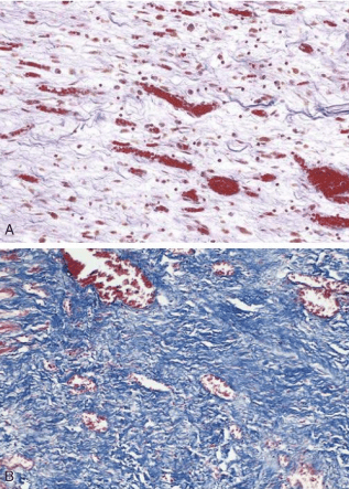
Histological Maturation: From Vascular Granulation Tissue to Collagenous Scar
7. Dynamics and Cellular Biology of Wound Contraction
Wound contraction is an active physiological process that significantly diminishes the volume of the original tissue defect:
- Temporal Profile: Centripetal inward margin movement commences around day 2 to day 3 following injury and achieves completion by approximately the 14th day.
- Magnitude: In open, gaping wounds, this process can reduce the surface area of the original defect by up to 80%, substantially minimizing the parenchymal volume requiring replacement by fibroplasia.
Evaluation of Proposed Mechanisms of Contraction
- Dehydration Theory (Historical): Posited that simple fluid loss and surface desiccation produced passive tissue shrinkage. Disproven as an active motor force.
- Collagen Contraction Theory (Historical): Suggested that progressive shortening of cross-linked collagen bundles pulled edges together. Disproven because peak wound contraction occurs between days 3 and 14, an interval when mature fibrillar collagen content remains very low.
- The Myofibroblast Model (Established): Contraction is executed by myofibroblasts. These cells establish dense cell-to-cell adherens junctions and intracellular actin-myosin bundles anchored to the extracellular fibronectin scaffold, applying coordinated mechanical traction to draw opposing wound margins toward the center.
8. Primary vs. Secondary Intention Cutaneous Repair
Cutaneous repair provides the classic paradigm of combined regeneration and scarring, bifurcating clinically into primary and secondary intention based on tissue loss and wound geometry:
| Clinical Parameter | Primary Intention (First Intention / Primary Union) | Secondary Intention (Secondary Union) |
|---|---|---|
| Wound Characteristics | Clean, uninfected surgical incision with minimal parenchymal cell loss; basement membrane disruption is restricted. | Extensive loss of cellular architecture, irregular margins, significant tissue gap; frequently complicated by microbial contamination. |
| Margin Approximation | Edges closely opposed and mechanically stabilized with surgical sutures, staples, or adhesive tape. | Wound margins are widely separated; defect cannot be primarily closed and must heal upward from the base. |
| Inflammatory Intensity | Mild, transient acute inflammation; minimal necrotic debris to clear. | Profound, protracted inflammatory phase due to copious necrotic slough, exudate, and microbial burden. |
| Granulation Quantity | Sparse volume of granulation tissue bridges the thin incisional slit. | Massive volume of granulation tissue required to slowly fill the extensive tissue cavity. |
| Role of Wound Contraction | Negligible to absent; margins are already in direct physical contact. | Crucial; myofibroblast-driven centripetal contraction reduces defect size by up to 80%. |
| Healing Time & Scarring | Rapid; complete epithelial continuity restored within 48 hours; results in a thin, linear, cosmetically neat scar. | Protracted, indolent course; yields a large, irregular, contracted, and cosmetically prominent scar. |
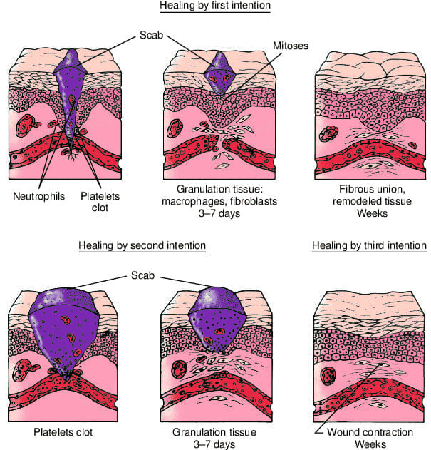
Mechanisms of Healing by First, Second, and Third Intention
9. Chronological Cascade of Primary Union
- Immediate Period (0–2 Hours): Blood rapidly extravasates into the incisional track. Fibrin polymerization creates a surface seal that shields against environmental pathogens and prevents desiccation.
- Within 24 Hours: Margins display active acute inflammation with early neutrophil infiltration. Mitotic activity accelerates within basal epidermal cells along both cut margins.
- 24 to 48 Hours: Basal epidermal cells mobilize and migrate horizontally across the wound gap beneath the non-viable surface eschar as dynamic epithelial spurs. In accurately approximated incisions, an intact, continuous sheet of epithelial cells covers the incision within 48 hours.
- Day 3: Recruited macrophages replace neutrophils as the dominant myeloid cell. Fibroblasts vigorously invade the provisional fibrin matrix along new capillary buds.
- Day 5: Neovascularization reaches peak density. Collagen fibrils bridge the incisional gap. Epidermal stratification restores normal multilayered cutaneous architecture.
- Day 7 (Suture Removal): Wound tensile strength measures approximately 10% of uninjured skin. Re-epithelialized suture puncture tracts close as sutures are extracted; retained epithelium within the track is typically resorbed.
- End of Week 4: Dermal collagen continues cross-linking, vascularity recedes, and an inactive, stable scar forms. Tensile strength reaches ~70% to 80% of normal skin by 3 months, but never fully regains 100% of pre-injury strength.
Pathology of Suture Tracks
Every puncture tract formed by a surgical needle represents an independent microscopic wound undergoing identical repair processes. If epidermal cells migrate down the suture track and fail to resorb following suture removal, they can persist and form an implantation epidermal inclusion cyst. Bacterial contamination along the foreign material can precipitate a localized, painful stitch abscess requiring drainage.
10. Chronological Cascade of Secondary Union
Wounds managed by secondary intention follow an extended timeline dictated by the physical dimensions of the parenchymal defect:
- Extensive Clotting: Initial extravasation generates a substantial blood and fibrin mass that dehydrates into a thick surface crust.
- Protracted Inflammatory Phase: Substantial quantities of necrotic tissue require extensive macrophage debridement, delaying the transition toward anabolism.
- Delayed Re-epithelialization: Although basal epidermal margins actively sprout, migrating epithelial spurs cannot bridge the chasm until substantial granulation tissue rises from the wound bed to provide a viable vascular substrate.
- Exuberant Granulation Synthesis: The crater fills with deep red, highly vascular, and friable granulation tissue that is susceptible to minor shear trauma.
- Robust Wound Contraction: Activated myofibroblasts contract along structural stress lines, physically reducing the wound surface area by up to three-quarters to four-fifths of its original dimensions.
- Impact of Microbial Contamination: Persistent bacterial colonization releases collagenases, elastases, and cytolytic toxins that induce secondary microvascular thrombosis and tissue necrosis. Healing halts until surgical debridement mechanically eliminates this devitalized tissue.
11. Extracellular Matrix (ECM) Components and Tensile Architecture
The extracellular matrix serves as more than passive structural glue; it functions as a dynamic biochemical reservoir regulating cell migration, proliferation, polarity, and structural remodeling. The ECM comprises five primary molecular components:
| ECM Constituent | Biochemical Subtypes & Structure | Primary Anatomical Location | Mechanical / Functional Role |
|---|---|---|---|
| Collagens | Fibrillar and non-fibrillar triple-helix glycoproteins (over 18 distinct isoforms described). | Type I Collagen represents 90% of total body collagen, distributed in dermis, bone, tendons, and mature scars. | Confers critical tensile framework and resistance to mechanical shear forces across tissues. |
| Adhesive Glycoproteins | Multifunctional cell-matrix adhesive ligands including fibronectin, tenascin (cytotactin), and thrombospondin. | Intercellular interstitial matrix and organizing granulation beds. | Functions as biological adhesive bridging cell-surface integrins with extracellular structural proteins; guides cell migration. |
| Basement Membranes | Amorphous, Periodic Acid–Schiff (PAS)-positive specialized matrices composed of non-fibrillar Type IV collagen and laminin. | Underlying all visceral/cutaneous epithelia and investing endothelial cell tubes. | Establishes cell polarity, regulates compartmental permeability, and serves as the obligate scaffold for parenchymal regeneration. |
| Elastic Fibers | Bicomponent protein networks consisting of an amorphous central elastin core surrounded by peripheral fibrillin microfibrils. | Large elastic arteries, pulmonary alveolar septa, dermis, and articular cartilage. | Endows tissues with passive recoil capacity following mechanical distension; degraded by neutrophil elastases during severe inflammation. |
| Proteoglycans & GAGs | Core proteins linked to glycosaminoglycan side chains: chondroitin sulfate, heparan sulfate, dermatan sulfate, keratan sulfate, and hyaluronic acid. | Widespread ground substance; abundant in dermis, cartilage, synovial fluid, and basement membranes. | Forms a highly hydrated gel conferring compressive resilience; their deposition strictly precedes collagen fibril laying during repair. |
12. Factors Influencing Healing Competency
Wound repair is sensitive to both local tissue microenvironmental conditions and systemic metabolic parameters:
Comprehensive Analysis of Wound Modifying Factors
- Local Modifying Factors:
- Microbial Infection: Quantitatively the single most common cause of delayed healing. It perpetuates destructive neutrophil activity and drives secondary tissue necrosis.
- Vascular Compromise: Diminished arterial perfusion (atherosclerosis, microvascular diabetic sclerosis) or impaired venous drainage (varicosities, chronic venous hypertension) starves reparative cells of oxygen and essential nutrients.
- Foreign Bodies: Retained surgical sutures, fragments of glass, wood splinters, or devitalized bone sequestra incite persistent foreign-body granulomatous inflammation.
- Mechanical Stress: Early physical motion, shear forces, or excessive tension across incision lines can disrupt immature microvessels and cause wound dehiscence.
- Electromagnetic Radiation: Exposure to ionizing therapeutic radiation damages endothelial and stromal progenitor cells; high-dose ultraviolet exposure retards repair.
- Wound Topography & Volume: Extensive deep wounds in poorly vascularized anatomic compartments heal with marked fibrosis and permanent functional impairment.
- Systemic Modifying Factors:
- Advanced Age: Reductions in cellular turnover rates, slower re-epithelialization, and diminished collagen cross-linking occur with aging.
- Nutritional Status: Severe protein depletion halts fibroplasia. Vitamin C deficiency (scurvy) abolishes prolyl and lysyl hydroxylase function, preventing stable collagen triple-helix assembly and causing immediate wound breakdown.
- Systemic Sepsis: Diverts metabolic substrate and produces generalized circulatory and immune dysregulation.
- Glucocorticoid Administration: High-dose exogenous corticosteroids exert potent anti-inflammatory effects, suppressing TGF-β transcription, decreasing fibroblast proliferation, and yielding mechanically frail, under-collagenized scars.
- Diabetes Mellitus: Multifactorial impairment characterized by microangiopathy, impaired leukocyte chemotaxis, diminished phagocytosis, and hyper-susceptibility to secondary infection.
- Hematological Disorders: Thrombocytopenia, severe neutropenia, and hereditary coagulopathies impair the foundational hemostatic and inflammatory phases of repair.
13. Clinical Pathology: Chronic Non-Healing Ulcerations
When systemic or local homeostatic disruptions persist, wounds fail to complete the physiological healing sequence, stagnating in a state of persistent chronic inflammation:
| Ulcer Category | Primary Pathophysiological Mechanism | Distinctive Clinical Characteristics | Histopathological Features |
|---|---|---|---|
| Venous Leg Ulcers (Stasis Ulcers) | Sustained ambulatory venous hypertension secondary to valvular incompetence, varicose veins, or congestive heart failure. | Typically located superior to the medial malleolus; features surrounding lipodermatosclerosis and brown skin discoloration from extravasated erythrocyte breakdown. | Dense dermal deposits of hemosiderin pigment, pericapillary fibrin cuffing, and chronic inflammatory infiltrate with hypoxic necrosis. |
| Arterial (Ischemic) Ulcers | Advanced peripheral arterial atherosclerotic occlusion (often compounded by diabetic vascular disease), producing critical tissue hypoperfusion. | Located over distal acral pressure points (toes, heel, lateral malleolus); sharply demarcated, punched-out pale base; intensely painful, exacerbated by elevation. | Severe epidermal and dermal ischemic coagulative necrosis with absent or minimal reparative granulation tissue. |
| Pressure Sores (Decubitus Ulcers) | Prolonged mechanical compression of soft tissue over bony prominences exceeding mean capillary closing pressure (~32 mmHg). | Common in bedridden, immobile individuals over the sacrum, ischial tuberosities, and greater trochanters. | Deep soft tissue ischemic necrosis progressing from deep fascial planes outward, accompanied by secondary suppurative inflammation. |
| Diabetic Foot Ulcers | Complex triad of peripheral sensorimotor neuropathy, micro/macrovascular occlusive ischemia, and systemic neutrophil impairment. | Typically plantar surface under metatarsal heads; painless due to loss of protective sensation; highly prone to deep osteomyelitis. | Epithelial ulcer crater with underlying thick, dysfunctional, non-progressing granulation tissue and vascular basement membrane hyalinization. |
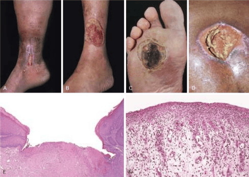
Clinical Manifestations and Histopathology of Chronic Cutaneous Ulcers
14. Pathologic Deviations in Repair and Scarring
Aberrations in the balance between extracellular matrix synthesis and matrix degradation yield characteristic pathological states:
Major Complications of Defective or Aberrant Repair
- Wound Dehiscence: Complete mechanical rupture and separation of previously closed wound margins. Most frequently follows major abdominal laparotomies secondary to acute surges in intra-abdominal pressure (e.g., intractable vomiting, severe coughing paroxysms, or paralytic ileus).
- Deficient Scar Formation: Insufficient collagen production or inadequate cross-linking, causing secondary mechanical breakdown and chronic ulceration.
- Incisional Herniation: Late mechanical breakdown of deep musculofascial scars in abdominal walls, allowing visceral extrusion under an intact cutaneous surface.
- Exuberant Granulation ("Proud Flesh"): Overgrowth of hyperplastic granulation tissue projecting above the surrounding epidermis. This mechanical shelf prevents epithelial migration until excised surgically or chemically cauterized with silver nitrate.
- Aggressive Fibromatosis (Desmoid Tumors): Rare, locally aggressive proliferation of well-differentiated fibroblasts and myofibroblasts arising within previous incisional sites. They represent low-grade, non-metastasizing fibroblastic neoplasms that frequently recur following wide local excision.
- Wound Contracture: Pathological exaggeration of wound contraction, culminating in structural distortion, cosmetic deformity, and functional limitation. Particularly common after deep dermal burns on palms, soles, and the anterior chest.
- Malignant Transformation (Marjolin Ulcer): Development of aggressive squamous cell carcinoma within chronically inflamed, unhealed scar tissue or longstanding sinus tracts.
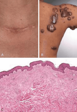
Gross Morphology and Histology of Excessive Dermal Scarring
| Diagnostic Feature | Hypertrophic Scar | Keloid |
|---|---|---|
| Spatial Boundaries | Raised and thickened, but remains confined strictly within the original margins of the wound. | Expands invasively beyond the borders of the original injury into surrounding normal dermis. |
| Predisposing Trauma | Deep thermal burns, surgical incisions traversing skin tension lines, or injuries involving deep reticular dermis. | Can arise from minimal cutaneous trauma (e.g., earlobe piercing, folliculitis, minor lacerations). |
| Demographic Predisposition | No strong ethnic predilection; related primarily to wound depth, location, and mechanical tension. | Marked genetic susceptibility, occurring with significantly higher frequency in individuals of African descent. |
| Clinical Evolution | Rapid initial proliferation followed by a spontaneous tendency to regress over several months. | Persists indefinitely without spontaneous regression; high rate of recurrence following simple surgical excision. |
| Microscopic Architecture | Organized, parallel arrays of predominantly Type III collagen accompanied by abundant active myofibroblasts. | Large, haphazardly arranged, thick, glassy, eosinophilic keloidal collagen bands (mainly Type I/III collagen). |
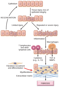
Cytokine Networks and Cellular Pathways of Persistent Tissue Fibrosis
15. Specialized Tissue Repair: Skeletal Bone Fracture Healing
Bone repair involves regeneration combined with fibrovascular reorganization. Fractures vary according to etiology and mechanical pattern:
- Traumatic: Arises from mechanical force exceeding the structural elasticity of the bone.
- Complete vs. Incomplete: Disruption traversing both cortices versus partial cortical fracture (e.g., pediatric green-stick fractures).
- Simple (Closed), Comminuted, or Compound: Contained within intact skin versus fragmented into multiple bony pieces, versus communicating directly with the external environment through breached skin.
Primary vs. Secondary Fracture Healing
- Primary Union (Direct Bone Healing): Occurs only when anatomical reduction is achieved with rigid internal fixation (e.g., compression plates) eliminating motion and interfragmentary gap. Osteonal remodeling occurs directly across the fracture plane via cutting cones, forming medullary callus without external periosteal callus. It permits early patient mobilization but carries risks of focal bone necrosis and delayed healing.
- Secondary Union (Indirect Bone Healing): The standard physiological sequence for non-rigidly fixed fractures, proceeding through organized stages of callus formation and remodeling.
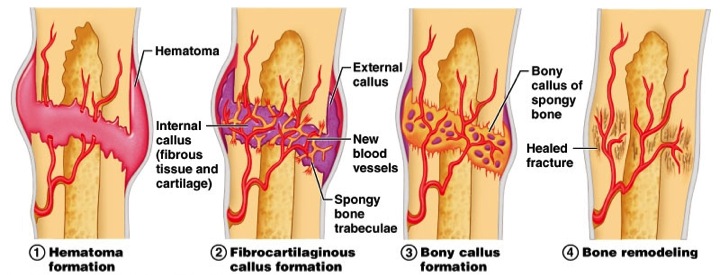
The Chronological Stages of Secondary Fracture Union
The Triphasic Sequence of Secondary Fracture Union
- Stage 1: Procallus Formation (Fibrocartilaginous / Soft Callus):
- Fracture ruptures intraosseous and periosteal vessels, generating a large, enclosed fracture hematoma.
- Local osteocyte necrosis occurs along sheared bone ends, inciting acute inflammation.
- Neovascular capillary sprouts and fibroblasts invade the hematoma, converting it into active granulation tissue.
- Periosteal and endosteal osteoprogenitor cells proliferate; under local microenvironmental hypoxia, pluripotential mesenchymal cells differentiate into chondrocytes, laying down cartilage that mingles with woven bone to create the procallus (soft callus) that stabilizes the bone ends.
- Stage 2: Osseous Callus Formation (Bony / Hard Callus):
- The soft procallus provides a structural scaffold for conversion into lamellar bone.
- Chondrocytes within the fibrocartilaginous callus undergo terminal hypertrophy and calcification; osteoclasts resorb calcified cartilage and woven bone.
- Invasive osteoblasts deposit unmineralized osteoid, which progressively calcifies into organized trabecular woven bone.
- Concentric osteon deposition constructs primitive Haversian systems around invading vascular loops.
- Stage 3: Bone Remodeling (Wolff's Law):
- Coordinated osteoblastic matrix deposition and osteoclastic bone resorption remodel the irregular bony callus along mechanical lines of weight-bearing stress.
- Excess external callus is resorbed, the central medullary cavity is recanalized, and the fracture site becomes anatomically and histologically indistinguishable from normal uninjured cortical bone.
Complications of Bone Fracture Repair
- Delayed Union: Bone bridging progresses at a substantially slower pace than clinically expected, often due to inadequate immobilization, poor vascularity, or low-grade infection.
- Non-Union: Complete cessation of reparative osteogenesis without bony bridge formation. If motion persists, the gap develops dense fibrous tissue and may form a synovial-lined fluid space, creating a false joint (pseudoarthrosis) with rounded, sclerotic bone ends and closed medullary cavities.
- Fibrous Union: Fracture gap is bridged by dense, pliable fibrous scar tissue rather than an osseous callus, resulting in mechanical instability.
- Malunion: Bone consolidation occurs in an abnormal, rotated, or deformed anatomical alignment, impairing biomechanical function.
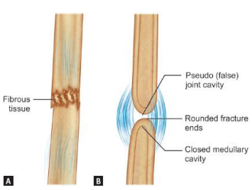
Structural Features of Fibrous Union and Pseudoarthrosis
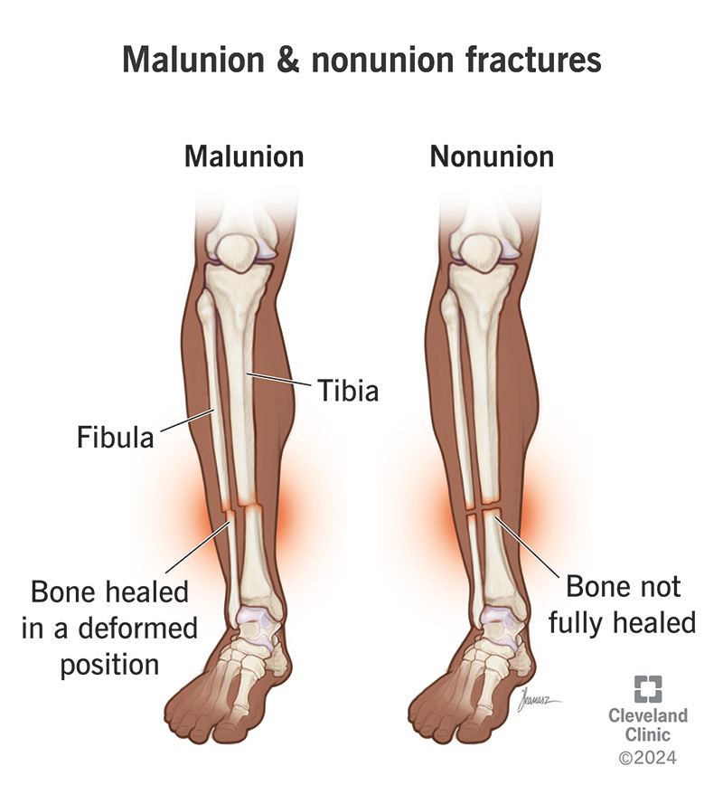
Clinical Distinction Between Bone Malunion and Non-Union
16. Specialized Tissue Repair: Nervous System Tissue
The cellular response to mechanical or ischemic trauma differs markedly between the central and peripheral nervous systems:
| Feature | Central Nervous System (CNS) | Peripheral Nervous System (PNS) |
|---|---|---|
| Neuronal Body Replacement | Destroyed neuronal soma in the cerebral cortex, spinal cord, and deep brain nuclei cannot replicate or be replaced (permanent post-mitotic cells). | Destroyed primary sensory or autonomic ganglion somas cannot be replaced; survival of the cell body is required for axonal regeneration. |
| Axonal Regeneration | Axons in CNS tracts do not regenerate due to local myelin-associated inhibitory proteins (e.g., Nogo) and absent basement membrane guidance tubes. | Axons exhibit robust, functional regeneration (growing ~1 mm/day) if the endoneurial sheath and Schwann cell basement membrane are intact. |
| Glial / Supporting Cell Response | Damaged neuroglia induce reactive proliferation and hypertrophy of astrocytes, forming an impenetrable glial scar (gliosis). | Schwann cells rapidly proliferate and align longitudinally within the basal lamina to construct guiding columns (Büngner bands). |
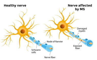
Comparison of Intact versus Demyelinated and Damaged Nerve Fibers
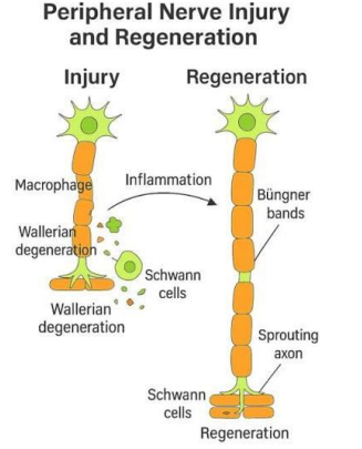
Cellular Dynamics of Wallerian Degeneration and Peripheral Axonal Sprouting
Stepwise Sequence of Peripheral Nerve Regeneration
- Acute Trauma & Neuronal Survival: Following transection, retrograde survival signals promote cell body survival, accompanied by central chromatolysis and upregulation of regenerative protein synthesis.
- Wallerian Degeneration: Distal to the transection, the axon and associated myelin sheath fragment within 24 to 48 hours. Recruited hematogenous macrophages and resident Schwann cells phagocytose axonal and lipid myelin debris.
- Schwann Cell Activation & Büngner Band Assembly: Denervated Schwann cells downregulate myelin gene expression, proliferate within intact endoneurial basement membrane cylinders, and arrange longitudinally into continuous cellular conduits termed bands of Büngner.
- Axonal Sprouting & Elongation: The proximal axon stump forms multiple microspikes and growth cones, advancing down the Büngner bands guided by neurotrophins (NGF, BDNF) and cell adhesion molecules.
- Target Re-innervation & Remyelination: Once the regenerating sprout contacts its motor or sensory target, Schwann cells arrest migration, differentiate, and synthesize a new myelin sheath to restore saltatory conduction.
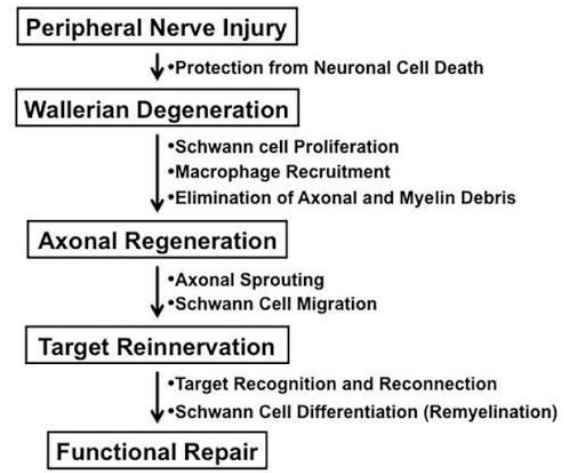
Sequential Pathway of Peripheral Nerve Injury, Degeneration, and Functional Re-innervation
17. Specialized Tissue Repair: Skeletal Muscle
Skeletal muscle trauma is common in sports medicine, accounting for 10% to 55% of all sustained athletic injuries. The most vulnerable muscle groups include the hamstrings, rectus femoris (quadriceps), adductor longus, and the gastrocnemius/soleus complex.
| Injury Pattern | Biomechanical Etiology | Structural Pathology | Microvascular Status |
|---|---|---|---|
| Shearing Injury (Strain, Contusion, Laceration) | Excessive intrinsic tensile stress (eccentric contractions) or direct blunt force. | Complete mechanical rupture of myofibers, disruption of the external basal lamina, and tearing of endomysial/perimysial sheaths. | Extensive capillary shearing causing hematoma formation; healing requires combined fibroblastic scar and myofiber regeneration. |
| In Situ Necrosis (Rhabdomyolysis) | Metabolic compromise, toxic exposure, prolonged ischemia, or crush trauma. | Partial sarcoplasmic necrosis while the surrounding basal lamina and mysial sheaths remain intact. | Adjacent capillary networks remain patent; provides an ideal architectural scaffold for complete, scarless regeneration. |
In strain injuries, rupture occurs predominantly near the myotendinous junction (MTJ). In pennate and complex muscles, numerous MTJs reside deep within the muscle belly where myofibers anchor to internal intramuscular fascial septa, rather than solely at discrete terminal tendons.
The Triphasic Sequence of Skeletal Muscle Repair
- 1. Destruction Phase:
- Mechanical shear tears the sarcoplasmic membrane, triggering a massive influx of extracellular calcium that activates proteases.
- Necrosis is restricted to the immediate zone of rupture. Progression along the fiber is halted by a "fire door" contraction band—a dense condensation of hyper-contracted cytoskeletal myofilaments formed within hours, across which a new sarcolemmal seal is synthesized.
- Retraction of the severed myofiber ends leaves a central gap that fills with a localized hematoma. An intense acute inflammatory infiltrate develops within 24 hours.
- 2. Repair Phase:
- Circulating blood monocytes infiltrate the injury bed and differentiate into active macrophages, phagocytosing necrotic sarcoplasmic debris and degraded myofilaments.
- Quiescent myogenic stem cells—satellite cells residing beneath the myofiber basal lamina—are activated.
- Committed satellite cells differentiate into mononucleated myoblasts. Simultaneously, undifferentiated stem satellite cells undergo asymmetric mitosis by 24 hours to renew the reserve stem pool.
- Myoblasts proliferate, align, and fuse to construct elongated multinucleated myotubes within 2 to 3 days.
- By days 5 to 6, regenerating myotubes extend within the preserved basal lamina cylinders and advance into the central intervening fibrovascular scar. Concurrently, capillary ingrowth establishes revascularization, with sprouts evident by day 3.
- 3. Remodeling Phase:
- Regenerating myofibers mature, synthesizing structured contractile sarcomeres with centralized nuclei that migrate to the periphery.
- Myofibers re-anchor to the extracellular matrix at newly formed myotendinous junctions.
- The intervening collagenous scar compacts and remodels. However, scar tissue in the repair zone leaves the healed muscle stiffer, with reduced range of motion and an increased risk of re-injury under sudden eccentric load.
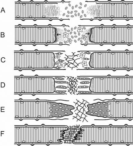
Microscopic Progression of Skeletal Muscle Repair Following Shearing Strain
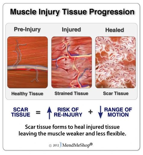
Functional Consequences of Fibrotic Scar Tissue in Healed Skeletal Muscle
18. Core Pathology Clinical Competencies
Pathological Curricular Review
- Mechanisms of Reversible and Irreversible Cell Injury: Contrast ATP depletion, cellular swelling (hydropic change), and fatty change with irreversible milestones: severe mitochondrial damage, profound membrane rupture, and nuclear degradation (pyknosis, karyorrhexis, karyolysis).
- Apoptosis versus Necrosis: Differentiate physiological, programmed, non-inflammatory single-cell clearance mediated by caspases from pathological, energy-independent, membrane-disruptive coagulative/liquefactive necrosis that elicits an acute inflammatory response.
- Vascular and Cellular Events of Acute Inflammation: Map transient vasoconstriction, sustained vasodilation, increased microvascular permeability, hemoconcentration, and endothelial marginations against the cellular sequence of selectin-mediated rolling, integrin-mediated firm adhesion, paracellular transmigration (diapedesis), and chemotaxis to the focus of injury.
- Pathogenesis of Granulomatous Inflammation: Characterize persistent, non-degradable antigens that elicit a cell-mediated Type IV hypersensitivity response: macrophage antigen presentation, CD4+ Th1 cell activation, interferon-gamma release, and the subsequent transformation of macrophages into epithelioid histiocytes and multinucleated giant cells (Langhans or foreign-body type), with clinical examples including Mycobacterium tuberculosis and foreign material.
- Primary vs. Secondary Intention Repair and Modifiers: Explain the biological criteria governing primary and secondary union, identifying how local factors (infection, ischemia, mechanical stress) and systemic parameters (diabetes, glucocorticoids, protein-calorie malnutrition, Vitamin C deficiency) alter fibrovascular repair, extracellular matrix synthesis, and functional recovery.