
Cytokines and Chemokines
Core Learning Objectives
- Foundational Principles: Defining cytokines, exploring their linguistic roots, and examining their core physiological traits.
- Taxonomy and Grouping: Classifying cytokine families based on functional behavior and structural origin.
- Physiological Roles: Analyzing how cytokines orchestrate immune defense, inflammation, hematopoiesis, and tissue repair.
- Chemokines: Understanding chemoattractant signaling, structural subfamilies, and cellular homing.
- Clinical Diagnostics and Therapeutics: Applying cytokine profiles to disease monitoring, recombinant drug therapies, and targeted receptor blockade.
1. Definition and General Properties of Cytokines
Key Definitions
- Etymology: The word cytokine comes from two Greek words: cyto (meaning cell) and kinos (meaning movement). Taken together, the name highlights their ability to drive cell migration and activity.
- Modern Clinical Terminology: Contemporary immunology refers to cytokines as immunomodulating agents. These are molecular mediators that fine-tune, amplify, or suppress immune responses.
- Functional Definition: Cytokines are compact, soluble, or membrane-bound protein-based signaling molecules. They enable cell-to-cell communication during immune responses. They also recruit white blood cells toward focal areas of injury, infection, and tissue trauma.
- Molecular Character: They are low-molecular-weight proteins that dictate the character, intensity, and duration of an immune reaction by modulating target lymphocytes and neighboring tissue cells.
Historical Evolution of Cytokine Terminology
Before scientists adopted the unified name cytokine, these signaling proteins were named according to the specific cells that produced them or their presumed target populations:
| Historical Term | Original Biological Basis | Modern Status |
|---|---|---|
| Lymphokines | Originally presumed to be produced only by activated lymphocytes. | Subsumed under the umbrella term cytokine; produced by many cell types. |
| Monokines | Originally identified as secretable products of mononuclear phagocytes (monocytes and tissue macrophages). | Subsumed under the general term cytokine. |
| Interleukins (ILs) | Named because they act as chemical messengers between different classes of leukocytes (white blood cells). | Maintained as a numbered naming system (IL-1 through IL-35+) for specific cytokines. |
| Chemokines | Specialized chemoattractant cytokines that guide leukocyte trafficking. | Classified as a major structural and functional subfamily of cytokines. |
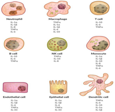
Cellular Sources of Cytokines
2. General Biological Attributes of Cytokines
- 1. Molecular Weight and Physical Structure: Most cytokines are low-molecular-weight polypeptides or glycoproteins ranging from 8 to 80 kDa. Most circulate and function as single-unit monomers.
- 2. Secretion by Activated Cells: Natural cytokines are not stored pre-formed inside resting cells. Instead, they are transcribed and secreted after cell activation. Key cellular sources include:
- Activated immune cells (T-helper lymphocytes, cytotoxic T cells, B cells, macrophages, dendritic cells, NK cells, granulocytes).
- Stromal matrix and structural cells (fibroblasts, vascular endothelial cells, epithelial barrier cells).
- Selected transformed or neoplastic tumor cells.
- 3. Shared Cellular Origins: A single cytokine can be synthesized and released by multiple distinct cell types. For example, both macrophages and endothelial cells produce IL-1 and IL-6.
- 4. Multi-Cytokine Secretory Capacity: A single cell type can synthesize and secrete many different cytokines simultaneously. For example, a single macrophage can release TNF-α, IL-1, IL-6, IL-8, and IL-10 depending on the stimulus.
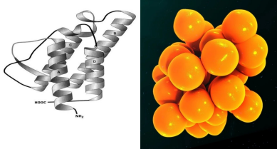
Monomeric Polypeptide Architecture of Cytokines
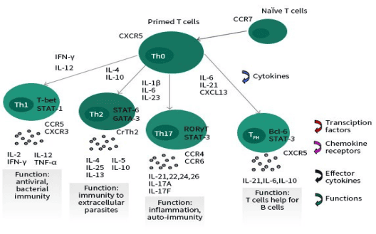
Polarization of Helper T-Cell Lineages Driven by Cytokine Signals
Mechanisms of Action: Delivery Pathways
Cytokines deliver regulatory signals through three distinct physiological routes:
| Signaling Route | Mechanism of Action | Practical Immunological Example |
|---|---|---|
| Autocrine Action | The cytokine binds to high-affinity surface receptors on the same cell that produced it. | An activated T lymphocyte secretes IL-2, which binds its own surface IL-2 receptors to trigger clonal proliferation. |
| Paracrine Action | The cytokine diffuses locally through the extracellular matrix to bind receptors on adjacent, nearby target cells. | A macrophage releases IL-1 and TNF-α, which activate neighboring vascular endothelial cells to express adhesion molecules. |
| Endocrine Action | The cytokine enters the bloodstream or lymphatic circulation and travels to distant target tissues throughout the body. | Elevated circulating levels of IL-1, IL-6, and TNF-α reach the hypothalamic thermoregulatory center to induce fever, and reach the liver to trigger acute-phase protein synthesis. |
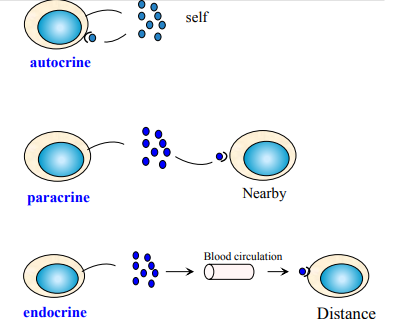
Modes of Cytokine Delivery: Autocrine, Paracrine, and Endocrine Action
High-Affinity Receptor Binding and Potency
- Picomolar Potency: Cytokines act at very low concentrations. Only small quantities, in the picomolar range (pmol/L), are needed to occupy high-affinity cell-surface receptors and trigger biological responses.
- Receptor-Restricted Selectivity: Cytokines only stimulate cells that express their specific, matching receptors. Cells without the appropriate receptor remain unaffected.
- Dynamic Receptor Regulation: The expression of both cytokines and their receptors is strictly regulated. For instance, resting T cells do not express the high-affinity alpha chain of the interleukin-2 receptor (IL-2Rα / CD25). They only express it after antigen presentation, ensuring that only antigen-specific clones proliferate.
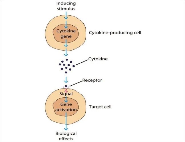
Receptor Activation and Intracellular Signaling Cascades of Cytokines
The Five Cardinal Properties of Cytokine Action
Cytokine networks use five classic signaling patterns that help the immune system mount coordinated, flexible defenses:
| Signaling Property | Definition & Operational Mechanism | Immunological Example |
|---|---|---|
| Pleiotropism (Pleiotropy) | The ability of a single cytokine to exert multiple different biological effects on diverse target cell types. | Interleukin-4 (IL-4): Induces B-cell activation, proliferation, and differentiation; drives thymocyte proliferation; and stimulates mast cell proliferation. |
| Redundancy | The phenomenon where two or more distinct cytokines share the same or overlapping biological functions. | IL-2, IL-4, and IL-5: All three can independently stimulate B-cell proliferation, providing functional backup if one cytokine is missing. |
| Synergy | A cooperative interaction where two or more cytokines act together to create a biological effect that is greater than the sum of their individual effects. | IL-4 plus IL-5: Working together, they effectively induce B cells to undergo immunoglobulin class switching to synthesize IgE. |
| Antagonism | The capacity of one cytokine to directly block, neutralize, or counter the biological action of another cytokine. | IFN-γ vs. IL-10: IFN-γ activates macrophages to enhance bacterial killing, whereas IL-10 inhibits macrophage activation to shut down inflammation. |
| Cascade Effect | A sequential signaling chain where one cytokine acts on a target cell to stimulate the production of one or more downstream cytokines. | An activated T-helper cell releases TNF, which binds to a macrophage. This stimulates the macrophage to secrete IL-1 and additional TNF, amplifying the inflammatory cascade. |
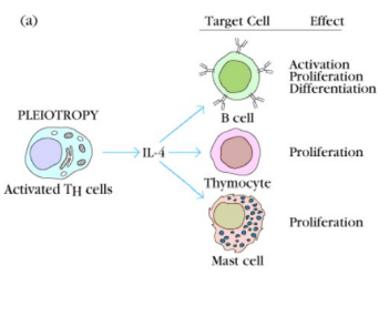
Pleiotropism: Diverse Biological Outcomes from a Single Cytokine
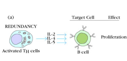
Redundancy: Overlapping Biological Functions of Distinct Cytokines
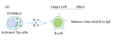
Synergy: Cooperative Amplification Leading to Immunoglobulin Class Switching
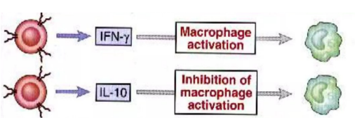
Antagonism: Reciprocal Counter-Regulation of Cellular Activation
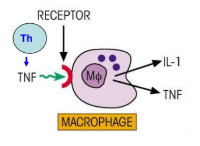
Cascade Effect: Sequential Amplification of Downstream Inflammatory Mediators
The Five Cardinal Features of Cytokines ("P-R-S-A-C")
Use the mnemonic "PR-SAC" to quickly recall how cytokines function:
- P → Pleiotropy: One cytokine produces multiple distinct effects on different cell types (e.g., IL-4 acting on B cells, mast cells, and thymocytes).
- R → Redundancy: Multiple cytokines carry out the same exact task (e.g., IL-2, IL-4, and IL-5 all driving B-cell proliferation).
- S → Synergy: Cytokines collaborate to create an amplified response (e.g., IL-4 + IL-5 driving IgE switching).
- A → Antagonism: One cytokine counters another to restore balance (e.g., IFN-γ activates macrophages, while IL-10 inhibits them).
- C → Cascade: One cytokine triggers a chain reaction that produces downstream cytokines (e.g., T-cell TNF stimulates macrophages to release IL-1 and TNF).
3. Classification of Cytokines
Cytokines can be categorized using two complementary systems: functional classification and structural/source family classification.
Functional Classification of Cytokines
| Functional Category | Primary Biological Role | Representative Cytokines |
|---|---|---|
| 1. Immunoregulatory | Directs antigen presentation, upregulates MHC molecules, and guides immune recognition. | IFN-γ (upregulates MHC Class I and II on antigen-presenting cells). |
| 2. Proinflammatory | Produced by B cells, T cells, and myeloid cells to initiate and sustain the inflammatory response. Highly active during acute and chronic infections. | IL-1, IL-6, TNF-α, IFNs, and various chemokines. |
| 3. Anti-inflammatory | Dampens, downregulates, or terminates an overactive immune response to prevent collateral tissue damage. | TGF-β, IL-10, IL-11, IL-1ra, IFN-β. |
| 4. Growth and Differentiation | Drives the renewal, expansion, and maturation of pluripotent hematopoietic stem cells and committed progenitor lineages. | Colony-stimulating factors (CSFs), stem cell factor (SCF), and erythropoietin (EPO). |
Cytokine Categorization by Specific Immune Target
| Functional Specialization | Associated Cytokines |
|---|---|
| Proinflammatory Cytokines | TNF, IL-1, IL-6, chemokines (many) |
| Anti-inflammatory Cytokines | IL-10, IL-1ra, TGF-β |
| Inhibition of Viral Replication | IFN-α, IFN-β |
| Macrophage-Activating Cytokines | IFN-γ |
| B Cell-Activating Cytokines | IL-4, IL-5, IL-6, IL-21 |
| T Cell-Activating Cytokines | IL-2, IL-4, IL-12, IFN-γ |
| Eosinophil- and/or Mast Cell-Activating Cytokines | IL-3, IL-4, IL-13, IL-5 |
Classification by Source and Structural Families
Cytokines are divided into six major structural and evolutionary families:
Family 1: Interleukins (ILs)
- Numbering and Discovery: Approximately 35 distinct interleukins have been characterized. They are numbered sequentially based on when they were discovered, spanning IL-1 through IL-35:
IL-1, IL-2, IL-3, IL-4, IL-5, IL-6, IL-7, IL-8, IL-9, IL-10, IL-11, IL-12, IL-13,IL-14, IL-15, IL-16, IL-17, IL-18, IL-19, IL-20, IL-21, IL-22, IL-23, IL-24, IL-25, IL-26 ... IL-35. - Primary Cellular Source: Synthesized primarily by T-helper (CD4+) cells, as well as monocytes, macrophages, and dendritic cells.
- Primary Function: Interleukins serve as specialized communication signals between white blood cells (leukocytes) to coordinate adaptive and innate immune responses.
Family 2: Interferons (IFNs)
- Biological Role: A family of antiviral glycoproteins produced by human and animal cells in response to viral invasion, intracellular parasites, and antigenic exposure.
- Naming Basis: They were named for their ability to interfere directly with viral replication and prevent pathogens from multiplying within host cells.
- Clinical Roles: Active in protecting against viral infections, modulating autoimmune diseases, and managing certain malignancies.
Major Interferon Types and Cellular Sources:
| Interferon Type | Primary Cellular Source | Key Biological Action |
|---|---|---|
| Interferon-alpha (IFN-α) | Leukocytes (especially plasmacytoid dendritic cells and monocytes) | Inhibits viral replication inside host cells; enhances MHC Class I expression. |
| Interferon-beta (IFN-β) | Fibroblasts and epithelial cells | Antiviral protection; modulates inflammatory responses. |
| Interferon-gamma (IFN-γ) | T lymphocytes (Th1, CD8+ cytotoxic T cells) and NK cells | Potent macrophage activator; enhances antigen presentation via MHC Class II. |
Family 3: Tumor Necrosis Factors (TNF)
- Historical Background: Originally discovered because they induced necrosis in solid tumor models. Today, they are recognized as master regulators of immune responses and inflammation.
- TNF-alpha (TNF-α): Synthesized primarily by monocytes and macrophages that have been activated by lipopolysaccharide (LPS) from Gram-negative bacterial cell walls.
- TNF-beta (TNF-β / Lymphotoxin, LT): Synthesized primarily by activated T lymphocytes, particularly Th0 and Th1 subsets.
- Diverse Biological Roles:
- Tumor-suppressive actions: Activates cytotoxic T cells and NK cells, damages tumor blood vessels, promotes dendritic cell maturation, increases MHC-I expression, and triggers apoptosis.
- Tumor-promoting actions: Can promote epithelial-to-mesenchymal transition (EMT) and enhance regulatory T cell (Treg) activity.
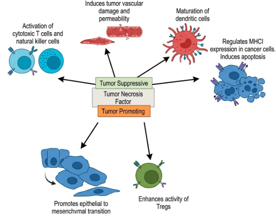
Pleiotropic Tumor-Suppressive and Tumor-Promoting Actions of TNF
Family 4: Colony-Stimulating Factors (CSFs)
Colony-stimulating factors drive the survival, proliferation, and differentiation of pluripotent hematopoietic stem cells into distinct blood cell lineages.
| Factor Name | Abbreviation | Target Cell Lineage and Function |
|---|---|---|
| Multi-Colony Stimulating Factor | Multi-CSF (IL-3) | Acts broadly on early multipotent hematopoietic progenitors to support diverse myeloid lineages. |
| Granulocyte-Macrophage CSF | GM-CSF | Stimulates bipotent progenitors to generate both granulocytes (neutrophils, eosinophils) and monocytes/macrophages. |
| Monocyte CSF | M-CSF | Directs committed precursor cells toward the monocyte and macrophage lineage. |
| Granulocyte CSF | G-CSF | Drives the commitment, proliferation, and release of neutrophils from the bone marrow. |
| Stem Cell Factor | SCF (c-kit ligand) | Maintains and expands primitive pluripotent hematopoietic stem cells. |
| Erythropoietin | EPO | Stimulates erythrocyte production from erythroid precursors in the bone marrow. |
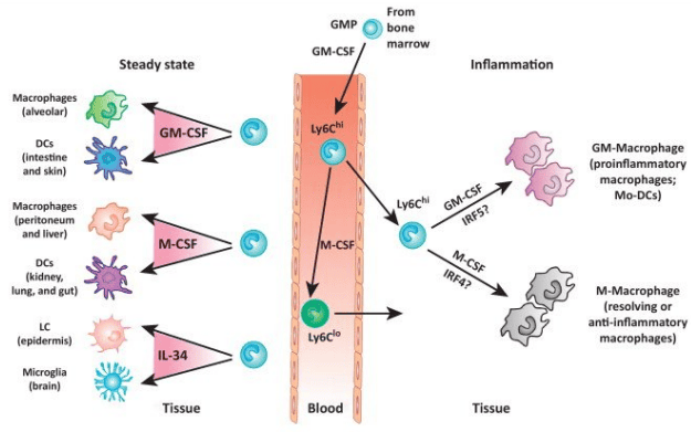
Colony-Stimulating Factors in Myeloid Homeostasis and Inflammation
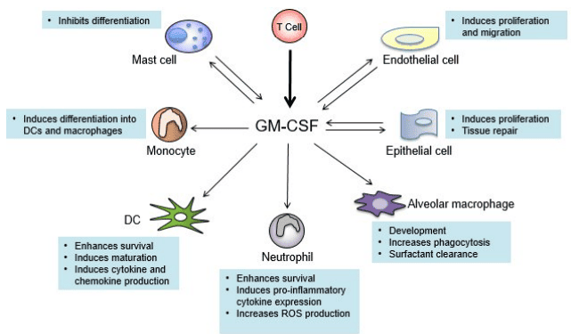
Target Spectrum and Functional Roles of GM-CSF
Family 5: Transforming Growth Factors and Growth Factors
Growth factors are specialized cytokines that stimulate cellular growth, division, and tissue regeneration:
- Transforming Growth Factor-beta (TGF-β): Promotes tissue repair and extracellular matrix deposition, while acting as a key anti-inflammatory mediator to resolve immune activity.
- Epidermal Growth Factor (EGF): Promotes epithelial cell proliferation and barrier re-epithelialization during wound healing.
- Vascular Endothelial Growth Factor (VEGF): Directly stimulates endothelial cell growth, survival, and new blood vessel formation (angiogenesis).
- Fibroblast Growth Factor (FGF): Stimulates fibroblast proliferation, wound healing, collagen production, and tissue repair.
4. Chemokines (Chemoattractant Cytokines)
Key Definitions & Biochemical Features
- Functional Definition: Chemokines (chemoattractant cytokines) are small proteins that recruit circulating monocytes, granulocytes, and lymphocytes into areas of inflammation and infection.
- Molecular Characteristics: Small secreted proteins ranging from 8 to 10 kDa. They bind to cell-surface heparin and extracellular matrix glycosaminoglycans, creating stable chemotactic gradients that guide cell movement.
Structural Subfamilies of Chemokines
Chemokines are classified into four distinct subfamilies based on the arrangement of their initial conserved cysteine (C) amino acid residues, separated by variable intervening amino acids (X):
| Subfamily | Structural Motif | Primary Representative | Target Cell Lineage & Role |
|---|---|---|---|
| CXC Chemokines (α subgroup) |
One variable amino acid separates the first two cysteines (C-X-C). | IL-8 (CXCL8) | Recruits and activates neutrophils to fight acute bacterial infections. |
| CC Chemokines (β subgroup) |
The first two cysteines sit directly next to each other (C-C). | MCP-1 (CCL2 / Monocyte Chemoattractant Protein-1) | Recruits monocytes, macrophages, memory T cells, and basophils. |
| C Chemokines (γ subgroup) |
Lacks two of the four canonical cysteines, keeping only a single terminal cysteine (C). | Lymphotactin (XCL1) | Recruits and homes T lymphocytes and NK cells. |
| CX3C Chemokines (δ subgroup) |
Three variable amino acids separate the first two cysteines (C-X-X-X-C). | Fractalkine (CX3CL1) | Acts both as a membrane-anchored adhesion molecule and a soluble chemoattractant for monocytes and T cells. |
Primary Biological Functions of Chemokines
- Inflammatory Cell Recruitment: Guides circulating white blood cells toward focal sites of active infection and tissue injury.
- Immune Homeostasis: Controls normal leukocyte trafficking through secondary lymphoid organs (lymph nodes, spleen) during everyday immune surveillance.
- Antimicrobial Host Defense: Recruits specialized immune cells to eliminate bacterial, parasitic, and viral infections.
- Wound Healing and Tissue Repair: Coordinates the migration of inflammatory cells, fibroblasts, and endothelial cells needed to rebuild damaged tissue.
- Hematopoiesis and Angiogenesis: Helps regulate bone marrow cell release and modulates blood vessel formation.
5. Clinical Applications of Cytokines
Disease Biomarkers and Septic Shock
Because cytokines are released into the circulation during systemic disease, their serum levels serve as valuable biomarkers for diagnosing and tracking illnesses:
- Septic Shock Triad: High serum levels of the proinflammatory cytokines TNF-α, IL-1, and IL-6 are detectable in patients with septic shock. They are major drivers of fever, profound hypotension, vascular leakage, and multi-organ failure.
- Immune Status and Diagnostic Testing: Measuring in vitro cytokine production provides a reliable assessment of cell-mediated immunity. For example, testing whether patient T cells release IFN-γ when exposed to mycobacterial antigens is the basis for Interferon-Gamma Release Assays (IGRAs) used to diagnose Mycobacterium tuberculosis infection.
Therapeutic Cytokines and Molecular Targets
In modern medicine, cytokines are used both as recombinant drug therapies and as targets for monoclonal antibody inhibition:
- Recombinant Cytokines as Medications:
- Interferon-alpha (IFN-α): Approved for treating chronic hepatitis C and hepatitis B viral infections, as well as selected malignancies (such as hairy cell leukemia, melanoma, and Kaposi sarcoma).
- Cytokines as Therapeutic Targets:
- TNF-α Antagonists: Anti-TNF biologics (e.g., infliximab, adalimumab, etanercept) neutralize excessive systemic inflammation. They are widely used to treat rheumatoid arthritis (RA), plaque psoriasis, ankylosing spondylitis, and inflammatory bowel disease.
- Interleukin-2 and Interleukin-15 Blockade: Inhibiting IL-2 and IL-15 signaling pathways prevents allograft rejection in organ transplantation and helps manage specific leukemias and lymphomas.
Comparison: Recombinant Cytokines vs. Cytokine-Targeted Antagonists
| Strategy | Mechanism | Clinical Agent Example | Primary Clinical Indication |
|---|---|---|---|
| Recombinant Cytokines | Administering synthetic cytokine to boost or restore a helpful immune response. | Recombinant IFN-α | Hepatitis C viral infection; selected oncology protocols. |
| Cytokine Antagonists | Using antibodies or soluble decoy receptors to block a cytokine or its receptor, halting destructive inflammation. | Anti-TNF-α biologics; IL-2 / IL-15 inhibitors | Rheumatoid arthritis (RA); organ transplant immunosuppression; autoimmune disorders. |
