
Cellular and Humoral Responses of Adaptive Immunity
Key Definitions
- Humoral Immunity: The arm of adaptive immunity mediated by B lymphocytes and circulating antibodies, which targets extracellular pathogens and soluble toxins.
- Cell-Mediated Immunity: The protective response directed by T lymphocytes, specialized in identifying and eliminating intracellular threats, such as viruses and mutated host cells.
- T-Dependent Antigens: Foreign substances, predominantly proteins, that require direct assistance from helper T cells to fully activate B cells.
- T-Independent Antigens: Complex molecules, typically polysaccharides and lipids, that stimulate B cells directly without assistance from T cells.
B Lymphocytes and the Antibody Response
Adaptive humoral defense begins when a matching foreign structure contacts a receptor anchored on the B lymphocyte surface. This binding event initiates an intracellular signal that prepares the cell to react. However, most responses require a second verification step. Before mounting a full immune attack, the B cell often must obtain confirmation from an activated helper T cell to ensure that the target is truly foreign and dangerous.
| Antigen Classification | Chemical Nature | T Helper Cell Requirement | Typical Immune Outcome |
|---|---|---|---|
| T-Dependent Antigens | Proteins and peptides | Mandatory (requires second confirmation signal) | Affinity maturation, antibody class switching, and durable memory formation |
| T-Independent Antigens | Polysaccharides, lipids, lipopolysaccharides (LPS) | Not required (direct B cell receptor cross-linking) | Primarily IgM production with minimal to no immunological memory |
Mechanisms of the Response to T-Dependent Antigens
When an intact protein antigen binds to a specific B cell receptor (BCR), the B cell engulfs the complex. This process moves through several distinct stages:
- Endocytosis and Internalization: The B cell wraps its outer membrane around the receptor-bound target, pulling it into an internal, membrane-enclosed vacuole or endosome.
- Proteolytic Processing: Inside this intracellular pocket, digestive enzymes break down the large foreign protein into short peptide fragments.
- MHC Class II Loading: The cell loads these processed fragments into the binding cleft of newly synthesized Major Histocompatibility Complex (MHC) class II molecules, which then travel to the cell surface for presentation.
- T-Cell Surveillance and Receptor Engagement: An effector helper T cell scans the displayed complex. Its T cell receptor (TCR) inspects the peptide to confirm whether it is an authentic foreign threat or harmless self-tissue.
- Cytokine Signaling: Upon confirming the foreign peptide, the helper T cell binds tightly and releases a focused stream of signaling proteins (cytokines) directly toward the B cell surface.
- Proliferation and Clonal Expansion: Driven by these cytokine instructions, the stimulated B cell enters rapid cell division, creating an army of identical daughter cells.
- Plasma Cell Differentiation: A significant portion of this progeny matures into plasma cells (effector B cells). These cells are biological factories specialized in manufacturing and releasing thousands of tailored antibody molecules every second.
- Terminal Apoptosis: After finishing their rapid antibody-producing burst, these short-lived plasma cells undergo programmed cell death.
Kinetic Properties of the Primary Immune Response
Initial contact with a new microbial threat does not produce antibodies right away. A noticeable delay (lag phase) lasting 10 days to 2 weeks passes before detectable antibody levels appear in the blood.
During this vulnerable window, the pathogen replicates unchecked by antibodies. As a result, the infected individual may develop clear clinical symptoms, which can sometimes become life-threatening. Even so, the immune system is working intensely behind the scenes. Clonal B cell selection is taking place, early plasma cells are beginning antibody synthesis, and activated lymphocytes are undergoing three vital evolutionary steps:
- Affinity Maturation: Fine-tuning the strength of antibody-antigen binding.
- Class Switching (Isotype Switching): Changing the constant region to direct antibodies to specific body compartments.
- Memory Cell Generation: Creating durable, long-lived surveillance cells for future defense.
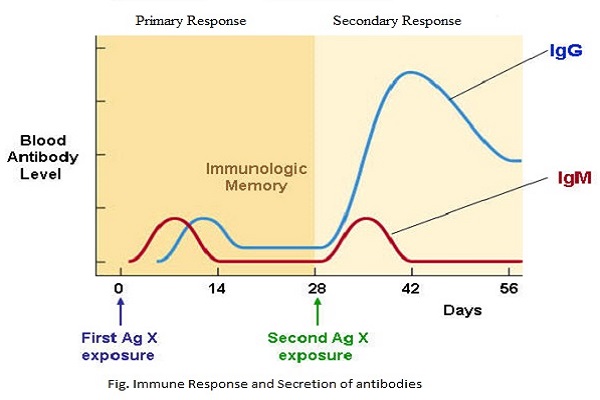
The Primary and Secondary Responses to Antigen
Affinity Maturation: Cellular Selection of High-Affinity Clones
Affinity Maturation is a process of natural selection within the body. It operates among proliferating B lymphocytes and selects for cells with the highest antibody binding accuracy.
As activated B cells replicate their genetic material prior to cell division, targeted hypermutations occur in the gene segments encoding the antibody variable domains. These spontaneous genetic alterations modify the shape and charge of the antigen-binding site on both the surface receptor and future secreted antibodies.
This process creates a competitive environment among new B cell clones:
- Clones with mutations that allow them to bind antigen most tightly and for the longest time capture survival signals and continue dividing.
- Clones with neutral, weakened, or self-reactive binding affinities fail to compete for antigen and are eliminated through apoptosis.
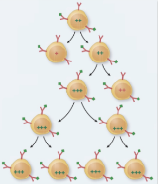
Affinity Maturation of Proliferating B Cells
Class Switching (Isotype Switching)
By default, immature, unactivated naive B cells are genetically wired to produce IgM antibodies because the IgM constant region gene segment sits directly adjacent to the assembled variable region DNA. However, different infection sites require different antibody forms.
Under the direction of specific helper T-cell cytokines, the B cell alters its genetic structure. It cuts out the intervening heavy chain constant genes, moving a downstream constant region gene (such as IgG, IgA, or IgE) right next to the original variable domain:
- Preserved Specificity: The variable region is completely untouched during this DNA recombination. Therefore, the antibody maintains its precise, original target specificity.
- Systemic Switching (IgG): B cells stimulated in peripheral lymph nodes and the spleen predominantly switch to IgG, the primary neutralizing antibody in the blood and tissue fluids.
- Mucosal Switching (IgA): B cells homing to Mucosa-Associated Lymphoid Tissue (MALT), such as the gut and respiratory tract (including Peyer's patches), switch to producing dimerized IgA to defend mucosal surfaces.
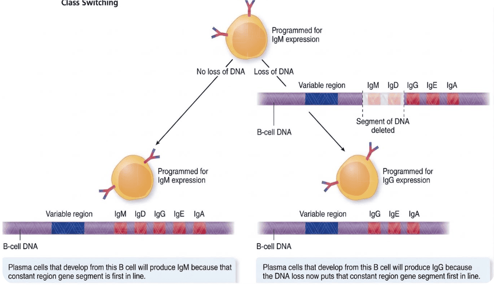
Genetic Mechanism of Immunoglobulin Class Switching
Memory Cell Generation and Secondary Immune Dynamics
A subset of stimulated B cells that successfully undergo affinity maturation and class switching do not turn into short-lived plasma factories. Instead, they become long-lived memory B cells. These cells survive for years, circulating quietly throughout the body in high numbers to provide ongoing immune surveillance.
Key Features of the Secondary Response
- Speed and Potency: The secondary response reacts significantly faster and with greater force than the initial exposure, often clearing invading microbes before symptoms can appear.
- Higher Starting Pool: A much larger population of memory B cells and memory helper T cells stands ready to identify the recurring antigen.
- Ultra-Sensitive Antigen Capture: Because their surface receptors were refined by earlier affinity maturation, memory B cells capture even tiny amounts of antigen that naive cells would miss entirely.
- Immediate Differentiation: Upon activation, memory B cells rapidly transform into plasma cells that produce high concentrations of protective IgG or IgA antibodies.
Mechanisms of T-Independent Antigen Activation
While fewer in number than protein antigens, T-independent antigens are clinically important targets found on many dangerous pathogens. Their distinct structure allows them to bypass the need for helper T cells:
- Repeating Polymeric Subunits: Polysaccharide capsules and lipopolysaccharide (LPS) molecules from Gram-negative bacteria feature identical, evenly spaced epitopes repeating across their surface.
- Receptor Cross-Linking: These repeating patterns allow the antigen to bind multiple adjacent B cell receptors simultaneously. This cross-linking generates an activation signal strong enough to trigger B-cell division without helper T-cell cytokines.
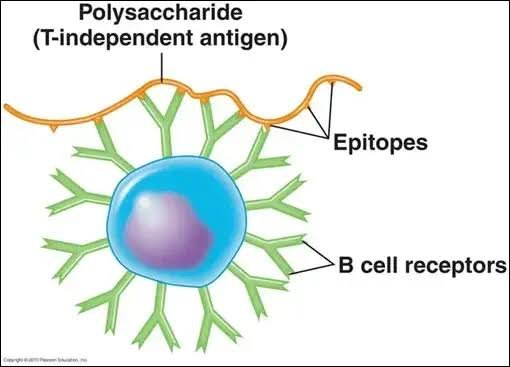
T-Independent B-Cell Activation via Receptor Cross-Linking
T Lymphocytes: Architecture, Receptors, and Processing
Unlike B cell antibodies, which can directly grab free-floating targets in body fluids, T cell receptors (TCRs) never interact with free, unattached antigens. A host cell must first process the antigen and present it on its surface.
Structural Anatomy of the T-Cell Receptor (TCR)
- Polypeptide Composition: Each membrane-bound TCR consists of two paired protein chains, most commonly an alpha (α) and beta (β) chain, or less frequently a gamma (γ) and delta (δ) chain.
- Domain Layout: Each chain contains an invariant constant region anchored through the membrane and an outer variable region that forms the antigen-binding site.
- Disulfide Linkage: A covalent disulfide bond tethers the two chains together into a functional unit.
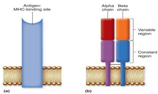
Structure of the Heterodimeric T-Cell Receptor (TCR)
Major Histocompatibility Complex (MHC): Class I vs. Class II
Before a T cell can recognize an antigen, presenting cells must break down foreign proteins into short peptide fragments and fit them into the surface grooves of Major Histocompatibility Complex (MHC) molecules.
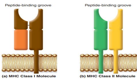
Peptide-Binding Architecture of MHC Class I and Class II Molecules
| MHC Type | Antigen Source | Cellular Distribution | Recognizing T-Cell Subset | Co-Receptor Required |
|---|---|---|---|---|
| MHC Class I | Endogenous (manufactured inside the cell, such as viral proteins or mutated tumor products) | Found on all nucleated cells throughout the body (absent on non-nucleated cells like red blood cells) | Cytotoxic T Cells (TC) | CD8 |
| MHC Class II | Exogenous (engulfed from outside the cell via phagocytosis or endocytosis) | Restricted to professional antigen-presenting cells (APCs): Dendritic cells, Macrophages, and B cells | Helper T Cells (TH) | CD4 |
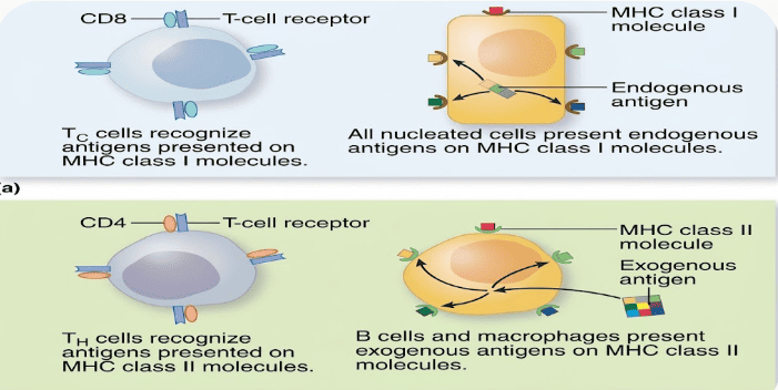
Antigen Recognition by Effector T Cells
Activation of Naive T Cells by Dendritic Cells
Dendritic cells are the primary coordinators of adaptive T-cell responses. They constantly patrol peripheral barrier tissues like the skin and mucous membranes:
- Antigen Sampling: Immature dendritic cells use phagocytosis to swallow whole particles and pinocytosis to drink extracellular fluids. In the digestive and respiratory tracts, they extend thin, tentacle-like projections directly between epithelial cells to sample the lumen.
- Pathogen Sensing: Surface pattern recognition receptors, particularly Toll-Like Receptors (TLRs), identify conserved microbial molecular signatures.
- Migration and Maturation: Recognizing a pathogen triggers maturation. The dendritic cell leaves the tissue and travels through lymphatic vessels to nearby secondary lymphoid organs (such as lymph nodes) to meet naive T cells.
- Co-stimulatory Expression (The Danger Signal): During migration, mature dendritic cells produce surface proteins called co-stimulatory molecules (such as B7-1/CD80 and B7-2/CD86). These act as biological "emergency lights," confirming to the T cell that the presented peptide represents genuine danger.
Activation Versus Peripheral Anergy
A naive T cell needs two signals to activate. Signal 1 is TCR binding to the antigen-MHC complex. Signal 2 is co-stimulation delivered by the mature dendritic cell.
If a naive T cell binds an antigen presented by a dendritic cell that lacks co-stimulatory molecules (indicating a harmless self-peptide or non-infectious debris), the T cell does not activate. Instead, it becomes anergic (unresponsive) and eventually undergoes apoptosis. This safeguard protects against autoimmune disease.
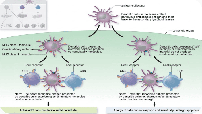
T-Cell Activation vs. Induction of Anergy
Cross-Presentation and Presentation Dynamics
- Cross-Presentation: Dendritic cells can process engulfed external antigens and present them on both MHC Class I and Class II molecules. This unique ability allows them to activate both cytotoxic CD8 and helper CD4 T cells against pathogens that do not directly infect the dendritic cell itself.
- Effector Homing: Activated T cells expand clonally, produce effector and memory subsets, leave the lymph node through efferent lymphatics, and enter the blood to reach infected tissues.
- Role of Macrophages and B Cells: Although B cells and macrophages can express MHC Class II and co-stimulatory signals, in vivo studies show that they primarily stimulate experienced memory T cells during secondary responses, rather than activating naive T cells.
Effector Cytotoxic T Lymphocyte (CD8) Operations
All nucleated cells continuously degrade internal cytoplasmic proteins and display the resulting fragments on surface MHC class I molecules. In healthy tissue, these fragments are normal self-proteins that cytotoxic T cells ignore.
However, when a virus infects a cell or a cell becomes cancerous, abnormal foreign peptides appear on its MHC Class I molecules. A cytotoxic T cell (CD8) recognizing these foreign fragments binds tightly to the target and delivers a targeted chemical attack:
- Perforin: A pore-forming protein that inserts itself into the target's lipid membrane, creating open channels that increase permeability.
- Proteases (Granzymes): Specialized enzymes that enter the target cell's cytoplasm through these perforin pores, activating internal caspases to trigger programmed cell death (apoptosis).
- Macrophage Clearance: The dying cell breaks apart into apoptotic bodies that macrophages quickly phagocytose, clearing the infection without releasing toxic contents that could inflame surrounding healthy tissue.
- Serial Killing: After delivering its cytotoxins, the unharmed CD8 T cell detaches to find and destroy additional infected or malignant cells.
- Alerting Cytokines: Effector CD8 cells also secrete cytokines that encourage neighboring cells to increase their own antigen processing and presentation, while activating local macrophages that have encountered microbes.
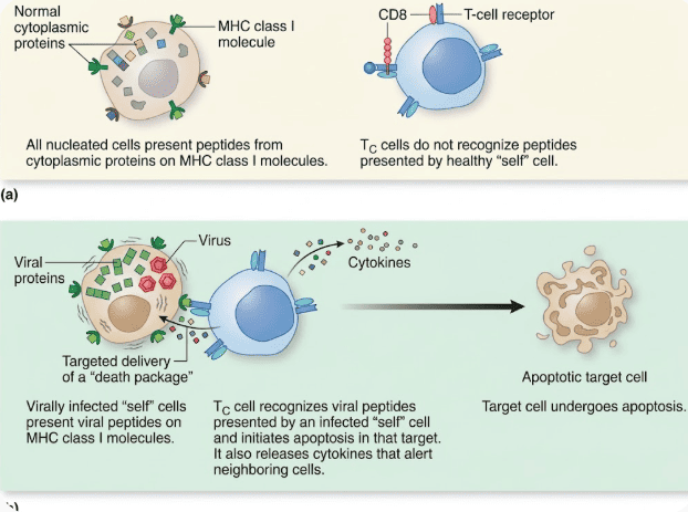
Functions of Effector Cytotoxic T (TC) Cells
Effector Helper T Lymphocyte (CD4) Subsets: Th1 vs. Th2
Effector CD4 helper T cells evaluate foreign peptides presented on MHC Class II molecules by professional antigen-presenting cells (APCs). They divide into two specialized functional subsets based on the type of threat:
| Helper Subset | Primary Target APC | Key Cytokine Effects | Main Immune Function |
|---|---|---|---|
| Th1 Cells | Macrophages displaying engulfed pathogen peptides | Releases cytokines that:
|
Clears intracellular pathogens, such as microbes that resist digestion inside phagosomes |
| Th2 Cells | Naive and Activated B Cells presenting endocytosed peptides | Delivers cytokines that drive:
|
Promotes humoral responses, antibody diversification, and defense against extracellular threats |
Macrophage Activation and Granuloma Formation
Certain resilient microorganisms can survive and replicate inside macrophage phagosomes by blocking normal digestive processes. Th1 cells provide the external signals needed to overcome this evasion:
- Antigen Processing: The macrophage engulfs the microbe into a phagosome, breaks its proteins into peptides, loads them onto MHC class II molecules, and displays them on its surface.
- Th1 Recognition: A matching Th1 cell binds the displayed peptide-MHC complex and delivers activating cytokines.
- Morphological Activation: The activated macrophage enlarges, develops a ruffled plasma membrane, and boosts its metabolic activity. It produces more lysosomes containing potent antimicrobial chemicals and reactive radicals that destroy the trapped invaders.
- Giant Cell Fusion: If activated macrophages cannot eliminate the pathogen on their own, multiple macrophages fuse together to form large multinucleated giant cells.
- Granuloma Encapsulation: When infection persists, these giant cells, surrounding macrophages, and T lymphocytes organize into a tightly packed wall called a granuloma. This structure walls off the microbes, starving them of nutrients and preventing them from spreading to neighboring tissue.
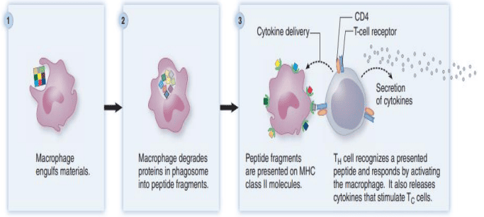
Helper T-Cell Mediated Macrophage Activation
Natural Killer (NK) Cells: Dual Surveillance Roles
Natural Killer cells belong to the innate immune system, but they work closely with adaptive responses to eliminate threats through two main mechanisms:
1. Antibody-Dependent Cellular Cytotoxicity (ADCC)
NK cells express high levels of surface Fc receptors (CD16) that bind the constant base (Fc portion) of IgG antibodies attached to target cells.
- Targeting Large Pathogens: When large parasitic organisms are too big for phagocytes to engulf, antibodies coat their surface. NK cells dock onto these antibodies via their Fc receptors and release perforin and proteases directly onto the parasite.
- Targeting Infected Host Cells: Viral proteins embedded in an infected host cell's membrane are flagged by antibodies. Binding of multiple Fc receptors triggers the NK cell to release its cytotoxic granules, destroying the infected cell via apoptosis.
2. The "Missing-Self" Hypothesis (Detection of MHC Class I Absence)
Cytotoxic T cells (CD8) can only kill infected cells that present foreign peptides on MHC Class I molecules. Because of this, some viruses evade detection by forcing the host cell to pull down or stop producing MHC class I molecules entirely.
- The Escape Problem: Cells lacking MHC class I molecules are invisible to cytotoxic T lymphocytes.
- The NK Solution: NK cells survey host tissues for normal MHC Class I expression, which serves as an inhibitory signal. If a cell lacks surface MHC Class I molecules, the inhibitory signal is lost. The NK cell identifies the cell as stressed or abnormal and releases perforin and proteases to trigger apoptosis.
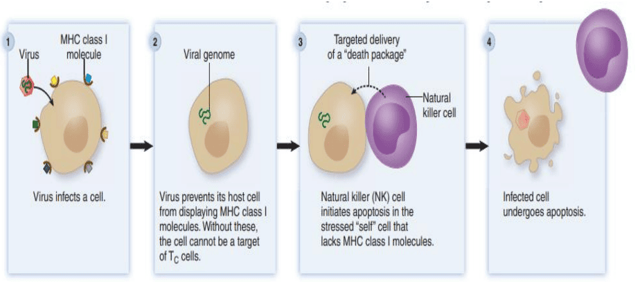
Natural Killer (NK) Cells Destroy Stressed "Self" Cells That Lack MHC Class I Molecules
Infection and Immunity: Classification of Acquired Protection
Immunity acquired throughout life is grouped into two main categories: naturally acquired (resulting from natural biological events) and artificially acquired (resulting from medical intervention like vaccination or antibody infusion). Both forms work through either active or passive mechanisms.
| Immunity Category | Mechanism of Acquisition | Onset & Memory | Clinical Examples |
|---|---|---|---|
| Natural Active Immunity | Direct infection by a live environmental pathogen stimulates the host's own B and T cells. | Delayed onset (1–2 weeks); generates long-lasting immunological memory. | Lifelong protection after recovering from wild varicella (chickenpox) or mumps infection. |
| Natural Passive Immunity | Direct transfer of maternal antibodies to the child across the placenta or through breast milk. | Immediate protection; no immunological memory (antibodies degrade over weeks to months). | Maternal IgG crossing placenta protects neonates for 3–6 months; secretory IgA in breast milk protects the infant gut. |
| Artificial Active Immunity | Deliberate exposure to non-pathogenic antigen preparations via vaccination. | Takes days to weeks to develop; builds long-term memory cells and antibody titers. | Immunization with MMR, hepatitis B subunit, or tetanus toxoid vaccines. |
| Artificial Passive Immunity | Injection of concentrated antibodies collected from immune human donors or hyperimmunized animals. | Immediate short-term defense; no memory formed (protection ends when donor proteins degrade). | Post-exposure prophylaxis using Rabies Immune Globulin (RIG), Tetanus Antitoxin, or Gamma Globulin for traveler protection. |
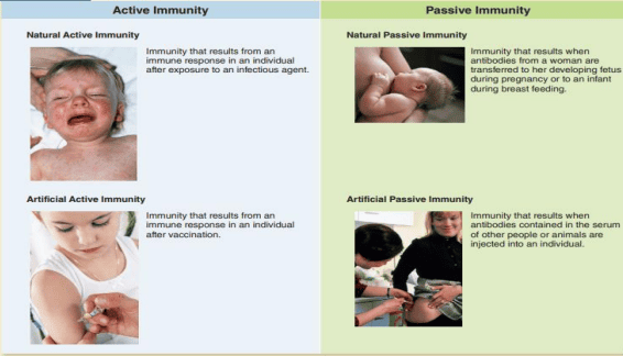
Classifications of Acquired Immunity
Preparations Used in Passive Immunization
Clinical Applications of Passive Therapy
- Antiserum: Any fluid serum preparation harvested from humans or animals that contains protective antibodies.
- Antitoxin: An antibody preparation that specifically binds and neutralizes dangerous bacterial toxins (such as botulinum or diphtheria toxins).
- Immune Serum Globulin (Gamma Globulin): A pooled fraction of IgG antibodies collected from many healthy blood donors. It provides broad, temporary protection to immunocompromised patients who cannot produce their own antibodies, or to individuals traveling to disease-endemic areas.
- Hyperimmune Globulin: Formulations prepared from donors with unusually high antibody levels against a specific pathogen. These are used for targeted treatment or emergency prevention. Common examples include:
- Hepatitis A Immune Globulin (HAIG)
- Hepatitis B Immune Globulin (HBIG)
- Rabies Immune Globulin (RIG)
- Human Tetanus Immune Globulin (HTIG)
Vaccinology Concepts and Population Protection
A vaccine is an immunogenic biological preparation that contains a weakened pathogen, an inactivated pathogen, or purified microbial components. Its purpose is to stimulate protective adaptive immunity and establish immunological memory without causing serious illness.
Herd Immunity (Community Protection)
Herd Immunity occurs when a critical proportion of a population becomes immune to an infectious agent, either through vaccination or prior infection.
This widespread immunity lowers the overall concentration of susceptible individuals. As a result, the pathogen cannot find enough hosts to sustain transmission chains, which protects vulnerable individuals who cannot be safely vaccinated (such as newborns and severely immunocompromised patients).
Major Human Vaccines: Formulations, Schedules, and Target Populations
| Pathogen / Disease | Formulation Type | Recommended Target Populations & Clinical Context |
|---|---|---|
| Anthrax | Subunit | Adults at high occupational risk of exposure, including specific military personnel and specialized laboratory workers. |
| Diphtheria | Toxoid (inactivated exotoxin) | Children (as the "D" in the pediatric DTaP series); adolescents and adults require booster doses every 10 years (as the "d" in Td or Tdap). |
| Haemophilus influenzae type b (Hib) | Polysaccharide-protein conjugate | Infants and children, protecting against invasive childhood meningitis, bacteremia, and epiglottitis. |
| Hepatitis A | Inactivated whole virus | Routine in infants and children; adolescents in designated regions; adults at high risk (including travelers to endemic nations, men who have sex with men, and close contacts of international adoptees). |
| Hepatitis B | Subunit (recombinant surface antigen, HBsAg) | Universal administration for newborns and children; adults at elevated risk (healthcare professionals with blood/fluid contact, multiple sexual partners, and household contacts of infected individuals). |
| Human Papillomavirus (HPV) | Virus-Like Particle (VLP) (recombinant capsid proteins covering 2, 4, or 9 oncogenic/wart serotypes) | Girls and boys, women and men typically aged 11 through 26 years to prevent cervical, anogenital, and oropharyngeal cancers. |
| Influenza | Three age-specific formulations:
|
All individuals aged 6 months and older, given annually due to frequent viral antigenic drift. When supplies are limited, administration prioritizes high-risk groups (elderly, immunocompromised, chronic pulmonary disease, pregnancy). |
| Measles | Attenuated live virus | Children (as the "M" in the combined MMR or MMRV childhood schedule); booster immunization for adults born after 1956 who lack evidence of immunity and have no medical contraindications. |
| Meningococcal Disease (Neisseria meningitidis) | Two age-specific preparations:
|
Adolescents; children and adults with medical vulnerabilities like anatomic/functional asplenia or terminal complement deficiencies; adults in high-risk environments (dormitory residents, military recruits, and travelers to the African meningitis belt). |
| Mumps | Attenuated live virus | Children (the second "M" in pediatric MMR/MMRV); adult boosters for post-1956 cohorts lacking confirmed immunity without medical contraindications. |
| Pertussis (Whooping Cough) | Subunit (acellular pertussis, "aP") | Infants and children (in DTaP); adolescents and pregnant women during each pregnancy (Tdap); adults aged 19–64 may receive a booster. |
| Pneumococcal Infection (Streptococcus pneumoniae) | Two distinct formulations:
|
Routine for all infants and children; adults aged 65 and older; individuals with chronic cardiopulmonary diseases, diabetes, asplenia, or other high-risk states. |
| Poliomyelitis | Two formulations:
|
Universal pediatric series. Attenuated OPV has been widely used in global eradication campaigns due to its low cost and ability to induce mucosal IgA, whereas IPV is standard in regions where vaccine-derived polio must be avoided. |
| Rabies | Inactivated whole virus | Pre-exposure prophylaxis for veterinarians, wildlife biologists, animal control staff, and travelers to high-risk areas; routine post-exposure prophylaxis following animal bites. |
| Rotavirus | Attenuated live virus (oral drops) | Young infants, protecting against severe dehydrating diarrheal gastroenteritis. |
| Rubella (German Measles) | Attenuated live virus | Pediatric population (the "R" in MMR/MMRV); non-immune women of childbearing age prior to conception to prevent congenital rubella syndrome (contraindicated during pregnancy). |
| Shingles (Herpes Zoster) | Attenuated live virus (or recombinant subunit) | Approved for adults aged 50 to 60 years and older to prevent latent varicella-zoster reactivation and postherpetic neuralgia. |
| Tetanus (Lockjaw) | Toxoid (inactivated tetanospasmin protein) | Children (the "T" in DTaP); routine booster dose every 10 years throughout adulthood (Td or Tdap). |
| Tuberculosis | Attenuated live bacterium (Bacillus Calmette-Guérin / BCG strain) | Widely administered to infants at birth in countries with high tuberculosis burdens; used only in exceptional, highly selective circumstances in the United States and low-incidence nations. |
| Typhoid Fever | Two distinct formulations:
|
Travelers journeying to endemic regions with poor water sanitation and individuals with close exposure to chronic Salmonella Typhi carriers. |
| Varicella-Zoster (Chickenpox) | Attenuated live virus | Pediatric populations (the "V" in MMRV); non-immune adolescents and adults without medical contraindications. |
| Yellow Fever | Attenuated live virus | Travelers visiting or residing in endemic tropical zones of South America and sub-Saharan Africa. |
Comparison of Attenuated vs. Inactivated Vaccines
All vaccines fall into two main categories depending on whether the immunizing component can replicate inside the recipient:
Attenuated (Live Weakened) Vaccines
These contain live microorganisms that have been weakened by deliberate mutation so they cannot cause severe illness in healthy hosts. The weakened pathogen replicates in small amounts, causing a mild or unnoticeable infection that stimulates a broad, long-lasting immune response.
- Advantages: They provide durable, often lifelong immunity with just one or two doses. If an orally vaccinated individual sheds the weakened virus, they can indirectly immunize close contacts.
- Disadvantages: In rare cases, they can mutate back to virulent forms. They can cause severe disease in immunocompromised patients, cross the placenta to harm a developing fetus, and require continuous refrigeration or freezing to remain viable.
Inactivated (Non-Replicating) Vaccines
These contain killed whole pathogens or specific purified parts that cannot replicate, while still presenting antigens to the immune system. Examples include toxoids, subunit vaccines, recombinant antigens, virus-like particles (VLPs), polysaccharide vaccines, and protein conjugates.
- Advantages: They cannot replicate, cause active infection, or revert to dangerous virulent strains. They are safe to administer to immunocompromised patients and pregnant women.
- Disadvantages: Because the dead or purified components do not replicate, they stimulate a weaker immune response. They require chemical adjuvants to boost immunogenicity and require multiple booster injections to maintain protective antibody levels over time.
| Feature / Characteristic | Attenuated Vaccines | Inactivated Vaccines |
|---|---|---|
| Humoral Antibody Response | Robust IgG; also induces mucosal secretory IgA if given orally or nasally. | Primarily circulating IgG; negligible mucosal IgA. |
| Cell-Mediated Immunity | Strong (Good); intracellular viral replication loads peptides onto MHC Class I, activating CD8 cytotoxic T cells. | Weak (Poor); exogenous dead antigens load mainly onto MHC Class II, providing little CD8 cytotoxic stimulation. |
| Duration of Protection | Long-term (often lifelong) | Short-term; antibody titers decline without booster doses |
| Need for Adjuvants | No (live replication naturally triggers innate pattern receptors) | Yes (requires aluminum salts or other additives to stimulate innate immunity) |
| Required Number of Doses | Usually a single dose (sometimes followed by a booster) | Multiple doses and regular periodic boosters |
| Risk of Reverting to Virulence | Very low (rare back-mutations can occur) | Absent (pathogen components cannot replicate) |
| Risk in Immunocompromised Hosts | Significant (weakened strains can cause severe, uncontrolled disease) | Absent (safe for immune-deficient and pregnant patients) |
| Administration Routes | Injection, oral drops, or nasal spray | Intramuscular or subcutaneous injection |
| Stability in Warm Environments | Poor (requires strict cold-chain refrigeration) | Good (stable at ambient temperatures) |
| Formulation Types | Attenuated viral strains; attenuated bacterial strains (e.g., BCG, Ty21a) | Inactivated whole pathogens, toxoids, subunit preparations, virus-like particles (VLPs), polysaccharide vaccines, and conjugate formulations |
