Doctors Revision

Biochemistry

proteins exam
Biochemistry

Proteins Lesson Exam

Biochemistry: Protein/Amino Acids Exam Biochemistry: Protein/Amino Acids Exam Test your knowledge with these 40 questions. Start Exam Protein/Amino Acids Exam Question 1/40 Submit Next Exam Complete! Here are your results, . Your Score 38/40 95% Download Full Report

sickle
Biochemistry

Abnormal Haemoglobin: Sickle Cell Scenario

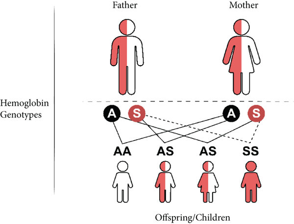
Abnormal Hemoglobin Objectives: Define Abnormal Hemoglobin: Understand what constitutes “abnormal” in the context of hemoglobin structure and function. Classify Abnormal Hemoglobins: Categorize the main types of abnormal hemoglobins based on their molecular defects. Explore Structural Hemoglobinopathies: Examine the molecular basis of common structural variants (e.g., HbS, HbC, HbE). Discuss the impact of specific amino acid substitutions on hemoglobin’s physical and chemical properties. Relate these molecular changes to the resulting clinical syndromes. Investigate Thalassemias (Quantitative Hemoglobinopathies): Differentiate between alpha (α) and beta (β) thalassemias. Elucidate the genetic defects leading to reduced or absent globin chain synthesis. Explain the pathogenic consequences of globin chain imbalance (e.g., ineffective erythropoiesis, hemolysis). Describe the clinical spectrum of thalassemia syndromes. Discuss Unstable Hemoglobins: Define unstable hemoglobin variants and their structural basis. Explain the mechanism of Heinz body formation and chronic hemolysis. Review Hemoglobins with Altered Oxygen Affinity: Explain the structural modifications that lead to increased or decreased oxygen affinity. Describe the clinical presentations associated with these variants (e.g., polycythemia, cyanosis). Summarize Diagnostic Approaches: Outline the key laboratory tests used to identify and characterize abnormal hemoglobins. Discuss Therapeutic Strategies: Briefly touch upon current and emerging treatments for common abnormal hemoglobin disorders. 1. Define Abnormal Hemoglobin Abnormal hemoglobin refers to any variant of the hemoglobin molecule that deviates from the normal adult hemoglobin (HbA) in its primary amino acid sequence, structure, or quantity, leading to impaired function or stability. These abnormalities can result in a range of clinical conditions, collectively known as hemoglobinopathies, affecting the red blood cells’ ability to effectively transport oxygen. 2. Classify Abnormal Hemoglobins Abnormal hemoglobins are broadly classified based on the nature of their underlying molecular defect: Structural Variants (Qualitative Defects): Involve a change in the amino acid sequence of a globin chain, often from a point mutation. This results in an abnormal protein. Examples: HbS, HbC, HbE. Thalassemias (Quantitative Defects): Involve reduced or absent production of a structurally normal globin chain due to gene deletions or mutations. This leads to a chain imbalance. Examples: α-thalassemia, β-thalassemia. Unstable Hemoglobins Structural variants where an amino acid substitution destabilizes the molecule, causing it to precipitate and lead to chronic hemolysis and Heinz body formation. Altered O₂ Affinity Structural variants where amino acid changes affect allosteric properties, altering the ability to bind and release oxygen, leading to polycythemia or cyanosis. 3. Explore Structural Hemoglobinopathies Structural hemoglobinopathies are characterized by the synthesis of an abnormal globin chain due to a mutation in the globin gene. a. Hemoglobin S (HbS) Molecular Basis: β6Glu→Val (Glutamate to Valine).Impact: Creates a hydrophobic patch, leading to polymerization of deoxygenated HbS.Syndrome: Sickle Cell Disease. Rigid sickled cells cause vaso-occlusion (pain crises) and chronic hemolytic anemia. b. Hemoglobin C (HbC) Molecular Basis: β6Glu→Lys (Glutamate to Lysine).Impact: Reduced solubility causes HbC to crystallize within RBCs.Syndrome: HbC Disease. Mild chronic hemolytic anemia, splenomegaly, and characteristic “target cells” on blood smear. c. Hemoglobin E (HbE) Molecular Basis: β26Glu→Lys (Glutamate to Lysine).Impact: Creates an alternative mRNA splice site, causing a mild quantitative defect (thalassemic effect).Syndrome: Mild microcytic anemia. Clinically significant when co-inherited with β-thalassemia. 4. Investigate Thalassemias (Quantitative Hemoglobinopathies) Thalassemias are characterized by a reduced rate of synthesis or absence of one or more of the globin chains, leading to an imbalance in the production of α and β globin chains. The individual globin chains produced are structurally normal. a. Alpha (α)-Thalassemia Genetic Defect: Deletion of one or more of the four α-globin genes on chromosome 16.Pathology: Excess β or γ chains form unstable tetramers (HbH, Hb Barts) that are poor oxygen carriers, leading to hemolysis and ineffective erythropoiesis.Spectrum: Severity depends on the number of genes deleted, ranging from a silent carrier (1 gene) to fatal hydrops fetalis (4 genes). b. Beta (β)-Thalassemia Genetic Defect: Point mutations in the two β-globin genes on chromosome 11, reducing (β+) or eliminating (β0) synthesis.Pathology: Excess α-chains are highly insoluble and precipitate in RBC precursors, causing severe ineffective erythropoiesis and hemolysis.Spectrum: Ranges from asymptomatic trait (minor) to transfusion-dependent anemic (major). 5. Discuss Unstable Hemoglobins Definition: These are structural hemoglobin variants that have amino acid substitutions, usually in the interior hydrophobic pocket or at the heme-globin contact points, which disrupt the stability of the hemoglobin molecule. Structural Basis: The mutations often expose heme or critical hydrophobic regions to the aqueous environment. This leads to conformational changes that loosen the binding of heme to the globin chain. Mechanism of Heinz Body Formation and Chronic Hemolysis: The unstable hemoglobin molecules readily denature (unfold) and precipitate into insoluble aggregates. These precipitated, denatured hemoglobin aggregates attach to the inner surface of the red blood cell membrane, forming characteristic intracellular inclusions called Heinz bodies. Heinz bodies make red blood cells rigid and susceptible to removal by the spleen (extravascular hemolysis), leading to chronic hemolytic anemia. Examples: Hb Zurich, Hb Köln. Clinical Presentation: Chronic hemolytic anemia, often exacerbated by oxidative stress (e.g., certain drugs). Splenomegaly is common. 6. Review Hemoglobins with Altered Oxygen Affinity These are structural hemoglobin variants where amino acid substitutions alter the allosteric regulation of oxygen binding and release. a. Increased Oxygen Affinity Mechanism: Mutations stabilize the R (oxygenated) state, making it harder to release O₂ to tissues.Presentation (Polycythemia): Tissue hypoxia stimulates erythropoietin, leading to increased red blood cell production (erythrocytosis).Examples: Hb Chesapeake, Hb Suresnes. b. Decreased Oxygen Affinity Mechanism: Mutations stabilize the T (deoxygenated) state, causing premature O₂ release.Presentation (Cyanosis): Higher levels of deoxygenated Hb in arterial blood cause a bluish discoloration of the skin, though O₂ delivery is adequate.Examples: Hb Kansas, Hb Beth Israel. 7. Summarize Diagnostic Approaches The diagnosis of abnormal hemoglobin disorders relies on a combination of clinical evaluation and specialized laboratory tests: Complete Blood Count (CBC) with Red Blood Cell Indices: Screens for anemia, microcytosis, or polycythemia. Peripheral Blood Smear: Crucial for morphological assessment (sickle cells, target cells, Heinz bodies). Hemoglobin Electrophoresis (Alkaline & Acid pH): Separates different hemoglobin types based on their electrical charge. High-Performance Liquid Chromatography (HPLC): A more sensitive and quantitative method for separating hemoglobin types. Genetic Testing (DNA analysis): Confirms specific mutations in globin genes, essential for

Bioenergetics (Thermodynamics and ATP)
Biochemistry

Bioenergetics (Thermodynamics and ATP)

Bioenergetics : Thermodynamics & ATP Bioenergetics: The Engine of Life – How Organisms Manage Energy Let’s shift our focus now to the fundamental concept of Bioenergetics. The term itself is quite descriptive: “Bio” means life. “Energetics” means the study of energy. Therefore, Bioenergetics is simply the study of how living organisms manage energy. It’s the exploration of the transfer and utilization of energy in biological systems. It delves into the intricate mechanisms that allow life to exist and thrive, from the smallest bacteria to the largest whales. This critical field encompasses several key aspects: How organisms obtain energy: This involves understanding the initial sources of energy. How organisms transform this energy from one form to another: Energy needs to be converted into a usable “currency”. How organisms use this energy to do all the things necessary for life: Fueling every biological process. At its core, Energy means the capacity or ability to do work. In biology, “work” means anything that requires effort or causes a change. It’s a much broader concept, encompassing all the dynamic processes that sustain life. Just like machines require energy to do work, all living organisms need a constant supply of energy to function. Examples of “Work” in Biological Systems Requiring Energy: Gross Motor Movement (Physical Work) Just as a car needs petrol, our muscles need energy to contract and allow us to walk, run, or lift objects. Our heart muscle continuously contracts to pump blood, and our diaphragm contracts to allow us to breathe. Growth and Development (Synthetic Work) A toddler growing into an adult requires a massive amount of energy to synthesize new cells, tissues, and complex molecules like proteins and DNA. This is “building work.” The process of reproduction also demands significant energy input. Maintaining Homeostasis (Maintenance Work) Even when resting, our body is still doing immense “work”! This basal metabolic activity includes our heart beating, lungs breathing, brain activity, and cells constantly repairing themselves and actively transporting ions across membranes. Where does this Energy Come From? The Sun For planet Earth, the main, original, and most abundant source of energy is sunlight. However, most living things, including humans, can’t directly use sunlight. It’s a fascinating journey through the food chain. The Journey of Sunlight Energy to You: Plants (Producers – The Solar Collectors): Plants capture light energy from the sun through a process called photosynthesis. They use simple molecules like CO₂ and water to convert this light energy into glucose, a form of stored chemical energy. Animals (Consumers – The Energy Transfer Agents): When you eat a plant product, you are directly consuming the glucose the plant made. When you eat an animal product, that animal likely consumed plants, so you are indirectly getting the energy that originally came from the sun. The Goal: Energy-Rich Molecules for Your Cells Regardless of what you eat, your body obtains these energy-rich molecules (carbohydrates, fats, proteins). Your cells then break them down through metabolic pathways to release the stored chemical energy. This released energy is then used to synthesize a special energy currency molecule called ATP (Adenosine Triphosphate), which is the direct fuel for almost all cellular work. Clinical Relevance for Nursing Students Understanding bioenergetics is foundational to many aspects of nursing: Nutrition and Energy Intake: Nurses assess patients’ nutritional status and energy needs for processes like wound healing and recovery. Malnutrition directly impacts a patient’s ability to heal. Metabolic Disorders: Diseases like diabetes mellitus are direct examples of impaired bioenergetics—the body’s inability to properly utilize glucose for energy. Exercise Physiology: The energy demands of physical activity are direct applications of bioenergetics. Pharmacology: Many drugs affect metabolic pathways, altering how cells produce or use energy. Patient Education: Explaining the importance of a balanced diet can empower patients to manage their health effectively. ATP: The Body’s Universal Energy Currency Once the body gets energy from food, it doesn’t directly use these complex food molecules to power every single tiny process. Instead, the body converts the chemical energy stored in these food molecules into a much more manageable and readily available form: a special molecule called ATP. ATP: Adenosine Triphosphate Why is ATP called the “Energy Currency”? Think of it like money. You don’t get paid in raw materials; you get paid in money, which you can use to buy whatever you need. Similarly, your body converts energy from diverse food sources into ATP (the “money”). Then, it uses ATP to “pay for” all its energy-requiring processes. ATP is the direct, usable form of energy for almost all cellular activities. How does ATP store and release energy? The key lies in the “high-energy” chemical bonds connecting its three phosphate groups. When your cells need energy, they break off one of the phosphate groups from ATP. This breaking of the bond releases a significant amount of free energy that the cell can immediately use. ATP → ADP + Pᵢ + Energy This reaction is reversible. When your body has excess energy, it can use it to reattach the phosphate group to ADP, converting it back into ATP, thus “recharging the battery.” Free Energy (Exergonic vs. Endergonic Reactions) Free energy (Gibbs Free Energy, G) is the amount of energy available to do work within a system. It helps us predict whether a chemical reaction will happen spontaneously (release energy) or require an input of energy. Analogy: A person moving down a hill is a spontaneous process that releases energy. A person lifting a weight up a hill is a non-spontaneous process that requires energy. Exergonic Reactions: Energy-Releasing These reactions release free energy and can happen spontaneously. The change in free energy (ΔG) is negative (ΔG < 0). Biological Examples: Cellular Respiration: The breakdown of glucose into CO₂ and water releases a lot of free energy used to make ATP. ATP Hydrolysis: The breakdown of ATP into ADP + Pᵢ is a classic exergonic reaction that releases usable energy for the cell. Endergonic Reactions: Energy-Requiring These reactions require an input of free energy and are non-spontaneous. The change in

Acids, Bases, pH and Buffer
Biochemistry

Acids, Bases, pH and Buffer

Acids, Bases, pH and Buffer : The Chemical Environment Objectives At the end of this section, you will be able to understand: Acids, Bases, and pH: What are acids and bases? What is pH? The pH scale. Buffers: Why they are crucial in living systems. Why it’s important: Biochemical reactions are very sensitive to pH. Maintaining the correct pH is vital for survival. Acids and Bases The environment within and around our cells is not static; it’s a dynamic chemical soup where countless reactions occur simultaneously. Just like a baker needs to precisely control oven temperature, the “chemical temperature” of our biological systems – its acidity or basicity – must be meticulously maintained within an incredibly narrow range. This control, measured by pH, is paramount for life. Even minor deviations can lead to catastrophic consequences, as the delicate structures of proteins and enzymes are exquisitely sensitive to pH changes. This maintenance of a stable internal pH is a cornerstone of homeostasis. What Makes Something Acidic or Basic? It’s All About the Proton (H⁺) At the heart of acidity and basicity is one tiny, yet profoundly powerful, particle: the hydrogen ion (H⁺). A hydrogen ion (H⁺) is essentially just a proton. The concentration of these free H⁺ ions in a solution is the ultimate determinant of whether that solution is acidic, neutral, or basic. Acids: The Proton Donors An acid is defined as any substance that, when dissolved in an aqueous solution, releases or donates hydrogen ions (H⁺), thereby increasing the concentration of free H⁺ in that solution. Strength: A strong acid dissociates almost completely in water, releasing nearly all its H⁺ ions. A weak acid only partially dissociates. Real-World and Physiological Examples: Hydrochloric Acid (HCl): A strong acid in your stomach, crucial for digestion. It undergoes almost complete dissociation: HCl(aq) → H⁺(aq) + Cl⁻(aq) Carbonic Acid (H₂CO₃): A crucial weak acid in your blood. It only partially dissociates, maintaining an equilibrium: H₂CO₃(aq) ⇌ H⁺(aq) + HCO₃⁻(aq)The double arrow (⇌) indicates the reaction is reversible. Bases: The Proton Acceptors A base (or alkali) is any substance that, when dissolved in an aqueous solution, decreases the concentration of H⁺ ions by “accepting” them or by releasing hydroxide ions (OH⁻). Strength: A strong base dissociates almost completely. A weak base only partially accepts H⁺ or releases OH⁻ ions. Real-World and Physiological Examples: Sodium Hydroxide (NaOH): A very strong base. It dissociates completely: NaOH(aq) → Na⁺(aq) + OH⁻(aq)The released OH⁻ then rapidly combines with H⁺ to form water: OH⁻(aq) + H⁺(aq) → H₂O(l) Bicarbonate (HCO₃⁻): The most important weak base in blood plasma. It can readily accept a free H⁺ ion to form carbonic acid, “soaking up” excess acid: HCO₃⁻(aq) + H⁺(aq) ⇌ H₂CO₃(aq) The Importance of “Aqueous Solution” The definition of acids and bases in this context relies on their behavior in aqueous solutions (where water is the solvent). Water itself can slightly dissociate: H₂O(l) ⇌ H⁺(aq) + OH⁻(aq). In pure water, the concentrations of H⁺ and OH⁻ are equal, making it neutral. Acids disturb this balance by increasing H⁺, and bases disturb it by decreasing H⁺. Clinical Significance for Nurses: Why Acid-Base Balance is Critical Understanding acids and bases is not just theoretical; it’s fundamental to clinical practice: pH Homeostasis: The body meticulously maintains the pH of arterial blood between 7.35 and 7.45. Even slight deviations (e.g., acidosis 7.45) can impair enzyme function, alter protein structures, disrupt electrolyte balance (e.g., potassium levels), and depress or overstimulate the central nervous system, potentially leading to organ failure and death. Buffer Systems: The body employs sophisticated buffer systems (like the bicarbonate buffer system, phosphate buffer system, and protein buffer system) to resist sudden changes in pH. These buffers are mixtures of weak acids and their conjugate bases (or weak bases and their conjugate acids) that can absorb excess H⁺ or release H⁺ as needed. Respiratory and Renal Regulation: The lungs regulate pH by controlling the exhalation of carbon dioxide (which forms carbonic acid in blood), while the kidneys regulate pH by reabsorbing bicarbonate and excreting H⁺ ions. Disease States: Many disease states, such as diabetic ketoacidosis, chronic obstructive pulmonary disease (COPD), renal failure, and sepsis, are characterized by severe acid-base imbalances that nurses must be able to recognize, monitor, and assist in managing. Medication Administration: The pH of intravenous fluids and medications must often be carefully considered to prevent local irritation or systemic acid-base disturbances. The pH Scale: A Precise and Powerful Ruler for Acidity While discussing “hydrogen ion concentration” ([H⁺]) is chemically precise, it’s cumbersome. To simplify this, scientists developed the pH scale – a brilliant shorthand that transforms these unwieldy numbers into an easy-to-use linear scale. What Does pH Stand For? pH literally stands for “potential of Hydrogen” or “power of Hydrogen.” It is a numerical scale that quantifies the concentration of hydrogen ions (H⁺) in a solution. The Mathematical Definition The pH is defined as the negative base-10 logarithm of the hydrogen ion concentration (in moles per liter, M): pH = −log₁₀[H⁺] The log₁₀ function makes large numbers manageable, and the negative sign (−) converts the negative results into the positive numbers we see on the scale. The pH Scale Range and Interpretations: The pH scale typically ranges from 0 to 14. Acidic (pH < 7) The lower the pH, the higher the [H⁺] concentration. Examples: Stomach acid (pH 1.5-3.5), lemon juice, coffee. Neutral (pH = 7) The concentration of H⁺ equals the concentration of OH⁻. Examples: Pure water, human tears. Basic/Alkaline (pH > 7) The higher the pH, the lower the [H⁺] concentration. Examples: Baking soda, ammonia, bleach. The Logarithmic Nature: A Crucial Detail for Nurses This is perhaps the most important concept about the pH scale. It is logarithmic, not linear. This means that a change of 1 pH unit represents a 10-fold (ten times) change in the actual concentration of H⁺ ions. Applying the Principle: A solution with a pH of 5 is 10 times more acidic than a solution with a pH of 6. A solution with
Water and Bonding
Biochemistry

Bonding and Water

Biochemistry Intro : Bonding & Water Biochemistry Intro : Bonding & Water Learning Objectives Chemical Bonds: Covalent bonds: Sharing electrons (the most important type in biochemistry). Ionic bonds: Transferring electrons (less common in biological molecules). Hydrogen bonds: Weaker bonds, but SUPER important for water and protein structure. Water: Its unique properties: Polarity, hydrogen bonding. Why it’s essential for life: How it dissolves things, its role in temperature regulation. Introduction to Chemical Bonds We’ve covered atoms, the fundamental building blocks. However, atoms rarely exist in isolation. The connection that holds atoms together is known as a chemical bond. Chemical bonds are the powerful attractive forces that hold atoms together to form molecules and compounds. They are entirely about the behavior and interactions of an atom’s outermost electrons, called valence electrons. The primary reason atoms form bonds is to achieve a more stable state, typically by having a full outer electron shell. Chemical Bonds We’ve explored atoms, the fundamental building blocks. However, in nature, atoms rarely exist in isolation. They are almost universally connected to other atoms. This crucial connection is known as a chemical bond. Definition: Chemical bonds are the powerful attractive forces that hold atoms together to form molecules and compounds. They are, in essence, the “glue” of chemistry, responsible for creating all the complex structures we see, from simple water molecules to intricate proteins and DNA. The Role of Electrons: Chemical bonds are not made of literal glue; rather, they are entirely about the behavior and interactions of an atom’s electrons, particularly its outermost electrons, which are called valence electrons. The Drive for Stability: The primary reason atoms form bonds is to achieve a more stable, “happy” state. For most atoms, this is attained by having a full outer electron shell, often with eight electrons (the “octet rule”). Atoms will gain, lose, or share valence electrons to reach this desired configuration. There are two main types of strong chemical bonds that are particularly important in biochemistry and healthcare: Covalent Bonds and Ionic Bonds. I. Covalent Bonds: The “Sharing is Caring” Bond A covalent bond forms when two atoms share one or more pairs of electrons. It’s a true partnership, and these are the strong bonds that form the backbone of the vast majority of molecules in our bodies. Mechanism: Each atom contributes one electron to the shared pair, and this shared pair is then mutually attracted to the nuclei of both atoms, effectively holding them together. Prevalence: Covalent bonds are the most common type of bond found in organic molecules – the fundamental molecules of life, including carbohydrates, lipids, proteins, and nucleic acids. Valence Electrons Revisited: Remember, valence electrons are the electrons in the outermost shell of an atom. They are the key players in chemical bonding and are the ones involved in the electron-sharing of covalent bonds. Analogy for Covalent Bonding Imagine two people who both deeply desire to own a dog, but neither can financially manage the full cost and responsibility alone. They decide to co-own one. They are now “bonded” together by their shared pet. The shared dog represents the shared pair of electrons, and the arrangement benefits both owners. The dog effectively belongs to both, much like the shared electrons are attracted to both atomic nuclei. Types of Covalent Bonds: Not All Sharing is Equal Even within covalent bonds, the sharing of electrons isn’t always perfectly equitable. This leads to two important subtypes: a) Nonpolar Covalent Bonds: Equal Sharing Definition: Nonpolar covalent bonds occur when electrons are shared equally between two atoms. Electronegativity Connection: This equal sharing happens when the two atoms have very similar electronegativity. Electronegativity is a measure of an atom’s intrinsic ability to attract shared electrons. If their “pull” is roughly equivalent, the electrons spend an equal amount of time around each nucleus. Result: There is no significant charge difference across the bond. The molecule remains electrically symmetrical. Examples: O=O (oxygen gas): Two identical oxygen atoms share electrons perfectly. C-C bonds: Carbon atoms share electrons equally. C-H bonds: Carbon and hydrogen have similar electronegativities, forming nearly nonpolar bonds found in fats and oils. Analogy: If two identical twins co-own a dog, the dog will spend exactly half its time at each house. The sharing is perfectly equal. b) Polar Covalent Bonds: Unequal Sharing Definition: Polar covalent bonds occur when electrons are shared unequally between two atoms. Electronegativity Difference: This arises when one atom is significantly more electronegative than the other, exerting a stronger “pull” on the shared electrons. Result: This creates a slight partial negative charge (δ−) on the more electronegative atom and a slight partial positive charge (δ+) on the less electronegative one. The molecule has distinct “poles” of charge. Examples: O-H bonds: Oxygen is much more electronegative than hydrogen. N-H bonds: Nitrogen is also more electronegative than hydrogen. Crucial for Water (H₂O): The highly electronegative oxygen pulls electrons from the hydrogen atoms, leaving the oxygen end partially negative (δ−) and the hydrogen ends partially positive (δ+). This polarity is essential for all life. Analogy: If a big, strong adult co-owns a dog with a small child, the dog will spend more time at the adult’s house. The sharing is unequal, creating a partial imbalance. Single, Double, and Triple Covalent Bonds: Degrees of Sharing Covalent bonds can also vary in the number of electron pairs shared: Single Bond: Atoms share one pair of electrons (2 electrons total). These bonds are generally flexible and allow rotation. Double Bond: Atoms share two pairs of electrons (4 electrons total). These are stronger and shorter and restrict rotation. Triple Bond: Atoms share three pairs of electrons (6 electrons total). These are the strongest and shortest and entirely prevent rotation. Why Understanding Covalent Bond Polarity is Vital in Nursing and Biology: The distinction between nonpolar and polar covalent bonds profoundly impacts biological systems: “Like Dissolves Like”: The Basis of Solutions Why IV Fluids Work: Polar water readily dissolves charged ions (like Na⁺ and Cl⁻ from salt), which explains why saline solution is the basis of

Model-of-the-atom
Biochemistry

Biochemistry Introduction to Atoms and Molecules

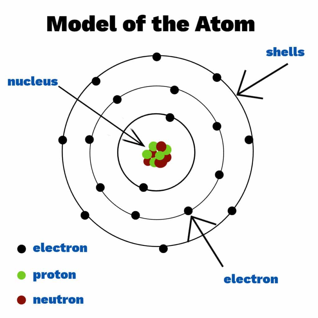
Biochemistry Intro : Atoms and Molecules Phase 1: Chemistry Fundamentals Objectives At the end of this section, you will be able to understand: Atoms and Molecules: What are atoms? Protons, neutrons, electrons. The periodic table (its organization, not memorization). What are molecules? How do atoms join together? Why it’s important: Biochemistry is all about molecules in living systems. You need to know what they are made of. Without a chemistry background, Let’s Review Chemistry. Chemistry is the study of matter and the ways in which different forms of matter combine with each other. You study chemistry because it helps you to understand the world around you. Everything you touch or taste or smell is a chemical, and the interactions of these chemicals with each other define our universe. Chemistry forms the fundamental basis for biology and medicine. Areas of Chemistry The study of modern chemistry has many branches, but can be broken down into five main disciplines, or areas of study: Physical chemistry The study of macroscopic properties, atomic properties, and phenomena in chemical systems. Organic chemistry The study of chemicals containing carbon. Inorganic chemistry The study of chemicals that, in general, are not primarily based on carbon. Analytical chemistry The study of the composition of matter. Biochemistry The study of chemical processes that occur in living things. Biology is the scientific study of life and living organisms, from the smallest single cells to entire ecosystems. It pays attention to the organization of life, their functions, patterns, growth, and development. Biochemistry, or biological chemistry, is the study of chemical processes within and relating to living organisms. Biochemistry may be divided into three fields: structural biology, enzymology, and metabolism. Carl Neuberg is considered the “father of modern biochemistry” for his work in the field, including discovering carboxylase and elucidating alcoholic fermentation. For a beginner, let’s go back in time and start from matter 😂😂 What is the matter? Matter is anything that occupies space and has weight. Literally. States of Matter Matter exists in various physical forms, each characterized by distinct particle arrangements and behaviors. Understanding these states is important for comprehending physiological processes and medication properties. 1. Solid State Particles are tightly packed in a fixed, orderly pattern, giving solids a definite shape and volume. Examples: Bone, pharmaceutical tablets, ice. 2. Liquid State Particles are close but can move past each other, allowing liquids to flow and take the shape of their container. They have a definite volume but an indefinite shape. Examples: Blood, intravenous fluids, water. 3. Gaseous State Particles are far apart and move randomly and rapidly. They have neither a definite shape nor volume and will expand to fill any container. Examples: Oxygen, anesthetic agents, air in the lungs. 4. Plasma State An ionized gas where some electrons have been stripped from atoms. It is the most abundant state in the universe. It has an indefinite shape and volume but can conduct electricity. Examples: Lightning, stars. Characteristics of matter Objects are distinguished from each other by their physical and chemical properties. Physical Properties Colour, taste and smell Density Melting point Boiling point Hardness Electric conductivity Thermal conductivity Chemical Properties Reactivity Flammability Acid and Basicity Corrosivity Toxicity Oxidation Radioactivity Physical Properties These are characteristics that can be observed or measured without changing the identity of the substance. A. Colour, Taste, and Smell Used to differentiate between substances like gold vs. iron, salt vs. sugar, and perfume vs. vinegar. B. Density The mass per unit volume of matter. Materials with a higher density than water sink, while those with lower density float. This is why water isn’t used for petrol fires and why helium balloons rise. C. Melting Point The temperature at which a solid changes to a liquid. Cooking pots are made of materials with high melting points. D. Boiling Point The temperature at which a liquid changes to a gas. The separation of petroleum oil components is based on their different boiling points. E. Hardness The resistance of a solid to being scratched or dented. Screwdrivers and building rods are made of extremely hard steel iron. F. Electric Conductivity The ability to allow electricity to flow. Electric wires are made of a conductor (copper) coated in an insulator (plastic). G. Thermal Conductivity The ability to allow heat to flow. Cooking pans are made of a good conductor (aluminum) while their handles are made of a bad conductor (wood or plastic). Chemical Properties These properties describe how a substance reacts with other substances to form new materials. 1. Reactivity The ability to undergo a chemical reaction. Antacids are used to neutralize stomach acid because their basic properties react with the acid. 2. Flammability The ability to burn or ignite when exposed to heat. Gasoline’s high flammability is used to power car engines. 3. Acidity and Basicity (pH) Describes whether a substance is an acid, a base, or neutral. Acidic cleaners are used to dissolve mineral buildup (which is basic). 4. Corrosivity The ability to damage or destroy another material through a chemical reaction. Bridges and cars are painted to prevent corrosion (rusting). 5. Toxicity The degree to which a substance can damage a living organism. Carbon monoxide detectors are installed in homes to protect against poisoning from this toxic gas. 6. Oxidation The tendency of a substance to lose electrons, often when combining with oxygen. Antioxidants are added to food to slow down the oxidation that causes spoilage. 7. Radioactivity The property of an unstable atomic nucleus to spontaneously decay, releasing energy as radiation. In medicine, radiation is used in cancer therapy and medical imaging. Atoms and Molecules The Atom: The Smallest Chemical Unit Imagine you have a piece of gold. If you keep cutting it into smaller and smaller pieces, eventually you’d reach the smallest possible piece that still retains the unique characteristics of gold. That irreducible particle is an atom. An atom is the smallest unit of matter that retains an element’s chemical identity. While it is the smallest chemical unit, it is composed of

Biochemistry

Batch 3: Biochemistry Introduction Quiz

Biochemistry Lesson Three: Acids, Bases, and Buffers Biochemistry Lesson Three Acids, Bases, and Buffers Test your knowledge with these 20 questions. Start Quiz Biochemistry: Acids & Bases Question 1/20 Submit Next Quiz Complete! Here are your results, . Your Score 18/20 90% Download Full Report

Scroll to Top