Doctors Revision

Biochemistry

Fatty Acid Metabolism
Biochemistry

Fatty Acid Metabolism

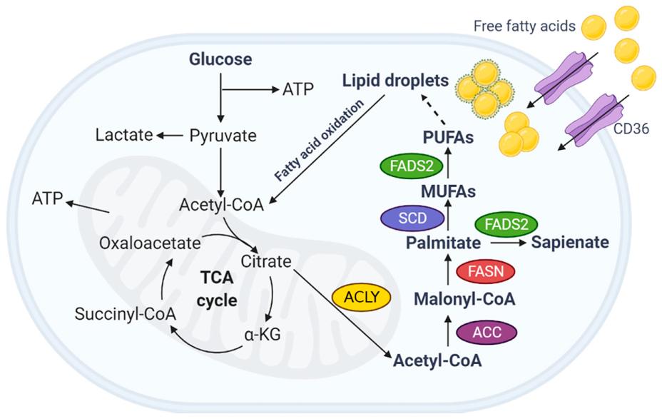
Fatty Acid : Metabolism Fatty Acid Metabolism Fatty acids are fundamental molecules in biology, playing roles in energy, structure, and signaling. Their metabolism is highly regulated and central to energy homeostasis in the body. For more details on Fatty Acids, Click Here. Briefly, What are Fatty Acids? Fatty acids are long hydrocarbon chains with a carboxyl group (-COOH) at one end. This makes them amphipathic molecules, meaning they have both hydrophobic (the hydrocarbon chain) and hydrophilic (the carboxyl group) regions. They are found esterified to glycerol in triacylglycerols (TAGs) or as components of phospholipids and sphingolipids. In their free form, they are called free fatty acids (FFAs). Classification of Fatty Acids A. Based on Saturation: Saturated Fatty Acids (SFAs): Contain no carbon-carbon double bonds. Examples: Palmitic acid (16:0), Stearic acid (18:0). Tend to be solid at room temperature. Unsaturated Fatty Acids (UFAs): Contain one or more carbon-carbon double bonds. Monounsaturated (MUFAs): Have one double bond (e.g., Oleic acid). Polyunsaturated (PUFAs): Have two or more double bonds (e.g., Linoleic acid). Properties: Tend to be liquid at room temperature. The double bonds usually have a cis configuration, causing kinks in the chain. B. Based on Chain Length: Short-Chain (SCFAs): 2 to 4 carbons. Medium-Chain (MCFAs): 6 to 12 carbons. Long-Chain (LCFAs): 14 to 20 carbons (most common). Very Long-Chain (VLCFAs): >20 carbons. C. Based on Essentiality: Non-Essential Fatty Acids: Can be synthesized by the body. Essential Fatty Acids (EFAs): Cannot be synthesized and must be obtained from the diet. Linoleic Acid (Omega-6): Precursor to arachidonic acid. ฮฑ-Linolenic Acid (Omega-3): Precursor to EPA and DHA. Major Physiological Roles of Fatty Acids Fatty acids are multifaceted molecules critical for life. Energy Storage Stored as triacylglycerols (TAGs), they are the body’s most concentrated and efficient form of long-term energy storage, yielding more ATP per gram than carbohydrates. Structural Components They are integral components of phospholipids and sphingolipids, which form the fundamental structure of all biological membranes. Signaling & Precursors Essential fatty acids are precursors to powerful local signaling molecules called eicosanoids (prostaglandins, thromboxanes, leukotrienes) involved in inflammation, pain, and blood clotting. Insulation & Absorption Adipose tissue provides thermal insulation and protection for organs. Dietary fats are also necessary for the absorption of fat-soluble vitamins (A, D, E, K). Primary Metabolic States: Fed vs. Fasted The body meticulously regulates fatty acid metabolism based on energy availability. Fed State (High Energy / Insulin Dominant) After a meal, excess carbohydrates and proteins are converted into fatty acids (Lipogenesis) and stored as TAGs in adipose tissue. The goal is to store energy. Fasted State (Low Energy / Glucagon Dominant) When nutrient intake is low, stored TAGs are broken down, releasing fatty acids. These are then broken down for energy (Beta-Oxidation). The goal is to release stored energy. Major Pathways Involved in Fatty Acid Metabolism Fatty Acid Synthesis (Lipogenesis): The process of building fatty acids from Acetyl-CoA. Occurs primarily in the cytosol. Fatty Acid Oxidation (Beta-Oxidation): The pathway that breaks down fatty acids into Acetyl-CoA to generate energy. Occurs primarily in the mitochondrial matrix. Triacylglycerol (TAG) Synthesis and Degradation: The processes of storing (esterification) and mobilizing (lipolysis) fatty acids. Ketone Body Metabolism: Ketogenesis: The liver converts excess Acetyl-CoA into ketone bodies during prolonged fasting. Ketolysis: Other tissues use ketone bodies as an alternative fuel source. Fatty Acid Mobilization and Transport When energy is needed, stored triacylglycerols (TAGs) in adipose tissue must be broken down, and the resulting fatty acids transported to other tissues for oxidation. 1. Triacylglycerol (TAG) Mobilization (Lipolysis) Lipolysis is the process of breaking down stored TAGs into fatty acids and glycerol, occurring in adipocytes. Stimuli: Hormones like epinephrine, norepinephrine, and glucagon signal a low-energy state and activate lipolysis. Insulin inhibits it. Key Players (Lipases): Hormone-Sensitive Lipase (HSL): The rate-limiting enzyme, activated by phosphorylation via a PKA-dependent pathway. Adipose Triglyceride Lipase (ATGL): Initiates the first step, converting TAGs to DAGs. Monoacylglycerol Lipase (MAGL): Catalyzes the final step. Products of Lipolysis: Free Fatty Acids (FFAs) and Glycerol are released into the bloodstream. Fate of Glycerol: Travels to the liver, where it can enter glycolysis or gluconeogenesis. Adipocytes lack the enzyme (glycerol kinase) to re-utilize it. 2. Transport of Free Fatty Acids (FFAs) in Blood Long-chain fatty acids are hydrophobic and require a carrier in the blood. Carrier Protein: Albumin, the most abundant plasma protein, serves as the primary carrier for FFAs. Mechanism: FFAs bind non-covalently to hydrophobic pockets on the albumin molecule. Delivery to Tissues: Fatty acid-albumin complexes deliver FFAs to tissues like muscle and heart, where they are taken up by specific fatty acid transporters. 3. Transport into Mitochondria (The Carnitine Shuttle) Long-chain fatty acids (LCFAs) cannot directly cross the inner mitochondrial membrane. They require the Carnitine Shuttle to enter the mitochondrial matrix for beta-oxidation. Steps of the Shuttle: Activation (Cytosol): The FFA is first activated to a fatty acyl-CoA by Fatty Acyl-CoA Synthetase, consuming 2 ATP equivalents. Transfer to Carnitine (Outer Membrane): The fatty acyl group is transferred from CoA to carnitine by Carnitine Palmitoyltransferase I (CPT-I), forming fatty acylcarnitine. CPT-I is the rate-limiting step and is inhibited by malonyl-CoA. Translocation (Inner Membrane): Carnitine-Acylcarnitine Translocase (CACT) transports fatty acylcarnitine into the matrix while simultaneously transporting a free carnitine out. Transfer Back to CoA (Matrix): Inside the matrix, Carnitine Palmitoyltransferase II (CPT-II) transfers the fatty acyl group back to a mitochondrial CoA, regenerating fatty acyl-CoA (now ready for beta-oxidation) and freeing carnitine for reuse. Now, with the fatty acyl-CoA ready in the mitochondrial matrix, we can move on to the actual breakdown process: Fatty Acid Oxidation (Beta-Oxidation). Fatty Acid Oxidation (Beta-Oxidation) Once long-chain fatty acids (as fatty acyl-CoA) have successfully entered the mitochondrial matrix via the carnitine shuttle, they are ready for a cyclic process called ฮฒ-oxidation. This pathway systematically cleaves two-carbon units from the carboxyl end of the fatty acyl-CoA, generating acetyl-CoA, NADH, and FADHโ‚‚, which then feed into the citric acid cycle and oxidative phosphorylation for ATP production. Primary Location: Mitochondrial matrix. Purpose: To generate energy (ATP) from stored fatty acids. The Sequential Steps of
Pentose Phosphate Pathway (1)
Biochemistry

Pentose Phosphate Pathway

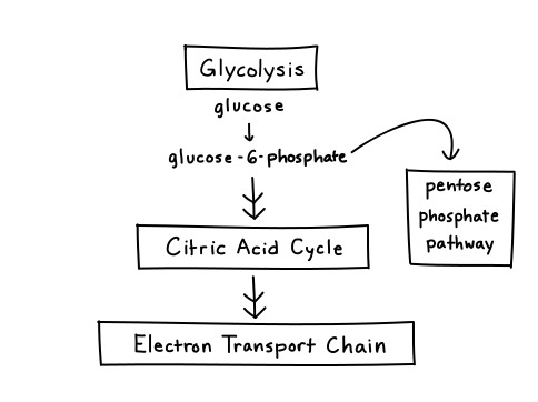
Pentose Phosphate Pathway: PPP Pentose Phosphate Pathway (PPP) The Pentose Phosphate Pathway (PPP), also known as the Hexose Monophosphate Shunt (HMP Shunt), is an alternative metabolic route for glucose metabolism that runs parallel to glycolysis. The HMP pathway is also known as the Warburg-Dickens pathway. About 10% of glucose entering this pathway per day. The liver & RBCs metabolise about 30% of glucose by this pathway. Unlike glycolysis, its primary purpose is not to generate ATP. Instead, its main functions are: Production of NADPH: Essential for reductive biosynthetic reactions and for protecting cells from oxidative stress. Production of Ribose-5-Phosphate: A vital precursor for the synthesis of nucleotides (DNA, RNA) and coenzymes. Think of the PPP as a “shunt” because it diverts glucose-6-phosphate away from glycolysis to serve these distinct purposes, and can then feed intermediates back into glycolysis. It primarily occurs in the cytosol of cells. Two Major Phases of the PPP The Pentose Phosphate Pathway is divided into two distinct phases: a) The Oxidative (Irreversible) Phase: Function: This phase is responsible for the generation of NADPH and the production of ribulose-5-phosphate (which is then converted to ribose-5-phosphate). Nature: It is largely irreversible. Key Reactions: Involves oxidative decarboxylation reactions where glucose-6-phosphate is oxidized, releasing COโ‚‚, and reducing NADPโบ to NADPH. b) The Non-Oxidative (Reversible) Phase: Function: This phase interconverts various sugar phosphates, primarily transforming pentose phosphates into glycolytic intermediates (fructose-6-phosphate and glyceraldehyde-3-phosphate). This allows carbon skeletons to be recycled back into glycolysis or used for gluconeogenesis. Nature: This phase is entirely reversible. Key Enzymes: Involves transketolase and transaldolase enzymes, which facilitate the transfer of two-carbon and three-carbon units, respectively. Products of the PPP The PPP is critically important because it provides two essential molecules: a) NADPH (Nicotinamide Adenine Dinucleotide Phosphate, reduced form) Structure: Similar to NADH, but with an additional phosphate group. Function: Unlike NADH (used in catabolism for ATP), NADPH is predominantly used in anabolic (biosynthetic) processes and as a reductant in antioxidant defense. Reductive Biosynthesis: Providing reducing power for the synthesis of fatty acids, cholesterol, and steroid hormones. Tissues actively involved in these syntheses (e.g., liver, adipose tissue, adrenal cortex) have a highly active PPP. Antioxidant Defense: Protecting cells from damage by reactive oxygen species (ROS) by maintaining the reduced state of glutathione. b) Ribose-5-Phosphate Structure: A five-carbon sugar phosphate. Function: This molecule is the direct precursor for the synthesis of: Nucleotides: The building blocks of DNA and RNA. Coenzymes: Such as ATP, NADH, FADHโ‚‚, and Coenzyme A. Demand: Cells that are rapidly dividing (e.g., bone marrow, skin, cancer cells) will have a high demand for ribose-5-phosphate. Location of the pathway The enzymes are located in the cytosol. The tissues such as liver, adipose tissue, adrenal gland, erythrocytes, testes & lactating mammary gland, are highly active in the HMP shunt. Most of these tissues are involved in the biosynthesis of fatty acids and steroids, which are dependent on the supply of NADPH. The Oxidative (Irreversible) Phase This phase consists of three main reactions, starting with glucose-6-phosphate and culminating in the production of NADPH and ribulose-5-phosphate. Key Concepts of the Oxidative Phase: Irreversible: The reactions in this phase are essentially unidirectional under physiological conditions. NADPH Production: This is the primary site of NADPH generation. Each molecule of glucose-6-phosphate entering this phase yields two molecules of NADPH. Substrate: Glucose-6-phosphate, which is also an intermediate in glycolysis. Location: Occurs in the cytosol. The Three Reactions of the Oxidative Phase: The oxidative phase involves the following sequential reactions: 1. Glucose-6-Phosphate Dehydrogenation (The Rate-Limiting Step) Enzyme: Glucose-6-Phosphate Dehydrogenase (G6PD) Reaction: Glucose-6-phosphate is oxidized, and NADPโบ is reduced to NADPH. A lactone (cyclic ester) intermediate, 6-phosphogluconolactone, is formed. Equation: Glucose-6-phosphate + NADPโบ โ†’ 6-Phosphogluconolactone + NADPH + Hโบ Significance: This is the rate-limiting and committed step of the entire Pentose Phosphate Pathway. The activity of G6PD is highly regulated. 2. Hydrolysis of 6-Phosphogluconolactone Enzyme: 6-Phosphogluconolactonase Reaction: The lactone ring is hydrolyzed to an open-chain carboxylic acid, 6-phosphogluconate. Equation: 6-Phosphogluconolactone + Hโ‚‚O โ†’ 6-Phosphogluconate Significance: This step prepares the molecule for the second oxidative reaction. 3. Oxidative Decarboxylation of 6-Phosphogluconate Enzyme: 6-Phosphogluconate Dehydrogenase Reaction: 6-phosphogluconate undergoes oxidative decarboxylation, meaning it is oxidized (another molecule of NADPโบ is reduced to NADPH) and a molecule of COโ‚‚ is released. The product is Ribulose-5-phosphate. Equation: 6-Phosphogluconate + NADPโบ โ†’ Ribulose-5-phosphate + NADPH + Hโบ + COโ‚‚ Significance: This reaction generates the second molecule of NADPH and the first pentose phosphate, which serves as the entry point into the non-oxidative phase. Summary of the Oxidative Phase: The net reaction for the oxidative phase is: Glucose-6-phosphate + 2 NADPโบ + Hโ‚‚O โ†’ Ribulose-5-phosphate + 2 NADPH + 2 Hโบ + COโ‚‚ Key Takeaways from the Oxidative Phase: Two molecules of NADPH are produced per molecule of glucose-6-phosphate. One molecule of COโ‚‚ is released. Ribulose-5-phosphate (a pentose sugar) is the final product. G6PD is the critical, rate-limiting enzyme. The Non-Oxidative (Reversible) Phase The non-oxidative phase is a series of reversible reactions that interconvert various sugar phosphates. Its primary functions are: Conversion of Ribulose-5-Phosphate: To other pentose phosphates, including ribose-5-phosphate (essential for nucleotide synthesis). Recycling of Carbon Skeletons: To produce glycolytic intermediates (fructose-6-phosphate and glyceraldehyde-3-phosphate) from excess pentose phosphates, linking the PPP back to glycolysis. Flexibility: The reversibility allows the cell to adjust the production of ribose-5-phosphate and NADPH according to its needs. Key Enzymes and Reactions of the Non-Oxidative Phase This phase involves three main enzymes: an isomerase, an epimerase, and two transketolases/transaldolases. 1. Interconversion of Pentose Phosphates The ribulose-5-phosphate generated in the oxidative phase needs to be converted into other pentose sugars. a) Ribulose-5-Phosphate Isomerase Enzyme: Ribose-5-phosphate Isomerase Reaction: Converts the ketose sugar ribulose-5-phosphate into the aldose sugar ribose-5-phosphate. This is crucial as ribose-5-phosphate is the direct precursor for nucleotide synthesis. Equation: Ribulose-5-phosphate โ‡Œ Ribose-5-phosphate b) Ribulose-5-Phosphate Epimerase Enzyme: Xylulose-5-phosphate Epimerase Reaction: Converts ribulose-5-phosphate into another ketose sugar, xylulose-5-phosphate, which is important for subsequent transketolase reactions. Equation: Ribulose-5-phosphate โ‡Œ Xylulose-5-phosphate 2. Transketolase and Transaldolase Reactions (The “Shunt” Part) These two enzymes are responsible for moving two-carbon and three-carbon units

GLUCONEOGENESIS
Biochemistry

Gluconeogenesis

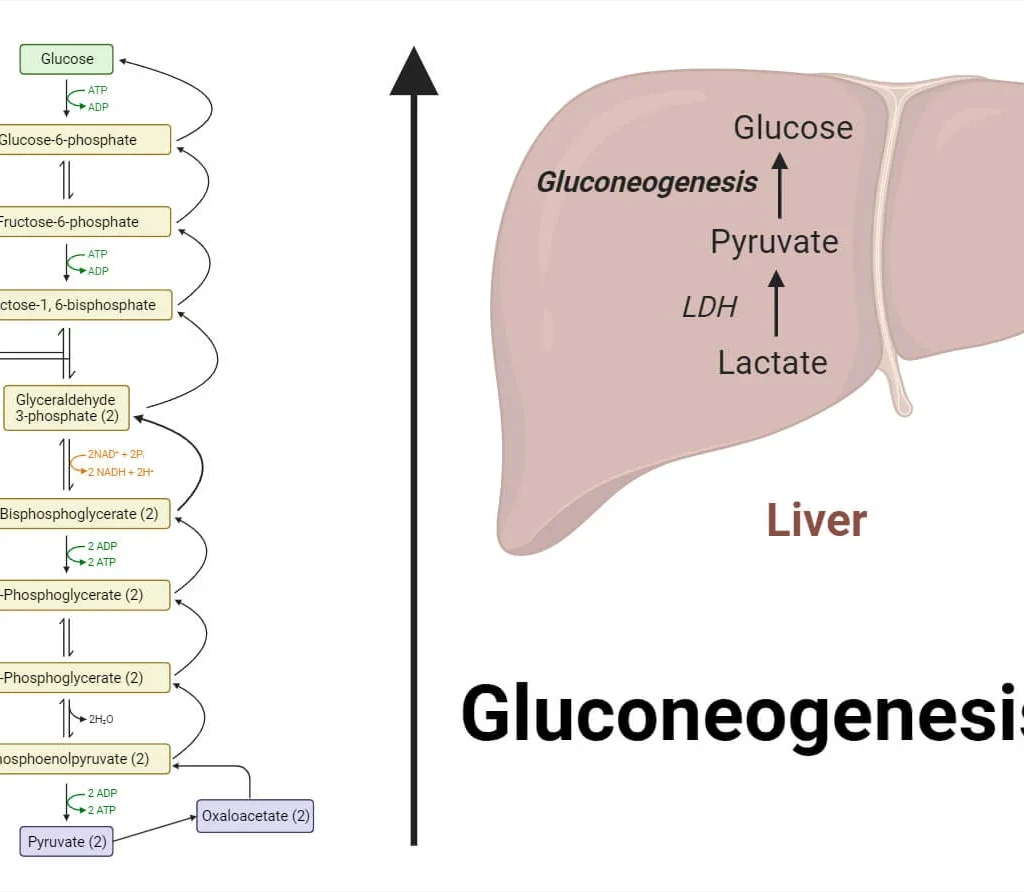
Gluconeogenesis: New Glucose GLUCONEOGENESIS The term “gluconeogenesis” literally means “new formation of glucose” (from Greek: glykys = sweet, neos = new, genesis = origin). It is a metabolic pathway that results in the generation of glucose from non-carbohydrate carbon substrates such as lactate, glycerol, and certain amino acids. Primary Purpose: The purpose of gluconeogenesis is to maintain blood glucose homeostasis, especially during periods when carbohydrate intake is insufficient (e.g., fasting, starvation, prolonged exercise). Why is this critical? The brain and red blood cells rely almost exclusively on glucose for their energy needs. Without gluconeogenesis, blood glucose levels would drop dangerously low (hypoglycemia) once glycogen stores are depleted, leading to severe physiological consequences. Primary Tissues/Organs: Gluconeogenesis primarily occurs in two organs: Liver (Hepatic Gluconeogenesis): This is the major site of gluconeogenesis. The liver can synthesize glucose and release it into the bloodstream for use by other tissues. Approximately 90% of all gluconeogenesis occurs in the liver. Kidney (Renal Gluconeogenesis): The kidneys also play a significant role, especially during prolonged fasting. The kidney can contribute up to 10% of glucose production during an overnight fast, and up to 40% during prolonged starvation. Key Precursors: Gluconeogenesis utilizes various non-carbohydrate molecules as starting materials. These precursors are ultimately converted into oxaloacetate, which then proceeds through the pathway. The three main classes are: 1. Lactate Origin: Produced by anaerobic glycolysis in actively contracting skeletal muscle and in red blood cells. Conversion: Lactate is transported to the liver, where it is converted back to pyruvate by lactate dehydrogenase. This cycle (muscle lactate to liver glucose) is known as the Cori Cycle. 2. Amino Acids (Glucogenic) Origin: Derived primarily from the breakdown of muscle protein, especially during fasting. Conversion: The carbon skeletons of many amino acids can be converted into pyruvate or TCA cycle intermediates (e.g., ฮฑ-ketoglutarate, succinyl-CoA). Alanine is particularly important, forming the Glucose-Alanine Cycle. Note: Fatty acids cannot be directly converted to glucose in animals because the conversion of acetyl-CoA (from fatty acid breakdown) to pyruvate or oxaloacetate is not possible. 3. Glycerol Origin: Released during the hydrolysis of triglycerides (fats) in adipose tissue. Conversion: Glycerol is transported to the liver, where it is phosphorylated and then oxidized to dihydroxyacetone phosphate (DHAP). DHAP is an intermediate in both glycolysis and gluconeogenesis, readily entering the pathway. Major Steps and Bypassing Irreversible Glycolysis Reactions Gluconeogenesis is NOT simply the reversal of glycolysis. While it shares many reversible steps, there are three highly exergonic (irreversible) steps in glycolysis that must be bypassed by different enzymes in gluconeogenesis. These bypasses are crucial for the pathway to be thermodynamically favorable and for regulatory control. Overview of the Pathway (from Pyruvate to Glucose) The overall process can be thought of as reversing glycolysis, but with four unique “bypass” reactions: Bypass 1: Pyruvate โ†’ Phosphoenolpyruvate (PEP) Bypass 2: Fructose-1,6-bisphosphate โ†’ Fructose-6-phosphate Bypass 3: Glucose-6-phosphate โ†’ Glucose Detailed Steps & Key Enzymes Let’s start from pyruvate, a common entry point for lactate and some amino acids. 1. Pyruvate to Phosphoenolpyruvate (PEP) – The First Bypass This bypass replaces the highly irreversible pyruvate kinase step of glycolysis. It requires two enzymes and crosses the mitochondrial membrane. Step 1a: Pyruvate to Oxaloacetate (in Mitochondria) Enzyme: Pyruvate Carboxylase (PC) Reaction: Pyruvate + COโ‚‚ + ATP โ†’ Oxaloacetate + ADP + Pi Cofactor: Biotin (carries COโ‚‚) Key Point: This enzyme is in the mitochondrial matrix and is allosterically activated by acetyl-CoA. High acetyl-CoA signals that pyruvate should be directed towards glucose synthesis. Step 1b: Oxaloacetate to PEP (Mitochondria and/or Cytosol) Oxaloacetate cannot directly cross the mitochondrial membrane. It must first be converted via one of two options, often involving a malate shuttle, to generate cytosolic NADH which is needed later. Enzyme: PEP Carboxykinase (PEPCK) Reaction: Oxaloacetate + GTP โ†’ PEP + GDP + COโ‚‚ 2. PEP to Fructose-1,6-bisphosphate From PEP, the pathway essentially reverses the reversible steps of glycolysis using the same enzymes, but in the reverse direction: PEP โ†’ 2-Phosphoglycerate โ†’ 3-Phosphoglycerate (via Enolase, Phosphoglycerate mutase) 3-Phosphoglycerate โ†’ 1,3-Bisphosphoglycerate (via Phosphoglycerate kinase, consuming ATP) 1,3-Bisphosphoglycerate โ†’ Glyceraldehyde-3-phosphate (via Glyceraldehyde-3-phosphate dehydrogenase, consuming NADH) Glyceraldehyde-3-phosphate โ†” Dihydroxyacetone phosphate (DHAP) (via Triose phosphate isomerase). DHAP from glycerol enters here. Glyceraldehyde-3-phosphate + DHAP โ†’ Fructose-1,6-bisphosphate (via Aldolase) 3. Fructose-1,6-bisphosphate to Fructose-6-phosphate – The Second Bypass This bypass replaces the irreversible phosphofructokinase-1 (PFK-1) step of glycolysis. Enzyme: Fructose-1,6-bisphosphatase (FBPase-1) Reaction: Fructose-1,6-bisphosphate + Hโ‚‚O โ†’ Fructose-6-phosphate + Pi Key Point: This is a hydrolysis reaction, releasing inorganic phosphate (Pi). It is a critical, reciprocally regulated point with PFK-1. 4. Fructose-6-phosphate to Glucose-6-phosphate Enzyme: Phosphohexose isomerase (reversible, same as glycolysis) Reaction: Fructose-6-phosphate โ†” Glucose-6-phosphate 5. Glucose-6-phosphate to Free Glucose – The Third Bypass This bypass replaces the irreversible hexokinase/glucokinase step of glycolysis. Enzyme: Glucose-6-phosphatase Reaction: Glucose-6-phosphate + Hโ‚‚O โ†’ Glucose + Pi Key Point: This enzyme is found primarily in the liver and kidney and is located in the endoplasmic reticulum membrane. It allows free glucose to be released into the bloodstream. Muscle cells lack this enzyme. Summary of the Bypasses and Key Enzymes: Glycolysis Irreversible Step (Enzyme) Gluconeogenesis Bypass Enzyme(s) Location Glucose โ†’ G6P (Hexokinase/Glucokinase) Glucose-6-phosphatase ER lumen (liver, kidney) F6P โ†’ FBP (PFK-1) Fructose-1,6-bisphosphatase (FBPase-1) Cytosol PEP โ†’ Pyruvate (Pyruvate Kinase) 1. Pyruvate Carboxylase2. PEP Carboxykinase (PEPCK) Mitochondria & Cytosol Energy Requirements Synthesizing glucose from non-carbohydrate precursors is an energy-intensive, anabolic process. Let’s calculate the ATP and GTP expenditure required to synthesize one molecule of glucose from two molecules of pyruvate. Here’s a breakdown of the energy-consuming steps: Pyruvate to Oxaloacetate (x2):Catalyzed by Pyruvate Carboxylase, this step consumes 1 ATP per pyruvate.Total cost: 2 ATP Oxaloacetate to Phosphoenolpyruvate (PEP) (x2):Catalyzed by PEP Carboxykinase, this step consumes 1 GTP per oxaloacetate.Total cost: 2 GTP 3-Phosphoglycerate to 1,3-Bisphosphoglycerate (x2):Catalyzed by Phosphoglycerate Kinase, this step consumes 1 ATP per 3-phosphoglycerate.Total cost: 2 ATP Total Energy Cost for Synthesizing one Glucose from two Pyruvates: 4 ATP + 2 GTP Important Considerations: NADH Requirement: In addition to ATP and GTP, the pathway consumes 2 NADH during the conversion of 1,3-bisphosphoglycerate to glyceraldehyde-3-phosphate. Energy Balance and

Glycogenolysis and Glycogenesis
Biochemistry

Glycogenolysis and Glycogenesis

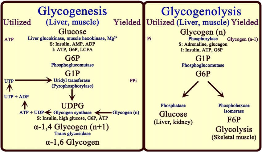
Glycogenolysis & Glycogenesis Glycogenolysis Glycogenolysis is the biochemical process by which glycogen, a stored form of glucose, is broken down into glucose-1-phosphate and then subsequently converted to glucose or glucose-6-phosphate. The suffix “-lysis” means “to break down,” so it literally means “breaking down glycogen.” Glycogen itself is a highly branched polysaccharide composed of glucose units. It serves as the primary storage form of glucose in animals. In humans, it is predominantly stored in the liver and skeletal muscles. Purpose The primary purpose of glycogenolysis is to mobilize stored glucose to meet the body’s immediate energy needs, particularly to maintain stable blood glucose levels and provide fuel for muscle contraction. Maintenance of Blood Glucose Homeostasis (Liver Glycogenolysis): The liver is crucial for regulating blood glucose. When blood glucose levels drop (e.g., during fasting or intense exercise), the liver breaks down its glycogen stores. The glucose-6-phosphate produced in the liver can be dephosphorylated to free glucose and then released into the bloodstream, supplying fuel to other tissues like the brain and red blood cells. Energy for Muscle Contraction (Muscle Glycogenolysis): Skeletal muscles also store glycogen, but unlike liver glycogen, it is primarily used to fuel the muscle’s own activity. During exercise, muscle glycogen is broken down to glucose-6-phosphate, which then enters glycolysis to produce ATP directly within the muscle cells. Muscle cells lack the enzyme glucose-6-phosphatase, so they cannot release free glucose into the bloodstream. Location Glycogenolysis primarily occurs in two major tissues in the human body: Liver Primary Role: The liver is the main organ responsible for maintaining blood glucose homeostasis. Capacity: The liver stores a significant amount of glycogen (up to 6-8% of its wet weight, or about 100-120 grams in an adult). This is important for absolutely glucose-dependent cells like neurons, RBCs, and the renal medulla. Mechanism: When blood glucose drops, liver glycogen is broken down. The resulting glucose-6-phosphate is dephosphorylated by the enzyme glucose-6-phosphatase to free glucose, which is then released into the bloodstream. Regulation: Liver glycogenolysis is highly regulated by hormones such as glucagon (released during low blood glucose) and epinephrine (released during stress). Skeletal Muscles Primary Role: Muscle glycogen serves as a readily available fuel source for the muscle itself during physical activity. Capacity: Skeletal muscles collectively store a larger total amount of glycogen than the liver (about 1-2% of muscle wet weight, or about 300-500 grams in an adult). Mechanism: During exercise, muscle glycogen is broken down to glucose-6-phosphate, which directly enters glycolysis within the muscle cell to produce ATP. Key Difference from Liver: Muscle cells lack the enzyme glucose-6-phosphatase. This means muscle glycogen cannot be used to directly replenish blood glucose. The glucose-6-phosphate is “trapped” within the muscle cell. Regulation: Muscle glycogenolysis is primarily regulated by epinephrine (during “fight or flight” responses) and by AMP (which signals a low energy state). Key Enzymes The breakdown of glycogen is a well-orchestrated process involving a few critical enzymes working in sequence. These enzymes ensure that glucose units are efficiently released from the glycogen molecule. The three main enzymes (or enzyme complexes) are: Glycogen Phosphorylase Debranching Enzyme (which has two enzymatic activities) Phosphoglucomutase Let’s look at each one: 1. Glycogen Phosphorylase Function: This is the primary enzyme responsible for breaking down glycogen. It catalyzes the phosphorolysis (breaking a bond using inorganic phosphate, not water) of the ฮฑ(1,4) glycosidic bonds that link glucose units. Mechanism: It removes glucose units one by one from the non-reducing ends of the glycogen molecule. The bond is broken by the addition of inorganic phosphate (Pแตข), yielding glucose-1-phosphate. Limitation: Glycogen phosphorylase cannot break the ฮฑ(1,6) glycosidic bonds at the branch points. It stops cleaving when it reaches about four glucose residues away from a branch point, leaving behind a “limit dextrin.” 2. Debranching Enzyme (Glycogen Debranching Enzyme) Since glycogen phosphorylase cannot handle the branch points, this specialized enzyme complex is required. It has two distinct catalytic activities: a. Oligo-ฮฑ(1,4)-ฮฑ(1,4)-glucantransferase activity (Transferase activity): Function: This activity transfers a block of three glucose residues from a branch to a non-reducing end of another chain. It forms a new ฮฑ(1,4) bond, making the chain longer and available for further action by glycogen phosphorylase. b. Amylo-ฮฑ(1,6)-glucosidase activity (Glucosidase activity): Function: After the transferase activity, this activity hydrolyzes the single remaining glucose residue at the ฮฑ(1,6) branch point, releasing it as free glucose (not glucose-1-phosphate). Significance: This is the only step in glycogenolysis that directly produces free glucose (about 8% of the glucose from glycogen is released this way). 3. Phosphoglucomutase Function: This enzyme is responsible for interconverting glucose-1-phosphate and glucose-6-phosphate. Mechanism: Glycogen phosphorylase produces glucose-1-phosphate. For this glucose to enter glycolysis (as glucose-6-phosphate) or to be released into the bloodstream (as free glucose in the liver), it first needs to be converted. Phosphoglucomutase catalyzes this reversible isomerization. Summary of Enzyme Action: Glycogen Phosphorylase removes glucose units as glucose-1-phosphate from the linear parts of glycogen. Debranching Enzyme “cleans up” the branch points: its transferase activity moves most of the branch, and its glucosidase activity releases the final branched glucose as free glucose. Phosphoglucomutase converts the glucose-1-phosphate into glucose-6-phosphate, which is the entry point for further metabolism. Steps of the Glycogenolysis Pathway Here’s a step-by-step breakdown of how glycogen is degraded to release glucose units, incorporating the enzymes we just discussed. Overall Goal: To convert glycogen into individual glucose units that can be used for energy or released into the bloodstream. Step 1: Phosphorolytic Cleavage of ฮฑ(1,4) Glycosidic Bonds Enzyme: Glycogen Phosphorylase Action: Begins acting on the non-reducing ends of the glycogen molecule, cleaving the ฮฑ(1,4) glycosidic bonds. Product: Each cleavage releases a molecule of glucose-1-phosphate (G1P). This process is called phosphorolysis because inorganic phosphate (Pแตข) is used to break the bond. Limitation: The enzyme stops when it reaches approximately four glucose residues away from an ฮฑ(1,6) branch point, leaving a “limit dextrin.” Step 2: Remodeling of the Glycogen Molecule at Branch Points Enzyme: Debranching Enzyme Action: The debranching enzyme resolves the limit dextrin structure: Transferase Activity (Oligo-ฮฑ(1,4)-ฮฑ(1,4)-glucantransferase): Transfers a block of three glucose residues from the branch

Tricarboxylic Acid (TCA) Cycle (Krebs Cycle / Citric Acid Cycle)
Biochemistry

Tricarboxylic Acid (TCA) Cycle (Krebs Cycle / Citric Acid Cycle)

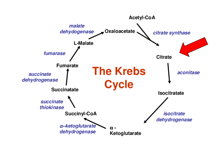
Tricarboxylic Acid (TCA) Cycle: (Krebs Cycle / Citric Acid Cycle) The Tricarboxylic Acid (TCA) Cycle (Krebs Cycle / Citric Acid Cycle) The Tricarboxylic Acid (TCA) cycle, also famously known as the Krebs cycle or the Citric Acid Cycle, is a central hub of metabolism. It’s a metabolic superhighway where the breakdown products of carbohydrates, fats, and proteins converge for final oxidation. Central Role in Aerobic Respiration: The TCA cycle is the second major stage of aerobic respiration. Unlike glycolysis, the TCA cycle requires oxygen indirectly to function, as its products (NADH and FADHโ‚‚) ultimately feed into the electron transport chain, which absolutely depends on oxygen. Without the ETC running, the NADโบ and FAD needed for the TCA cycle would not be regenerated, and the cycle would grind to a halt. Main Function: Complete Oxidation of Acetyl-CoA: The primary catabolic function of the TCA cycle is the complete oxidation of acetyl-CoA to carbon dioxide (COโ‚‚). This acetyl-CoA is primarily derived from: Carbohydrates: Pyruvate (from glycolysis) is converted to acetyl-CoA. Fats: Fatty acids are broken down into acetyl-CoA via beta-oxidation. Proteins: Certain amino acids are degraded into acetyl-CoA or other TCA cycle intermediates. As acetyl-CoA is oxidized, the cycle captures the released energy in the form of high-energy electron carriers (NADH and FADHโ‚‚) and a small amount of GTP (which is interconvertible with ATP). These carriers are then channeled into the Electron Transport Chain (ETC) to drive the synthesis of the vast majority of cellular ATP. Amphibolic Nature (Both Catabolic and Anabolic Roles): One of the most fascinating aspects of the TCA cycle is its amphibolic nature, meaning it serves both catabolic (breakdown) and anabolic (synthesis) roles. Catabolism: It catabolizes acetyl-CoA to COโ‚‚, generating ATP, NADH, and FADHโ‚‚. Anabolism: Many of the intermediates are precursors for various biosynthetic pathways. For example: Citrate can be used for fatty acid and cholesterol synthesis. ฮฑ-Ketoglutarate is a precursor for several amino acids (e.g., glutamate). Succinyl-CoA is used in the synthesis of porphyrins (like heme). Oxaloacetate is a precursor for amino acids and glucose (via gluconeogenesis). Because these intermediates are often “siphoned off” for synthesis, the cell has mechanisms (called anaplerotic reactions) to replenish them. Location: The location of the TCA cycle is critical to its function and regulation. Mitochondrial Matrix: In eukaryotic cells, the entire TCA cycle takes place within the mitochondrial matrix, the innermost compartment of the mitochondrion. Why is this significant? Proximity to ETC: NADH and FADHโ‚‚ are produced directly where they are needed, ensuring efficient energy transfer to the ETC on the inner membrane. Isolation and Concentration: Confining the cycle within the matrix allows for the concentration of substrates and enzymes. Coupling with Oxidative Phosphorylation: This spatial arrangement is essential for the effective coupling of the TCA cycle with ATP production. In prokaryotic cells, which lack mitochondria, the TCA cycle enzymes are found in the cytosol. Entry Point: Pyruvate Dehydrogenase Complex (PDC) The Pyruvate Dehydrogenase Complex (PDC) is a critical bridge between glycolysis and the TCA cycle. It’s not part of the TCA cycle itself, but it’s an absolutely essential prerequisite for aerobic glucose metabolism to proceed. Irreversible Conversion of Pyruvate to Acetyl-CoA: The PDC catalyzes an irreversible oxidative decarboxylation of pyruvate to form acetyl-CoA. This reaction is a metabolic crossroads: once pyruvate is converted to acetyl-CoA, it cannot be converted back to glucose. The fate of glucose is committed to complete oxidation or fatty acid synthesis. Location: This reaction also takes place in the mitochondrial matrix. Pyruvate from glycolysis is transported into the matrix by a specific translocase protein. Overall Reaction and Coenzymes Involved: The PDC is a complex of three distinct enzymes and five different coenzymes. The overall reaction is: Pyruvate + CoA + NADโบ โ†’ Acetyl-CoA + COโ‚‚ + NADH + Hโบ The Five Coenzymes (or Prosthetic Groups): Thiamine Pyrophosphate (TPP): From vitamin B1 (thiamine). Decarboxylates pyruvate. Lipoate (Lipoamide): Transfers the acetyl group to CoA. Flavin Adenine Dinucleotide (FAD): From vitamin B2 (riboflavin). Re-oxidizes the reduced lipoamide. Nicotinamide Adenine Dinucleotide (NADโบ): From vitamin B3 (niacin). Re-oxidizes FADHโ‚‚. Coenzyme A (CoA): From vitamin B5 (pantothenic acid). Accepts the acetyl group. Regulation of the PDC: Because this step is irreversible, the PDC is a crucial point of regulation. Allosteric Regulation: Inhibitors (high energy signals): Acetyl-CoA, NADH, ATP. Activators (low energy signals): CoA, NADโบ, AMP. Covalent Modification (Phosphorylation/Dephosphorylation): This is the primary long-term regulatory mechanism. A PDC Kinase adds a phosphate group to INACTIVATE the PDC. The kinase is activated by high energy signals (ATP, NADH, Acetyl-CoA). A PDC Phosphatase removes the phosphate group to ACTIVATE the PDC. The phosphatase is activated by Caยฒโบ and insulin. In summary, when the cell has plenty of energy, the PDC is turned off. When energy is needed, the PDC is activated. The Cycle Itself (Key Steps, Enzymes, and Products) The cycle consists of eight enzymatic steps, leading to the complete oxidation of the two carbons from acetyl-CoA and the regeneration of oxaloacetate. Overall Summary of One Turn of the Cycle: A four-carbon oxaloacetate condenses with a two-carbon acetyl unit (from acetyl-CoA) to yield a six-carbon citrate. Citrate is isomerized and then oxidatively decarboxylated to form five-carbon ฮฑ-ketoglutarate, releasing one molecule of COโ‚‚ and producing NADH. ฮฑ-ketoglutarate is oxidatively decarboxylated to yield four-carbon succinyl-CoA, releasing another COโ‚‚ and producing NADH. Succinyl-CoA is converted to succinate, generating one GTP (or ATP). Oxaloacetate is regenerated from succinate through a series of steps involving fumarate and malate, producing one FADHโ‚‚ and one NADH. In total, two carbon atoms (from acetyl-CoA) enter the cycle, and two carbon atoms leave as COโ‚‚. Energy is captured as 3 NADH, 1 FADHโ‚‚, and 1 GTP (ATP). The Reactions of the Citric Acid Cycle Now that Acetyl-CoA has been generated in the mitochondrial matrix, it enters the eight-step cyclical pathway. Each turn of the cycle processes one molecule of Acetyl-CoA, systematically oxidizing its two carbons to COโ‚‚ while capturing high-energy electrons in the form of NADH and FADHโ‚‚. Let’s examine each step. Step 1: Formation of Citrate This is the entry point for

Biochemistry

Krebs Cycle for Slow learners

TCA Cycle Adventure – Complete Game (Stages 1-10) 1 2 3 4 5 6 7 8 9 10 ๐ŸŽฏ Score 0 โญ High Score 0 ๐Ÿ”ฅ Streak 0 0 NADH Produced 0 FADHโ‚‚ Produced 0 GTP Produced 0 COโ‚‚ Released Stage 1: The Great Entry ๐Ÿšช Acetyl-CoA + Oxaloacetate โ†’ Citrate: Committing to the Cycle! Acetyl-CoA (Cโ‚‚) CHโ‚ƒ C=O S-CoA O 2-Carbon fuelHigh-energy thioester ๐Ÿ”— Citrate Synthase Oxaloacetate (Cโ‚„) C=O CHโ‚‚ CHโ‚‚ C=O O O O 4-Carbon acceptorRegenerated later โšก Energy Investment: Thioester bond hydrolysis The high-energy thioester bond in Acetyl-CoA powers this condensation ๐ŸŽฎ INSERTING YOUR TOKEN! Acetyl-CoA is your “game token” – you insert it into the machine (citrate synthase) where it merges with the existing 4C platform. This forms a 6C citrate molecule and commits you to playing through the entire cycle! ๐ŸŽฏ Key Points: โœ… C-C bond formation – only step that builds a bond โœ… Irreversible – commitment step to the cycle โœ… Oxaloacetate acts as a catalyst (regenerated) โœ… Energy from thioester bond makes reaction favorable ๐Ÿ“ Challenge Questions – Stage 1 Q1: Why is this step irreversible? A) It uses too much ATP B) High-energy thioester bond drives it forward C) It releases COโ‚‚ Q2: What happens to the CoA group? A) It stays attached to citrate B) It is released and can be reused C) It is broken down for energy Q3: Why is oxaloacetate called a catalyst? A) It’s consumed in the reaction B) It’s regenerated by the end of the cycle C) It inhibits the reaction ๐ŸŽ‰ Stage 1 Complete! Score: 0/30 Proceed to Stage 2 โ†’ Stage 2: The Isomerization Shuffle ๐Ÿ”„ Citrate โ†’ Isocitrate: Getting Ready for Oxidation! Citrate (Cโ‚†) COOH CHโ‚‚ COH CHโ‚‚ COOH CHโ‚‚ O O O O 6-CarbonTertiary alcohol (unstable) ๐ŸŽฒ Aconitase Isocitrate (Cโ‚†) COOH CHโ‚‚ CH CHโ‚‚ COOH CHโ‚‚ O O O O OH 6-CarbonSecondary alcohol (oxidizable!) โšก Energy Status: Near equilibrium Two-step process via cis-aconitate intermediate ๐Ÿ”ง ADJUSTING THE SPARK PLUG! Citrate’s hydroxyl group is in the wrong position (tertiary). Aconitase “moves” it via dehydration then rehydration, converting it to isocitrate’s secondary alcohol – now perfectly positioned for oxidation! ๐ŸŽฏ Key Points: โœ… Isomerization: Same formula, different structure โœ… Moves OH group from tertiary โ†’ secondary carbon โœ… Involves dehydration then rehydration โœ… Activates molecule for the first oxidation step ๐Ÿ“ Challenge Questions – Stage 2 Q1: Why is this rearrangement necessary? A) To release COโ‚‚ B) To make the OH group oxidizable C) To add energy Q2: What is the intermediate? A) cis-Aconitate B) ฮฑ-Ketoglutarate C) Fumarate Q3: This reaction involves… A) Adding water then removing it B) Removing water then adding it back C) No water exchange ๐ŸŽ‰ Stage 2 Complete! Score: 0/60 Proceed to Stage 3 โ†’ Stage 3: First Oxidation Strike โšก Isocitrate โ†’ ฮฑ-Ketoglutarate: First NADH & COโ‚‚! Isocitrate (Cโ‚†) COOH CHโ‚‚ CH(OH) CHโ‚‚ COOH CHโ‚‚ O O O O OH 6-CarbonReady to oxidize โšก Isocitrate Dehydrogenase ฮฑ-Ketoglutarate (Cโ‚…) COOH C=O CHโ‚‚ COOH CHโ‚‚ O O O O 5-Carbonฮฑ-keto acid +1 NADH + Hโบ โ†‘ COโ‚‚ Released ๐ŸŽ‰ FIRST ENERGY HARVEST! โœ… Oxidation: Isocitrate loses 2 electrons โœ… Decarboxylation: One carbon leaves as COโ‚‚ โœ… NADโบ reduced: To NADH + Hโบ (energy carrier) โœ… Product: ฮฑ-Ketoglutarate (5 carbons) ๐ŸŽฏ FIRST PRIZE CLAW! You’re playing a claw machine (isocitrate dehydrogenase). You grab the electron prize (NADH) and drop the waste carbon (COโ‚‚) into the chute. You walk away with one less carbon but an energy-rich NADH battery! ๐Ÿ“ Challenge Questions – Stage 3 Q1: What is the carbon count change? A) Cโ‚† โ†’ Cโ‚„ B) Cโ‚† โ†’ Cโ‚… (lost as COโ‚‚) C) No change Q2: What is the energy product? A) 1 NADH + Hโบ B) 2 ATP C) 1 FADHโ‚‚ Q3: What type of reaction is this? A) Oxidative decarboxylation B) Substrate-level phosphorylation C) Dehydration ๐ŸŽ‰ Stage 3 Complete! NADH: 0 | COโ‚‚: 1 Proceed to Stage 4 โ†’ Stage 4: Second Oxidation Blast ๐Ÿ’ฅ ฮฑ-Ketoglutarate โ†’ Succinyl-CoA: Second NADH & COโ‚‚! ฮฑ-Ketoglutarate (Cโ‚…) COOH C=O CHโ‚‚ COOH CHโ‚‚ O O O O 5-Carbon ฮฑ-keto acidNext to be oxidized ๐Ÿ’ฅ ฮฑ-Ketoglutarate Dehydrogenase Complex Succinyl-CoA (Cโ‚„) COOH CHโ‚‚ CHโ‚‚ COOH S-CoA O O O O 4-CarbonHigh-energy thioester +1 NADH + Hโบ โ†‘ COโ‚‚ Released + CoA-SH Added ๐ŸŽ‰ SECOND ENERGY CAPTURE! โœ… Oxidative decarboxylation (like Stage 3) โœ… Uses same enzyme complex as pyruvate dehydrogenase โœ… Produces high-energy thioester (succinyl-CoA) โœ… 2nd NADH and 2nd COโ‚‚ per acetyl-CoA ๐Ÿ—‘๏ธ SECOND TRASH COMPACTOR! Another carbon gets kicked out as COโ‚‚ waste, and you grab another NADH energy prize! The enzyme complex is identical to the one that converts pyruvate โ†’ acetyl-CoA. ๐Ÿ“ Challenge Questions – Stage 4 Q1: Total COโ‚‚ released so far? A) 1 COโ‚‚ B) 2 COโ‚‚ (both carbons from acetyl-CoA) C) 3 COโ‚‚ Q2: What makes succinyl-CoA special? A) High-energy thioester bond for next step B) It’s the final product C) It contains nitrogen Q3: This enzyme is similar to… A) Pyruvate dehydrogenase complex B) Citrate synthase C) Malate dehydrogenase ๐ŸŽ‰ Stage 4 Complete! NADH: 0 | COโ‚‚: 2 Proceed to Stage 5 โ†’ Stage 5: Energy Cash-Out ๐Ÿ’ฐ Succinyl-CoA โ†’ Succinate: The TCA Cycle’s Only Direct ATP! Succinyl-CoA (Cโ‚„) COOH CHโ‚‚ CHโ‚‚ COOH S-CoA O O O O High-energy thioesterReady to release energy ๐Ÿ’ธ Succinyl-CoA Synthetase Succinate (Cโ‚„) COOH CHโ‚‚ CHโ‚‚ COOH O O O O Standard acidLower energy +1 GTP โ†’ GDP + Pi โ†’ CoA-SH Released ๐ŸŽ‰ SUBSTRATE-LEVEL PHOSPHORYLATION! โœ… Thioester bond energy transferred to GTP โœ… GTP can phosphorylate ADP โ†’ ATP (nucleoside diphosphate kinase) โœ… Only direct energy currency produced in TCA cycle โœ… Equivalent to 1 ATP per acetyl-CoA ๐Ÿฆ CASHING A CHECK! Your succinyl-CoA is a cashier’s check (high-energy thioester). The bank (succinyl-CoA synthetase) cashes it into GTP cash. You can immediately convert GTP to ATP at the currency exchange! ๐Ÿ“ Challenge Questions – Stage 5 Q1: What is unique about this step? A) Only substrate-level phosphorylation in TCA B) It uses ATP C) It produces FADHโ‚‚ Q2: GTP

Carbohydrate Metabolism and Glycolysis
Biochemistry

Carbohydrate Metabolism and Glycolysis

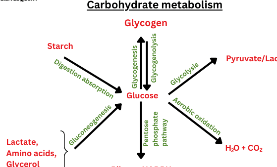
Carbohydrate Metabolism: Glycolysis Glycolysis: The Embden-Meyerhof Pathway Building upon our understanding of bioenergetics, we now delve into Glycolysis, also known as the Embden-Meyerhof Pathway (EMP). This fundamental metabolic pathway is the initial step in breaking down glucose to generate energy in nearly all living organisms. Glycolysis (from Greek “glykys” = sweet, “lysis” = splitting) is the process where one molecule of glucose (a 6-carbon sugar) is broken down into two molecules of pyruvate (a 3-carbon compound). This process releases a small amount of energy, which is captured as ATP and NADH. Location: All the reactions of glycolysis occur in the cytoplasm of the cell. This means it doesn’t require mitochondria. Key Roles: Energy for Mitochondria-Lacking Tissues: It’s the primary way tissues without mitochondria (like red blood cells, cornea, and lens) produce ATP. Brain’s Energy Source: The brain relies heavily on glucose, and glycolysis is its initial step in energy extraction. Anaerobic vs. Aerobic Fates: Without Oxygen (Anaerobic): Pyruvate is converted to lactate, providing a quick, albeit limited, energy supply (2 net ATP per glucose). With Oxygen (Aerobic): Pyruvate enters the mitochondria for further breakdown in the Citric Acid Cycle and Oxidative Phosphorylation, which yields a much larger amount of ATP. Energy Yield and Thermodynamics of Glycolysis Glycolysis is an energy-releasing (exergonic) pathway. Overall Chemical Transformation: Glucose + 2 NADโบ โ†’ 2 Pyruvate + 2 NADH + 2Hโบ This reaction generates energy that is used to produce ATP: 2 ADP + 2 Pi โ†’ 2 ATP + 2 Hโ‚‚O Free Energy Changes (Standard Biological Conditions, ฮ”Gยฐ’): Energy released from glucose conversion to pyruvate: ฮ”Gยฐ’ = -146 kJ/mol Energy required to form 2 ATP: 2 ร— (30.5 kJ/mol) = 61 kJ/mol Overall Net Free Energy Change: ฮ”Gยฐ’ (overall) = -146 kJ/mol + 61 kJ/mol = -85 kJ/mol Thermodynamic Summary: The significantly negative overall ฮ”Gยฐ’ indicates that glycolysis is an exergonic reaction that proceeds spontaneously and is largely irreversible under standard conditions. Glycolysis releases only a small fraction of the total potential energy stored in glucose. Specifically: “5.2% of the total free energy that can be released by glucose is released in glycolysis.” The complete oxidation of glucose yields much more energy (ฮ”Gยฐ’ = -2840 kJ/mol), meaning the majority of glucose’s energy remains in pyruvate and NADH, awaiting further aerobic processing. Fates of Glucose in Living Systems Once glucose enters a cell, it has 4 primary metabolic fates, depending on the organism’s immediate needs: Storage: Glucose can be linked to form large storage polymers like Glycogen in animals or Starch in plants. Structural Synthesis: Glucose derivatives can be used to synthesize polysaccharides that form structural components, such as the extracellular matrix. Oxidation via Pentose Phosphate Pathway (PPP): Glucose is converted to Ribose 5-phosphate. This pathway is vital for producing NADPH (for biosynthesis and protecting from oxidative damage) and Ribose 5-phosphate (for synthesizing nucleotides like DNA and RNA). Oxidation via Glycolysis (Energy Production): Glucose is broken down to Pyruvate, serving as the initial step for ATP production. Historical Discovery of Glycolysis The elucidation of glycolysis was a monumental achievement, marking it as one of the first and “oldest” metabolic pathways to be fully understood. Louis Pasteur (1854-1864): Observed that fermentation was caused by microorganisms. His “Pasteur effect” noted that organisms use less glucose in the presence of oxygen because aerobic respiration is far more efficient. Eduard Buchner (1897): Revolutionized biochemistry by demonstrating that yeast extracts, even without living cells, could carry out fermentation, proving that enzymes were responsible. Harden and Young (1905): Made two key discoveries: inorganic phosphate is essential for fermentation, and yeast extracts could be separated into heat-stable small molecules (“Co-zymase,” later identified as NAD+, ATP, ADP) and heat-labile protein enzymes (“Zymase”). By 1940: Through the combined efforts of many scientists, including Gustav Embden, Otto Meyerhof, and Jacob Parnas, the complete step-by-step pathway of glycolysis was definitively established. Click here for the Glycolysis Game Digestion and Absorption of Dietary Carbohydrates Before carbohydrates can be used by the body, complex forms (polysaccharides and disaccharides) must be broken down into monosaccharides for absorption. This process begins in the mouth and continues in the small intestine. 1. Digestion Digestion involves the enzymatic hydrolysis of glycosidic bonds. In the Mouth: Mechanical digestion (chewing). Salivary alpha-amylase (Ptyalin): Begins the breakdown of starch into smaller polysaccharides (dextrins) and some maltose. It’s inactivated by stomach acid. In the Stomach: No significant carbohydrate digestion occurs here due to the acidic environment. In the Small Intestine: This is where the bulk of carbohydrate digestion takes place. Pancreatic alpha-amylaseme: Continues breaking down starch and dextrins into maltose and other small polymers. Brush Border Enzymes: Located on the microvilli of intestinal cells, these are responsible for the final breakdown into monosaccharides. Maltase: Hydrolyzes maltose โ†’ two glucose molecules. Sucrase: Hydrolyzes sucrose โ†’ one glucose and one fructose. Lactase: Hydrolyzes lactose โ†’ one glucose and one galactose. Alpha-dextrinase (Isomaltase): Hydrolyzes alpha-1,6 bonds in limit dextrins, releasing glucose. The end products are almost exclusively monosaccharides: glucose, fructose, and galactose. 2. Absorption Monosaccharides are absorbed by intestinal epithelial cells (enterocytes) and then transported into the bloodstream. Glucose and Galactose Absorption: Primarily absorbed by secondary active transport via the SGLT1 (Sodium-Glucose Cotransporter 1) protein. This requires energy and co-transports Naโบ ions. From the enterocyte, they exit into the bloodstream via facilitated diffusion through the GLUT2 transporter. Fructose Absorption: Absorbed solely by facilitated diffusion via the GLUT5 transporter. This does not require energy. From the enterocyte, it also exits into the bloodstream via the GLUT2 transporter. 3. Transport to the Liver Once absorbed, these monosaccharides enter the hepatic portal vein, which carries them directly to the liver. The liver is the primary site for fructose and galactose metabolism, converting them into glucose or its intermediates. Clinical Significance: Lactose Intolerance: A deficiency of the enzyme lactase, leading to maldigestion of lactose. Pancreatic Insufficiency: Conditions like cystic fibrosis can lead to maldigestion of starch. SGLT1 Deficiency: A rare genetic disorder where glucose and galactose cannot be absorbed. Fates of Absorbed Monosaccharides (Especially Glucose) After absorption, our monosaccharides (primarily glucose)

Biochemistry

Glycolysis for Slow learners

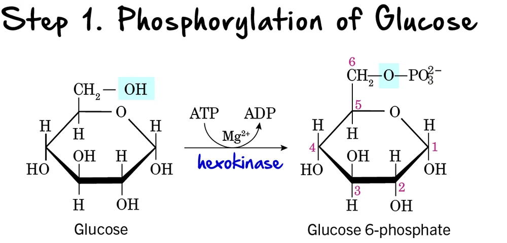
Glycolysis Adventure – Complete Game (Stages 1-10) 1 2 3 4 5 6 7 8 9 10 ๐ŸŽฏ Score 0 โญ High Score 0 ๐Ÿ”ฅ Streak 0 0 ATP Produced 0 NADH Produced 2 ATP Invested Stage 1: Glucose Activation ๐Ÿš€ The First Investment – Adding a Phosphate Group Glucose (Cโ‚†Hโ‚โ‚‚Oโ‚†) C C C C C O O O O O O Starting moleculeFree glucose in cell ๐Ÿ”„ Hexokinase Glucose-6-Phosphate C C C C C O O O O O O P Activated moleculeTrapped in cell โšก ๐Ÿ’ฐ Energy Investment: Uses 1 ATP โ†’ ADP + Pi ๐ŸŽฎ GAME ANALOGY Theme park ticket activation! You pay $1 (ATP) to stamp your ticket (phosphate) so you can’t lose it outside the park. Now you’re trapped inside and ready for rides! ๐ŸŽฏ Key Points: โœ… Traps glucose inside cell (phosphate can’t cross membrane) โœ… Investment: 1 ATP โœ… Enzyme: Hexokinase โœ… Rate-limiting step ๐Ÿ“ Challenge Questions – Stage 1 Q1: Why does the cell “spend” ATP at the start? A) To create energy immediately B) To trap glucose inside by adding phosphate C) To break glucose into pieces Q2: What if hexokinase was missing? A) Glucose would still enter normally B) Entire glycolysis would STOP C) Only last stage affected Q3: The phosphate group is like… A) A key unlocking the membrane B) A luggage tag preventing bag from leaving C) A ticket to buy food ๐ŸŽ‰ Stage 1 Complete! Score: 0/30 Proceed to Stage 2 โ†’ Stage 2: The Isomerization Shuffle ๐Ÿ”„ Rearranging Atoms – Preparing for the Big Split! Glucose-6-Phosphate (Aldose) C1 C2 C3 C4 C5 O O O P 6-membered ringCarbonyl at C1 ๐ŸŽฒ Phosphoglucose Isomerase Fructose-6-Phosphate (Ketose) C2 C3 C4 C5 C=O O O O P 5-membered ring + tailCarbonyl at C2 ๐ŸŽฎ PARTY SETUP! Same furniture, but you rearrange to make space for dancing. The carbonyl group moves from C1 to C2 – crucial for the upcoming split! ๐Ÿ“ Challenge Questions – Stage 2 Q1: Why is isomerization necessary? A) To create energy B) Only fructose form can be split in half C) To use another ATP Q2: What does “isomerization” mean? A) Adding/removing atoms B) Rearranging same atoms C) Breaking in half Q3: Difference between G6P and F6P? A) Different atoms B) Carbonyl moves C1โ†’C2 C) More phosphates ๐ŸŽ‰ Stage 2 Complete! Score: 0/60 Proceed to Stage 3 โ†’ Stage 3: The Power Boost โšก Second ATP Investment – The Commitment Step! Fructose-6-Phosphate C2 C3 C4 C5 C=O O O O P ONE phosphateAt carbon #6 โšก Phosphofructokinase-1 (PFK-1) COMMITMENT ENZYME Fructose-1,6-Bisphosphate C2 C3 C4 C5 C=O O O O P P TWO phosphates!At #1 AND #6 ๐Ÿ’ฐ Energy Investment – Second Payment ATP โšกโ†’ ADP + Pi Total invested: 2 ATP ๐ŸŽฏ What is a “Commitment Step”? โš ๏ธ Irreversible – Cannot go backward โš ๏ธ Rate-limiting – Most controlled step โš ๏ธ Point of no return – Glycolysis MUST finish ๐ŸŽฎ FINAL BOSS UPGRADE! You’re about to face the boss! You spend gold (2nd ATP) to activate “Mega Weapon”. PFK-1 is the shopkeeper who controls this upgrade. No refund = you’re committed to the fight! ๐Ÿ“ Challenge Questions – Stage 3 Q1: Why is this the “Commitment Step”? A) Uses most ATP B) Irreversible – once F1,6BP forms, no turning back C) It’s the fastest step Q2: What makes PFK-1 special? A) It’s the biggest enzyme B) It’s the main control point – inhibited by high ATP C) It works first Q3: If PFK-1 was blocked? A) Glycolysis would speed up B) Glycolysis STOPS – no ATP production C) Only first steps work ๐ŸŽ‰ Stage 3 Complete! Score: 0/90 Proceed to Stage 4 โ†’ Stage 4: The Great Split ๐Ÿ’ฅ From 6-Carbon to Two 3-Carbon Molecules! Fructose-1,6-Bisphosphate C2 C3 P C4 C=O O O C5 P C6 6 CARBONSReady to cleave! โš”๏ธ Aldolase “The Splitter” Glyceraldehyde-3-P (G3P) P C1 C2 C3 O O O 3-CarbonWill become G3P Dihydroxyacetone-P (DHAP) P C4 C5 C6 O O O 3-CarbonWill convert to G3P โšก Energy Status: No gain/loss Just breaking in half – like splitting a candy bar! ๐Ÿซ SPLITTING A CANDY BAR! You have a 6-piece candy bar (F1,6BP). You break it exactly in half โ†’ two 3-piece halves (G3P & DHAP). Same total pieces, just separated! ๐ŸŽฏ Key Points: โœ… C-C bond broken between C3 and C4 โœ… Creates TWO 3-carbon molecules โœ… DHAP must convert to G3P (next step) โœ… No energy change – just cleavage ๐Ÿ“ Challenge Questions – Stage 4 Q1: What happens in the aldolase reaction? A) A phosphate is added B) 6-carbon molecule splits into two 3-carbon molecules C) ATP is produced Q2: Where is the bond broken? A) Between C1 and C2 B) Between C3 and C4 C) Between C5 and C6 Q3: Energy outcome of this step? A) 2 ATP produced B) 1 ATP used C) No energy change – just cleavage ๐ŸŽ‰ Stage 4 Complete! Score: 0/120 Proceed to Stage 5 โ†’ Stage 5: Twin Conversion ๐Ÿ”„โ†’โšก DHAP โ†’ G3P: Getting Two for the Price of One! Dihydroxyacetone-P (DHAP) P C=O CHOH CHโ‚‚OH O O O DHAPKetone structure ๐Ÿ”€ Triose Phosphate Isomerase “The Converter” Glyceraldehyde-3-P (G3P) P CHO HC-OH CHโ‚‚OH O O O G3PAldehyde structure โšก Energy Status: Near equilibrium Very fast reaction – both DHAP and G3P are present ๐ŸŽฏ CRITICAL INSIGHT Now we have TWO G3P molecules! Even though glycolysis splits F1,6BP into one G3P and one DHAP, DHAP quickly converts to G3P. So effectively, we have TWO identical 3-carbon molecules moving forward. This means everything from now on happens TWICE per glucose! ๐ŸŽฏ TWO FOR ONE DEAL! You split a candy bar and get one milk chocolate half (G3P) and one dark chocolate half (DHAP). But you only like milk chocolate! So you trade the dark piece with a friend who converts it to milk chocolate. Now you have two identical pieces! ๐ŸŽฏ Key Points: โœ… DHAP and G3P are isomers โœ… DHAP converts to G3P (not much G3P โ†’ DHAP) โœ… Reaction is fast and

Biochemistry

Bioenergetics and Metabolism Intro

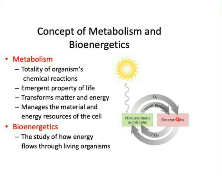
Bioenergetics &: Metabolism Bioenergetics Bioenergetics is the specialized field that studies how living organisms acquire, transform, and utilize energy. It’s essentially the application of the principles of thermodynamics โ€“ the study of energy and its effects on matter โ€“ to biological processes. Concepts of Energy Energy: At its core, energy is the capacity to do work. In biological systems, “work” can encompass a vast array of activities: muscle contraction, nerve impulse transmission, synthesizing complex molecules, maintaining body temperature, and transporting substances across cell membranes. Forms of Energy: Kinetic Energy: The energy of motion. Examples include heat energy, light energy, and mechanical energy (e.g., a moving muscle). Potential Energy: Stored energy, or the energy of position. This is the energy that could do work. In biology, the most crucial form is chemical energy, stored within the bonds of molecules like ATP, glucose, and fats. Thermodynamics and Chemical Reactions Thermodynamics provides the framework for understanding energy changes during chemical reactions. Exergonic vs. Endergonic Reactions: Exergonic Reactions (Energy-Releasing): These reactions release energy into the surroundings. They are spontaneous. Endergonic Reactions (Energy-Consuming/Requiring): These reactions require an input of energy to proceed. They are non-spontaneous. Free Energy (ฮ”G): The “Usable” Energy Free Energy (G): Represents the portion of a system’s energy that is available to do work. Change in Free Energy (ฮ”G): The difference in free energy between the products and reactants. A negative ฮ”G: The reaction is exergonic and spontaneous. A positive ฮ”G: The reaction is endergonic and non-spontaneous. ฮ”G = 0: The reaction is at equilibrium. Enthalpy and Entropy: The change in free energy (ฮ”G) is determined by two other thermodynamic quantities: Enthalpy and Entropy. The relationship is expressed by the Gibbs Free Energy Equation: ฮ”G = ฮ”H โˆ’ Tฮ”S Where: ฮ”G = Change in Free Energy ฮ”H = Change in Enthalpy T = Absolute Temperature (in Kelvin) ฮ”S = Change in Entropy Enthalpy (ฮ”H): Represents the change in heat content. Negative ฮ”H (Exothermic): Heat is released. Positive ฮ”H (Endothermic): Heat is absorbed. Entropy (ฮ”S): Represents the change in randomness or disorder. The universe tends towards maximum entropy (Second Law of Thermodynamics). Positive ฮ”S: The system becomes more disordered. Negative ฮ”S: The system becomes more ordered. In biological systems, reactions often lead to a temporary decrease in entropy locally (e.g., building a complex protein). However, this is always accompanied by a greater increase in disorder in the surroundings, maintaining the Second Law of Thermodynamics overall. Standard Free Energy (ฮ”Gยฐ) and Biological Standard Free Energy (ฮ”Gยฐ’) Standard Free Energy (ฮ”Gยฐ): The change in free energy under standard conditions (1 M concentration, 25ยฐ C, 1 atm pressure). Biological Standard Free Energy (ฮ”Gยฐ’): In biology, a modified standard condition is used to better reflect physiological conditions: pH 7.0 (neutral). All other conditions remain the same as ฮ”Gยฐ. The actual free energy change (ฮ”G) in a living cell will depend on the actual concentrations of reactants and products. However, ฮ”Gยฐ’ is a useful reference point. Biological Energy Transformations: Coupled Reactions Living organisms power endergonic reactions through coupled reactions, where the liberation of energy from an exergonic reaction is used to drive an endergonic one. ATP Hydrolysis: A Classical Example of an Exergonic Reaction: The hydrolysis of Adenosine Triphosphate (ATP) to Adenosine Diphosphate (ADP) and inorganic phosphate (Pi) is a highly exergonic reaction, releasing a significant amount of free energy (approx. -7.3 kcal/mol or -30.5 kJ/mol under standard biological conditions, ฮ”Gยฐ’). This released energy is then used to fuel various endergonic processes in the cell. Additive Nature of Free Energy Changes in Pathways Biochemical pathways consist of a series of sequential reactions. The overall free energy change (ฮ”G) for an entire pathway is the sum of the ฮ”G values of all individual reactions. Crucially, even if some individual reactions are endergonic (positive ฮ”G), the pathway can still proceed as long as the sum of all ฮ”G values for the entire pathway is negative. This is achieved by coupling endergonic steps with highly exergonic steps, effectively “pulling” the pathway forward. Cellular Metabolic Reactions Metabolism refers to all the chemical reactions that occur in living organisms to maintain life. These processes allow organisms to grow, reproduce, maintain their structures, and respond to their environments. Cellular metabolism is a highly organized and interconnected network of reactions that takes place within the cell. Types of Metabolism There are two main types of metabolic reactions: Catabolism (Breakdown): The process of breaking down complex molecules into simpler ones, usually releasing energy in the process. This energy is then captured and stored in molecules like ATP (adenosine triphosphate). Analogy: Demolition โ€“ taking apart a complex building to get raw materials and energy. Example: The breakdown of glucose into carbon dioxide and water to produce ATP. Anabolism (Build-up/Synthesis): The process of building complex molecules from simpler ones, which typically requires an input of energy (often supplied by ATP). Analogy: Construction โ€“ using raw materials and energy to build a complex structure. Example: The synthesis of proteins from amino acids, or DNA from nucleotides. These two processes are linked: the energy released during catabolism fuels anabolism, creating a continuous cycle of energy transformation and matter recycling within the cell. Characteristics of Cellular Metabolic Reactions Enzyme-Catalyzed: Almost all metabolic reactions are catalyzed by specific enzymes. Enzymes allow reactions to occur quickly and efficiently at physiological temperatures and pH. Highly Regulated: Metabolic pathways are tightly controlled to ensure that cells only produce what they need, when they need it. Regulation occurs at various levels: Enzyme activity: Allosteric regulation, feedback inhibition, covalent modification (e.g., phosphorylation). Enzyme synthesis: Gene expression can be turned on or off. Substrate availability: The presence or absence of reactants can dictate reaction rates. Occur in Pathways: Metabolic reactions are rarely isolated events. Instead, they occur in a series of sequential, interconnected steps called metabolic pathways. The product of one reaction often serves as the substrate for the next. Linear pathways: A โ†’ B โ†’ C โ†’ D Branched pathways: A โ†’ B โ†’ C and A โ†’ B โ†’ D Cyclic pathways: A โ†’ B

Biochemical Techniques
Biochemistry

Biochemical Techniques in Biochemistry

Biochemical Techniques: Lab Advanced Techniques in Clinical Chemistry Clinical chemistry laboratories are at the forefront of medical diagnostics, utilizing sophisticated instrumentation and methodologies to analyze biological samples. The goal is to provide accurate, precise, and timely results that aid in disease diagnosis, prognosis, treatment monitoring, and prevention. The advent of computerization and automation has revolutionized these labs, dramatically increasing productivity and improving the quality of services. A deep understanding of the underlying principles and instrumental theories is paramount for laboratory professionals to effectively operate and troubleshoot these systems, ensuring the highest standard of patient care. A diverse range of analytical techniques are employed in clinical chemistry, each tailored to specific analytes and diagnostic needs. The most fundamental and widely used methods include: Electrophoresis Chromatography Spectrophotometry Mass Spectrometry Fluorometry Nephelometry Turbidimetry Biochip (Protein and DNA Chip/Array) Biosensor Let’s embark on a detailed exploration of each of these techniques, starting with Electrophoresis. Electrophoresis: Principles and Applications in Clinical Chemistry Electrophoresis refers to the migration of charged solutes or particles in a liquid or a porous supporting medium, such as cellulose acetate sheets or agarose gel film, under the influence of an electrical field. This fundamental biophysical technique is widely used for separating and analyzing macromolecules, primarily proteins and nucleic acids, based on their charge, size, and shape. Theory of Electrophoresis: The Driving Forces The movement of charged particles in an electric field is governed by fundamental electrochemical principles. Key Definitions: Anode: The positively charged electrode. Negatively charged molecules (anions) migrate towards the anode. Cathode: The negatively charged electrode. Positively charged molecules (cations) migrate towards the cathode. Isoelectric Point (pI) of a Molecule: This is the specific pH at which a molecule carries no net electrical charge. At its pI, a molecule will not move in an electrical field. Ampholyte or Zwitterion: A molecule that possesses both acidic and basic functional groups (e.g., proteins with NHโ‚‚ and COOH groups). These molecules can carry a net positive, net negative, or zero charge depending on the pH. Mechanism of Migration: In a solution more acidic than its pI, a protein will take on a net positive charge and migrate toward the cathode (negative electrode). In a solution more alkaline (basic) than its pI, a protein will take on a net negative charge and migrate toward the anode (positive electrode). Factors Influencing the Rate of Migration: The velocity (v) of a charged molecule is influenced by several factors: The Net Electrical Charge of the Molecule: The primary determinant. Molecules with a greater net charge will migrate faster. The Size and Shape of the Molecule: Larger and more irregularly shaped molecules experience greater frictional resistance and migrate slower. The Electric Field Strength: A stronger electric field (higher voltage) leads to faster migration but also generates more heat. The Characteristics of the Supporting Medium: The type, concentration, and pore size of the medium (e.g., agarose gel) create a sieve-like effect that impacts migration. The Operation Temperature: Higher temperatures decrease buffer viscosity, which increases migration rates, but excessive heat can cause sample denaturation and band distortion. Description of an Electrophoresis System Schematic Diagram Components: Two Buffer Boxes with Baffle Plates: These reservoirs hold the buffer, which maintains a constant pH and conducts the current. Electrodes: Made of inert materials like platinum, these are connected to the power supply to create the electric field. Electrophoresis Support: The medium where separation occurs (e.g., agarose gel, cellulose acetate). Wicks (Strips): Porous materials that connect the support to the buffer, ensuring continuous electrical contact. Cover: Minimizes evaporation, maintains stable temperature, and protects the system. Direct Current (DC) Power Supply: This component provides the electrical energy and can be set to constant voltage, constant current, or constant power (often preferred as it controls heat generation). Automated Electrophoresis Systems Highly automated systems have revolutionized clinical labs by improving throughput and reproducibility. Evolution: From labor-intensive manual techniques, electrophoresis has evolved with prepackaged gels and integrated platforms. Example: Analyzers like the Rapid Electrophoresis (REP) Analyzer feature automated sample application, programmed running conditions, automated staining, and integrated densitometry for quantitative analysis, streamlining the entire workflow. Different Types of Electrophoresis 1. Starch Gel Electrophoresis Principle: Separates macromolecules based on both surface charge and molecular size, using a gel matrix made from potato starch. Limitations in Clinical Labs: Preparation is technically difficult and gels are opaque, hindering visualization. Reproducibility is poor. Current Status: Rarely used in modern clinical labs, largely superseded by agarose and polyacrylamide methods. 2. Agarose Gel Electrophoresis Principle: A convenient method using agarose, a purified polysaccharide, as the supporting medium. The gel forms a porous matrix. For proteins, separation is based on charge; for nucleic acids, it’s primarily size. Advantages: Lower affinity for proteins (clearer separations), optically clear after drying (excellent for densitometry), easy preparation, and a wide range of pore sizes. Successful Applications in Clinical Chemistry: Serum Proteins Electrophoresis (SPEP): The most common application, separating serum proteins (albumin, ฮฑโ‚, ฮฑโ‚‚, ฮฒ, and ฮณ-globulins). Hemoglobin Variants: Separation of normal and abnormal hemoglobins (e.g., HbA, HbS, HbC). Isoenzymes: Separation of different forms of enzymes like LDH and CK. Lipoprotein Fractions: Separation of VLDL, LDL, and HDL. Nucleic Acids: Fundamental for DNA and RNA analysis. 3. Cellulose Acetate Electrophoresis (CAE) Principle: Uses a highly porous membrane made from cellulose acetate. Separation is based on net charge and size. Advantages of CAE: Speed of Separation: Relatively rapid (20 minutes to 1 hour). Transparency and Storage: Membranes become transparent after treatment, allowing for easy densitometric scanning, and can be stored as a permanent record. Small Sample Volumes: Requires relatively small amounts of sample. Applications: Similar to agarose, used for rapid screening of serum proteins and hemoglobin variants. Comparison to Agarose: While CAE is faster, agarose often provides better resolution. However, for quick, routine separations, CAE remains a viable option. Chromatography: Principles of High-Resolution Separation Chromatography is a family of laboratory techniques for the separation of mixtures. The mixture is dissolved in a fluid called the mobile phase, which carries it through a structure holding another material called the stationary phase.

Scroll to Top