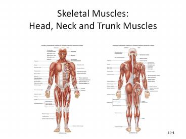
Muscles of the Head, Neck and Trunk
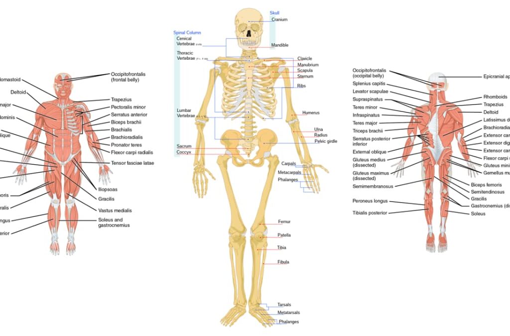
Axial Skeleton Muscles: The Footress. Muscles of the Axial Skeleton A. Muscles of the Head and Face The muscles of the head can be broadly categorized into muscles of facial expression and muscles of mastication (chewing). 1. Muscles of Facial Expression These unique muscles insert into the skin or other muscles, allowing us to show a wide range of emotions. They are all innervated by the Facial Nerve (Cranial Nerve VII). a. Occipitofrontalis (Epicranius) A broad muscle covering the top of the skull with two bellies. The Frontal belly raises the eyebrows and wrinkles the forehead, while the Occipital belly pulls the scalp posteriorly. b. Orbicularis Oculi A ring-like muscle encircling the eye. Its primary action is to close the eye (blinking, winking) and squint. c. Orbicularis Oris A complex muscle encircling the mouth. It closes and protrudes the lips, as in puckering or kissing. d. Zygomaticus Major and Minor Extend from the cheekbone to the corner of the mouth. They are the primary “smiling” muscles, raising the lateral corners of the mouth upward. e. Buccinator A thin, flat muscle of the cheek. It compresses the cheek for whistling or sucking and holds food between the teeth during chewing. f. Platysma A broad, superficial sheet of muscle in the neck. It tenses the skin of the neck, depresses the mandible, and pulls the lower lip down. 2. Muscles of Mastication (Chewing) These four pairs of muscles are responsible for moving the mandible for chewing. They are all innervated by the Mandibular division of the Trigeminal Nerve (Cranial Nerve V3). a. Masseter A powerful muscle on the side of the jaw. It is the primary elevator of the mandible (closes the jaw). b. Temporalis A fan-shaped muscle in the temporal fossa. It elevates and retracts the mandible. c. Medial Pterygoid Located deep to the mandible. It elevates the jaw and assists in side-to-side grinding movements. d. Lateral Pterygoid Located deep in the jaw. It protracts the mandible (pulls it forward), moves it side-to-side, and is the only muscle of mastication that helps open the jaw. Summary Table of Head & Face Muscles Muscle Origin Insertion Action FACIAL EXPRESSION (CN VII) Occipitofrontalis Galea aponeurotica (Frontal); Occipital bone (Occipital) Skin of eyebrows; Galea aponeurotica Raises eyebrows, wrinkles forehead, pulls scalp Orbicularis Oculi Frontal and maxillary bones Tissue of eyelid Closes eye, squints, blinks Orbicularis Oris Maxilla and mandible Skin and muscle at angles of mouth Closes and protrudes lips (puckering) Zygomaticus Major/Minor Zygomatic bone Skin and muscle at angle of mouth Raises lateral corners of mouth (smiling) Buccinator Molar region of maxilla and mandible Orbicularis oris Compresses cheek (whistling, sucking) Platysma Fascia of chest Base of mandible; skin at corner of mouth Tenses skin of neck, depresses mandible MASTICATION (CN V3) Masseter Zygomatic arch Angle and ramus of mandible Elevates mandible (closes jaw) Temporalis Temporal fossa Coronoid process of mandible Elevates and retracts mandible Medial Pterygoid Sphenoid and palatine bones Medial surface of ramus of mandible Elevates mandible, moves side-to-side Lateral Pterygoid Sphenoid bone Condylar process of mandible; TMJ capsule Protracts and depresses (opens) jaw B. Muscles of the Neck The muscles of the neck are diverse, responsible for moving the head, stabilizing the cervical spine, assisting in breathing, and facilitating swallowing and speech. They are categorized here based on location and primary actions. 1. Superficial Anterior Neck Muscles a. Sternocleidomastoid (SCM) A large, two-headed muscle on each side of the neck. When acting alone (unilaterally), it rotates the head to the opposite side and flexes it to the same side. When both act together (bilaterally), they flex the neck (chin to chest). 2. Suprahyoid Muscles (Above the Hyoid Bone) These muscles form the floor of the mouth and are primarily responsible for elevating the hyoid bone during swallowing and speaking. a. Digastric Two-bellied muscle that elevates the hyoid or depresses the mandible (opens the mouth). b. Mylohyoid Forms the floor of the mouth; elevates hyoid and floor of mouth. c. Geniohyoid Elevates and protracts the hyoid bone. d. Stylohyoid Elevates and retracts the hyoid bone. 3. Infrahyoid Muscles (Strap Muscles – Below the Hyoid) These “strap-like” muscles primarily depress the hyoid bone and larynx during swallowing and speaking. a. Sternohyoid Depresses the hyoid bone and larynx. b. Omohyoid Two-bellied muscle that depresses and retracts the hyoid. c. Sternothyroid Depresses the larynx and hyoid bone. d. Thyrohyoid Depresses the hyoid bone but elevates the larynx. 4. Deep Lateral Neck Muscles (Scalenes) The Anterior, Middle, and Posterior Scalene muscles are important for lateral flexion of the neck. They also act as accessory muscles of inspiration by elevating the first two ribs. Summary Table of Neck Muscles Muscle Origin Insertion Innervation Action Sternocleidomastoid Manubrium & Clavicle Mastoid process CN XI, C2-C3 Unilateral: Rotates head opp., flexes same side. Bilateral: Flexes neck. Digastric Mandible & Mastoid process Hyoid bone CN V3 & CN VII Elevates hyoid, depresses mandible. Mylohyoid Mandible Hyoid bone CN V3 Elevates hyoid & floor of mouth. Sternohyoid Manubrium & Clavicle Hyoid bone Ansa cervicalis Depresses hyoid and larynx. Omohyoid Scapula Hyoid bone Ansa cervicalis Depresses and retracts hyoid. Sternothyroid Manubrium Thyroid cartilage Ansa cervicalis Depresses larynx and hyoid. Thyrohyoid Thyroid cartilage Hyoid bone C1 via CN XII Depresses hyoid, elevates larynx. Scalenes (Ant, Mid, Post) Cervical vertebrae (C2-C7) First & Second ribs Cervical spinal nerves Flexes neck, elevates ribs for inspiration. C. Muscles of the Torso (Trunk) The muscles of the trunk are vital for maintaining posture, protecting internal organs, facilitating respiration, and enabling a wide range of movements. 1. Muscles of the Back These complex, layered muscles move and stabilize the vertebral column, head, and shoulders. a. Superficial Back Muscles Primarily act on the upper limbs. Includes the large Trapezius (moves scapula), Latissimus Dorsi (extends and adducts arm), and the deeper Rhomboids and Levator Scapulae (retract and elevate scapula). b. Intermediate Back Muscles Respiratory muscles. The Serratus Posterior Superior elevates ribs for inspiration, while the Serratus Posterior Inferior depresses ribs for expiration. c. Deep (Intrinsic) Back Muscles Responsible for posture