Doctors Revision

Author name: doctorsrevision@gmail.com

Spleen and Pancreas
Anatomy

Hepatobiliary System: Spleen and Pancreas

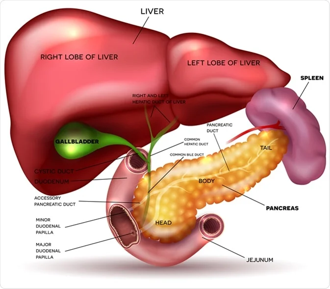
Hepatobiliary System Spleen & Pancreas The Pancreas: Anatomy and Physiology GASTROINTESTINAL SYSTEM The Spleen The spleen is a vital secondary lymphoid organ located in the upper left quadrant of the abdomen, playing crucial roles in immune surveillance and blood filtration. Anatomical Summary (“Odd Numbers Rule of 1,3,5,7,9,11”): This mnemonic is a useful way to remember key splenic facts: 1 inch thick, 3 inches wide, 5 inches long. Weighs approximately 7 ounces (approx. 200 grams). Lies between the 9th and 11th ribs. Development: Of mesodermal origin, similar to blood components it processes. Shape: Often described as having the size and shape of a clenched fist. Borders: Typically has an anterior notched border. Location and Relations: Location: Intraperitoneal organ situated in the left upper part of the abdomen, in the left hypochondrium. Diaphragmatic Surface: Convex and smooth, molded to the concavity of the diaphragm. Visceral Surface: Irregular, with impressions from adjacent organs. Hilum: Located on the visceral surface, usually between the stomach and the left kidney impressions. Long Axis: Lies along the line of the 10th rib. Lower Pole: Under normal circumstances, the lower pole does not extend beyond the midaxillary line, making it generally non-palpable in healthy adults. Functions The spleen is analogous to a lymph node in that it filters fluid and facilitates immune responses, but it filters blood, not lymph. 1. Immunological Responses Filter Blood-borne Antigens: Acts as a large filter for the blood, exposing blood-borne antigens to lymphocytes and macrophages. Production of Antibodies: Crucial site for initiating primary immune responses against encapsulated bacteria (e.g., Streptococcus pneumoniae, Haemophilus influenzae), producing IgM antibodies. Storage of Lymphocytes: Contains a large reserve of lymphocytes. 2. Filtration and Hemocatheresis (Removal of Aged/Defective Cells) Removes Particulate Matter: Phagocytoses bacteria, parasites, and cellular debris from the blood. Removes Aged/Defective Blood Cells: Primarily removes old, rigid, or damaged red blood cells (erythrocytes) and platelets. This process is crucial for maintaining a healthy circulation and preventing the accumulation of non-functional cells. Iron Recycling: Extracts iron from heme of senescent red blood cells for reuse in erythropoiesis. 3. Hematopoiesis (in Fetus and Disease States) Fetal Role: In the human fetus, the spleen is an important site of extramedullary hematopoiesis until the bone marrow takes over later in gestation. Adult Resumption: This function may be resumed in adults in certain pathological conditions, such as severe bone marrow failure (myelofibrosis). 4. Blood Reservoir Can store a significant volume of blood, particularly red blood cells and platelets, which can be released into circulation when needed (e.g., hemorrhage, stress), though its role as a reservoir is less pronounced in humans compared to some other mammals. Splenectomy (Surgical Removal of the Spleen) Despite its important functions, the spleen is not essential for survival. Compensatory Mechanisms: Its functions are largely taken over by other organs: Liver: Compensates for the filtration and removal of old red blood cells. Bone Marrow: Takes over hematopoietic functions. Lymph Nodes and other Lymphoid Tissues: Compensate for immune functions. Clinical Considerations Post-Splenectomy: Individuals without a spleen (asplenic or functionally asplenic) are at increased risk of overwhelming post-splenectomy infection (OPSI), particularly from encapsulated bacteria. Vaccination against these bacteria (pneumococcus, meningococcus, H. influenzae type B) and prophylactic antibiotics are crucial. Blood Supply of the Spleen Arterial Supply Splenic Artery: A large and tortuous branch of the celiac trunk. Course: Runs along the superior border of the pancreas, posterior to the stomach, within the splenorenal ligament (which attaches the spleen to the posterior abdominal wall near the kidney). Branches: Gives off branches to the pancreas, short gastric arteries (supplying the fundus of the stomach), and the left gastro-omental (gastroepiploic) artery before reaching the splenic hilum. At the hilum, it typically divides into 4-5 (or more) segmental branches that enter the spleen. Venous Drainage Splenic Vein: Formed by several tributaries from the splenic hilum. Course: Runs posterior to the tail and body of the pancreas. Confluence: Posterior to the neck of the pancreas, the splenic vein joins with the superior mesenteric vein (SMV) to form the hepatic portal vein. It also receives the inferior mesenteric vein (often). Lymphatic Drainage: Lymphatic vessels typically follow the splenic artery back to the pancreaticosplenic lymph nodes located along the splenic artery. From there, lymph drains into the celiac lymph nodes. Nerve Supply: Supplied by autonomic nerves originating from the celiac plexus. Primarily sympathetic innervation, which causes vasoconstriction and contraction of the splenic capsule (if present and muscular enough, as in some animals, to expel blood). Its role in humans is mainly to regulate blood flow. Parasympathetic innervation is less clearly defined or functionally significant. Supports of the Spleen (Ligaments) The spleen is supported and held in place by several peritoneal folds, or ligaments, derived from the embryonic dorsal mesentery. Gastrosplenic Ligament Description: A double layer of peritoneum that extends from the greater curvature of the fundus of the stomach to the splenic hilum. Contents: Contains the short gastric arteries and veins (from the splenic artery/vein) and the left gastro-omental (gastroepiploic) artery and vein. Splenorenal (Lienorenal) Ligament Description: A double layer of peritoneum that extends from the splenic hilum to the anterior aspect of the left kidney and posterior abdominal wall. Contents: Contains the splenic artery and vein and the tail of the pancreas. This is a key anatomical relationship; injury to the splenic vessels or damage to the pancreas during splenectomy is a concern. Phrenicosplenic Ligament Often considered a part of the splenorenal ligament or a separate fold connecting the superior aspect of the spleen to the diaphragm. Splenocolic Ligament Description: Connects the inferior aspect of the spleen to the left colic (splenic) flexure of the colon. Hilum of the Spleen The hilum is the area on the visceral surface of the spleen where structures enter and leave the organ. Contents of the Hilum: Splenic Artery (dividing into segmental branches) Splenic Vein (formed by tributaries) Lymphatic Vessels Nerves (from the celiac plexus) Histology of the Spleen The spleen has a characteristic macroscopic and microscopic structure optimized for its functions. Macroscopic Appearance: When sectioned, the spleen consists of
Hepatobiliary System: Liver, Gallbladder
Anatomy

Hepatobiliary System: Liver, Gallbladder

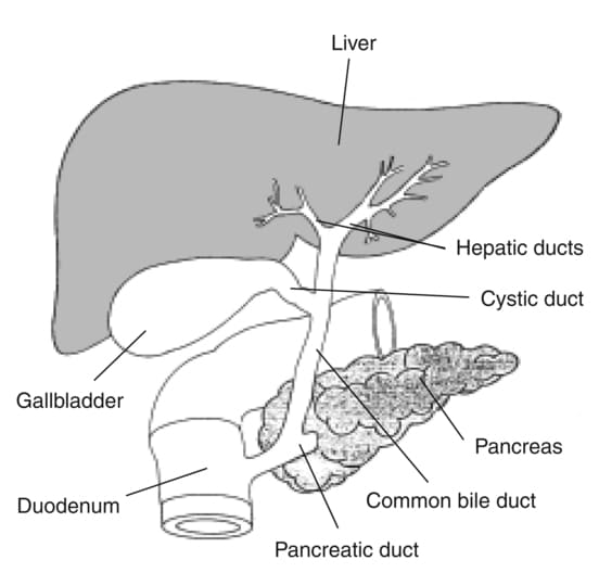
Hepatobiliary System Liver & Gallbladder The Hepatobiliary System SYSTEMS PHYSIOLOGY The Hepatobiliary System The hepatobiliary system is a vital part of the digestive apparatus, comprising the liver, gallbladder, and the bile ducts. Its primary functions revolve around the production, transport, concentration, and regulated secretion of bile, which is crucial for lipid digestion and the excretion of waste products. Main Functions: Bile Production: The liver continuously produces bile. Bile Transport: Bile ducts transport bile from the liver to the duodenum or to the gallbladder for storage. Bile Concentration: The gallbladder stores and concentrates bile. Bile Secretion: Bile is released into the duodenum to aid in digestion. Bile Composition and Function: Bile is a complex, alkaline fluid (pH 7.6-8.6) typically yellowish-green in color, produced by hepatocytes. It consists of: Bile Salts Derived from cholesterol, these are the most functionally important component. They emulsify dietary fats (lipids and fatty acids) in the small intestine, breaking large fat globules into smaller ones. This increases the surface area for lipase enzymes to act upon, facilitating fat digestion and absorption (especially of fat-soluble vitamins A, D, E, K). Bilirubin A yellowish-green pigment, it is the primary waste product of heme metabolism (from the breakdown of old red blood cells). The liver conjugates bilirubin (makes it water-soluble) and excretes it into bile. Its presence gives feces their characteristic brown color (after further modification by gut bacteria). Other Components Cholesterol: A significant component of bile, primarily excreted this way. Phospholipids (e.g., lecithin): Aid in emulsification. Water: The largest component. Electrolytes: Bicarbonate, sodium, potassium, chloride. Trace metals, heavy metals, drugs: Excreted via bile. The Liver The liver is the largest gland in the body and also the largest internal organ, weighing approximately 1.3-1.5 kg (around 2% of adult body weight). It is a metabolically diverse and highly vascularized organ with an astounding array of functions, far beyond just bile production. Location and Structure Location: Occupies most of the upper right quadrant of the abdomen, extending into the epigastric region and even into the upper left quadrant. It lies immediately inferior to the diaphragm, largely protected by the lower ribs. Surfaces: Diaphragmatic (Anterior/Superior) Surface: This large, convex surface is molded by the diaphragm. Visceral (Postero-inferior) Surface: This irregular, concave surface is molded by the various abdominal organs it contacts (e.g., stomach, duodenum, colon, right kidney, adrenal gland). Basic Functions (Beyond bile production): While bile production is a key excretory function, the liver’s metabolic roles are paramount: Metabolic Activities: Central to the metabolism of: Carbohydrate Metabolism: Maintenance of blood glucose homeostasis Glycogenesis (glucose to glycogen), glycogenolysis (glycogen to glucose), gluconeogenesis (non-carbohydrate sources to glucose), regulation of blood glucose. Lipid Metabolism (Fats): Synthesis and degradation of fatty acids, cholesterol, triglycerides, and lipoproteins. Synthesis of cholesterol, lipoproteins, triglycerides; fatty acid oxidation; ketone body formation. Protein Metabolism: Synthesis of most plasma proteins (e.g., albumin, clotting factors), deamination of amino acids, urea formation (detoxification of ammonia). Deamination of amino acids, leading to the formation of urea (detoxification of ammonia). Detoxification and Filtration: Detoxification: Metabolizes and detoxifies various endogenous substances (e.g., hormones) and exogenous substances (e.g., drugs, toxins, alcohol). Filtration: Its extensive vascular network (hepatic sinusoids) and specialized macrophages (Kupffer cells) act as a reticuloendothelial system, filtering blood to remove bacteria, foreign particles, old red blood cells, and cellular debris that have gained entry through the gastrointestinal tract via the portal circulation. Storage: Stores glycogen, vitamins (A, D, B12), iron (ferritin), and copper. Synthesis of Blood Components: Plasma Proteins: Albumin (maintains oncotic pressure), globulins, fibrinogen, prothrombin, and other clotting factors. Heparin: While often listed, heparin is primarily produced by mast cells and basophils. The liver’s role is more in the synthesis of other anticoagulants and factors involved in coagulation. Production and Secretion of Bile: Essential for fat digestion and absorption, and for the excretion of waste products like bilirubin. Bilirubin is a breakdown product of heme (from worn-out red blood cells’ hemoglobin), which the liver processes and excretes into bile. Immunological Role: Contains Kupffer cells (macrophages) which phagocytose foreign material and play a role in immune surveillance. Lobes of the Liver Traditionally, the liver is divided into four anatomical lobes based on external landmarks: Right Lobe: The largest lobe, occupying the right side of the liver. It is demarcated from the left lobe by the falciform ligament on the anterior/superior surface. On the visceral surface, it includes the gallbladder fossa and groove for the inferior vena cava. Left Lobe: Smaller than the right lobe, located to the left of the falciform ligament. Caudate Lobe: A small, quadrilateral lobe located on the visceral surface, superior to the porta hepatis, between the fissure for the ligamentum venosum (left) and the groove for the inferior vena cava (right). Quadrate Lobe: A small, quadrilateral lobe located on the visceral surface, inferior to the porta hepatis, between the fissure for the ligamentum teres (left) and the gallbladder fossa (right). Note: The caudate and quadrate lobes are functionally considered part of the left functional lobe of the liver. This functional division is based on the intrahepatic branching of the portal triad (hepatic artery, portal vein, bile duct). From a surgical perspective (Couinaud classification), the liver is divided into 8 functionally independent segments, each with its own vascular and biliary supply and drainage. The caudate lobe often has independent vascular supply and venous drainage, making it a unique “lobe.” Porta Hepatis (Liver Hilum) Location: Found on the visceral (postero-inferior) surface of the liver. It is a deep transverse fissure situated between the caudate lobe superiorly and the quadrate lobe inferiorly. Attachment: The lesser omentum (specifically, the hepatoduodenal ligament part) attaches to its margins. Contents (Portal Triad and associated structures): The porta hepatis is the entry and exit point for all structures forming the “portal triad” and other associated elements: Proper Hepatic Artery: Divides into right and left hepatic arteries, which supply oxygenated blood to the liver parenchyma. Hepatic Portal Vein: Divides into right and left portal veins, carrying nutrient-rich, deoxygenated blood from the gastrointestinal tract, spleen, and pancreas to the liver for

arterial and nerve supply of palate
Anatomy

Teeth, Tongue & Salivary Glands

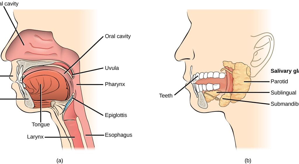
Teeth, Tongue & Salivary Glands Anatomy: Teeth, Tongue, and Salivary Glands GROSS ANATOMY Teeth Teeth are specialized, calcified, whitish structures securely anchored in the jaws of many vertebrates, primarily adapted for the mechanical breakdown of food (mastication). Beyond digestion, they play vital roles in speech articulation and facial aesthetics. Distinctive Properties: Teeth differ significantly from bone in several key aspects: Hardness: They are considerably harder than bone, primarily due to their higher mineral content and the unique structure of enamel and dentin. Regeneration: Unlike bone, which can continuously remodel and repair itself, mature teeth (specifically enamel and dentin) have a very limited capacity for self-repair or regeneration. Mineral Content: Teeth possess a significantly higher mineral content (mainly hydroxyapatite) than bone, contributing to their superior hardness and durability. Parts of a Tooth Each tooth is anatomically divided into three principal parts: the crown, the neck, and the root. 1. The Crown Location: This is the visible portion of the tooth that projects into the oral cavity above the gingiva (gum line). Protection: It is entirely covered and protected by enamel, the hardest substance in the human body. Bulk: The main bulk of the crown is composed of dentin, which lies immediately deep to the enamel. 2. The Neck (Cervix) Location: This is the constricted junction where the crown meets the root. It is typically encircled by the gingival margin. Covering: At the neck, the enamel of the crown meets the cementum of the root. 3. The Root Location: This is the portion of the tooth that is embedded within the alveolar bone of the jaw. Attachment: It serves as the anchor for the tooth within its socket. Covering: The entire surface of the root is covered by a thin layer of specialized bone-like tissue called cementum. The cementum, in turn, is connected to the alveolar bone of the tooth socket by the periodontal ligament, a fibrous connective tissue that acts as a shock absorber and provides sensory feedback. Internal Structures of a Tooth: Beyond the superficial layers, teeth have vital internal components: Enamel: The outermost layer of the crown, composed of approximately 96% mineral (hydroxyapatite), making it the hardest biological substance. It is translucent and provides the protective covering for the crown. Formed by ameloblasts during tooth development, which are lost upon eruption, hence its inability to regenerate. Dentin: Composition: Constitutes the bulk of the tooth, both in the crown and the root. It is a calcified connective tissue, but less mineralized than enamel (around 70% mineral, 20% organic matrix, and 10% water). Structure: It contains microscopic channels called dentinal tubules, which radiate outwards from the pulp cavity towards the enamel or cementum. These tubules contain cellular extensions of odontoblasts and dentinal fluid, making dentin somewhat permeable and sensitive. Dentinogenesis: The formation of dentin begins prior to enamel formation and is an ongoing process throughout the life of the tooth. It is initiated and maintained by specialized cells called odontoblasts. Odontoblasts: The cell bodies of the odontoblasts are aligned along the inner aspect of the dentin, forming the peripheral boundary of the dental pulp. Their processes extend into the dentinal tubules. Pulp: Location: The innermost cavity of the tooth, centrally located within the dentin. Contents: It contains the tooth’s living tissues: nerves (providing sensation), blood vessels (providing nutrients), and connective tissue. Functions: Provides vitality to the tooth, nourishing the odontoblasts and maintaining the dentin. Alveolar Ridge and Tooth Sockets: Alveolar Ridge: The bony projection of the maxilla and mandible that houses the teeth. Alveolus (Tooth Socket): The specific depression or socket within the alveolar ridge where each tooth root is embedded. Dentition: Sets of Teeth Humans are diphyodont, meaning they develop two sets of teeth during their lifetime: deciduous (primary or “milk”) teeth and permanent (secondary) teeth. 1. Deciduous Teeth (Primary Dentition): Number: There are 20 deciduous teeth in total. Distribution per Jaw (Maxilla and Mandible): 4 Incisors (2 central, 2 lateral) 2 Canines 4 Molars (first and second molars) Note: Deciduous dentition does not include premolars. Eruption: Typically begin to erupt around 6 months after birth. By the end of the second year (approximately 24-30 months), all deciduous teeth have usually erupted. Sequence: Generally, the teeth of the lower jaw appear before those of the upper jaw, and incisors erupt first, followed by molars and then canines. 2. Permanent Teeth (Secondary Dentition): Number: There are 32 permanent teeth in total. Distribution per Jaw (Maxilla and Mandible): 4 Incisors (2 central, 2 lateral) 2 Canines 4 Premolars (first and second premolars) 6 Molars (first, second, and third molars) Eruption: Begin to erupt around the 6th year of life, replacing the deciduous teeth and adding new molars. “Wisdom Teeth” (Third Molars): The third molars are the last teeth to erupt, typically between the ages of 17 to 30 years. They are commonly referred to as “wisdom teeth” due to their late eruption. They often cause problems (impaction, pain) due to insufficient space in the jaws. Types of Teeth and Their Functions: Each tooth type is specialized for a particular function: Incisors: (Front teeth, 4 per jaw in permanent dentition). Characterized by thin, sharp cutting edges, primarily used for biting and cutting food. Canines: (Pointed teeth, 2 per jaw). Possess a single, prominent, conical cusp, designed for tearing food. Premolars (Bicuspids): (Behind canines, 4 per jaw in permanent dentition). Each typically has two cusps, used for crushing and grinding food. (Absent in deciduous dentition). Molars: (Most posterior teeth, 6 per jaw in permanent dentition). Characterized by three or more broad cusps and a large occlusal (chewing) surface, highly efficient for grinding and pulverizing food. Neurovascular Supply of the Teeth and Gingiva The teeth and their supporting structures receive a rich blood supply and sensory innervation crucial for their health and function. Blood Supply: Arteries: Superior Alveolar Arteries: Branches of the maxillary artery (a terminal branch of the external carotid artery) supply the maxillary (upper) teeth and gingiva. These include the anterior, middle, and posterior superior alveolar arteries. Inferior Alveolar Artery: A branch

Oral Cavity Mouth Cavity
Anatomy

Oral Cavity/Mouth Cavity

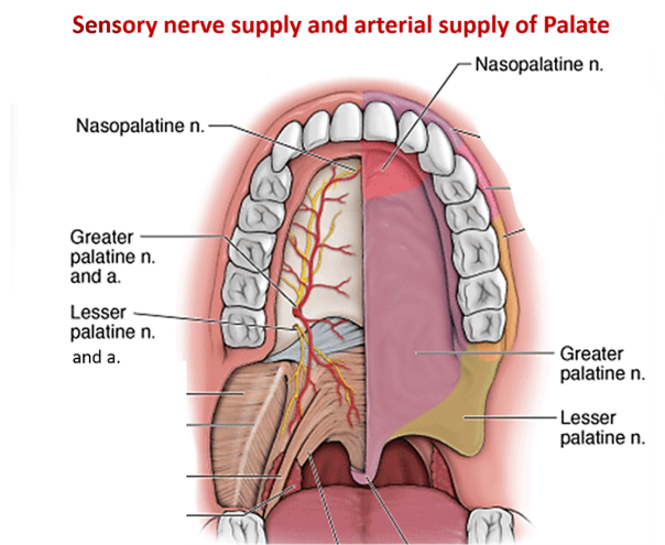
Oral Cavity / Mouth Cavity Anatomy: The Oral Cavity & Palate GROSS ANATOMY & EMBRYOLOGY The Oral Cavity/Mouth Cavity The oral cavity, often simply referred to as the mouth, serves as the initial segment of the digestive system and plays crucial roles in respiration and speech. Boundaries: It extends from the external opening, formed by the lips, posteriorly to the oropharyngeal isthmus. The oropharyngeal isthmus represents the critical junction where the oral cavity transitions into the pharynx (throat). Primary Divisions: The oral cavity is distinctly divided into two main components: The Oral Vestibule The Oral Cavity Proper (or Mouth Proper) The Oral Vestibule The oral vestibule is a peripheral, slit-like space, serving as the entranceway to the main oral cavity. Communication: It directly communicates with the external environment through the oral fissure, which is the opening defined by the lips. Defining Boundaries: Externally: Its outer limits are established by the mobile structures of the lips (anteriorly) and the cheeks (laterally). Internally: Its inner boundary is formed by the fixed structures of the gingivae (gums) and the teeth. Lining: The entire vestibule is lined by a specialized mucous membrane. This membrane seamlessly covers the internal surfaces of the lips and cheeks and then reflects (folds back) onto the superior and inferior gingivae and the adjacent teeth. Detailed Structures of the Vestibule: A. The Cheeks (Buccae) The cheeks constitute the primary lateral walls of the oral vestibule and are integral to various oral functions. Core Structure: The foundation of each cheek is the buccinator muscle. This muscle is essential for maintaining the shape of the cheek and facilitating mastication. Layers: External Surface: Covered by the skin of the face and a superficial connective tissue layer known as the buccinator fascia. Internal Surface: Lined by the smooth, moist mucous membrane of the oral vestibule. Muscular Function: The buccinator muscle is classified as a muscle of facial expression. Its contractions are vital for: Pressing the cheeks against the teeth, which prevents food from accumulating in the vestibule during chewing (mastication). Aiding in the expulsion of air (e.g., blowing). Contributing to facial expressions. Innervation: The motor innervation to the buccinator muscle is provided by the buccal branch of the facial nerve (Cranial Nerve VII). (Note: The provided text had “fascial nerve,” which is a common typo for “facial nerve.”) B. The Lips (Labia) The lips are highly mobile, musculofibrous folds that encircle the oral fissure. They are crucial for both oral function and communication. Extent and Location: Superiorly: They extend to the nasolabial sulci (the grooves running from the sides of the nose to the corners of the mouth) and the base of the nares (nostrils). Inferiorly: They are bounded by the mentolabial sulcus (the groove separating the lower lip from the chin). Laterally: They merge with the cheeks at the oral commissures (corners of the mouth). Composition: The lips are complex structures containing: Muscles: Primarily the orbicularis oris muscle, which acts as a sphincter to close the mouth, along with superior and inferior labial muscles that assist in lip movement. Connective Tissue: Fibrous tissue providing support and structure. Glands: Numerous small labial salivary glands (mucous and serous). Vessels and Nerves: An extensive network to supply the rich functions of the lips. Lip Functions and Control: Diverse Functions: The lips perform a wide array of functions: Grasping Food: Facilitating the intake of food and fluids. Sucking Fluids: Essential for actions like drinking from a straw or nursing. Maintaining Oral Seal: Crucially, they keep food and liquids within the oral cavity proper, preventing spillage into the vestibule during chewing and swallowing. Speech Articulation: Modulating sounds for clear speech. Facial Expression and Communication: Conveying emotions and participating in social interactions (e.g., kissing). Control of the Oral Opening: The opening and closing of the mouth, as well as the intricate movements of the lips, are orchestrated by a group of circumoral muscles. These include: Orbicularis Oris: The primary sphincter of the mouth. Buccinator: Assists in pressing the lips against the teeth. Risorius: Draws the corner of the mouth laterally. Various depressor muscles (e.g., depressor anguli oris, depressor labii inferioris) that pull the lips downwards. Various elevator muscles (e.g., levator anguli oris, levator labii superioris) that pull the lips upwards. Blood Supply to the Lips: The arterial supply to the lips is highly vascular, arising from branches of the facial artery. Upper Lip: Primarily supplied by the superior labial artery (a branch of the facial artery) and contributions from the infraorbital artery (a branch of the maxillary artery). Lower Lip: Primarily supplied by the inferior labial artery (a branch of the facial artery) and contributions from the mental artery (a terminal branch of the inferior alveolar artery). Innervation of the Lips: Sensory innervation to the lips is derived from branches of the trigeminal nerve (Cranial Nerve V). Upper Lip: Receives sensory innervation from the superior labial branches of the infraorbital nerve. The infraorbital nerve is itself a branch of the maxillary nerve (CN V2). Lower Lip: Receives sensory innervation from the inferior labial branches of the mental nerve. The mental nerve is a terminal branch of the inferior alveolar nerve, which stems from the mandibular nerve (CN V3). Lymphatic Drainage of the Lips: The lymphatic drainage patterns of the lips are important for understanding the spread of infections or malignancies. Upper Lip and Lateral Aspects of the Lower Lip: Lymph from these regions typically drains into the submandibular lymph nodes. Medial Part of the Lower Lip: Lymph from this specific area drains into the submental lymph nodes. C. The Gingivae (Gums) The gingivae are the specialized mucous membranes that surround the necks of the teeth and cover the alveolar processes of the jaws. Composition: They are composed of dense fibrous tissue richly covered by a specialized mucous membrane. Types of Gingiva: Gingiva Proper (Attached Gingiva): This portion is characteristically firmly attached to the underlying periosteum of the alveolar processes of the jaws and also directly to the cementum at the necks of the teeth. Visually, it presents as

Stomach and Intestines Anatomy
Anatomy

Stomach and Intestines Anatomy

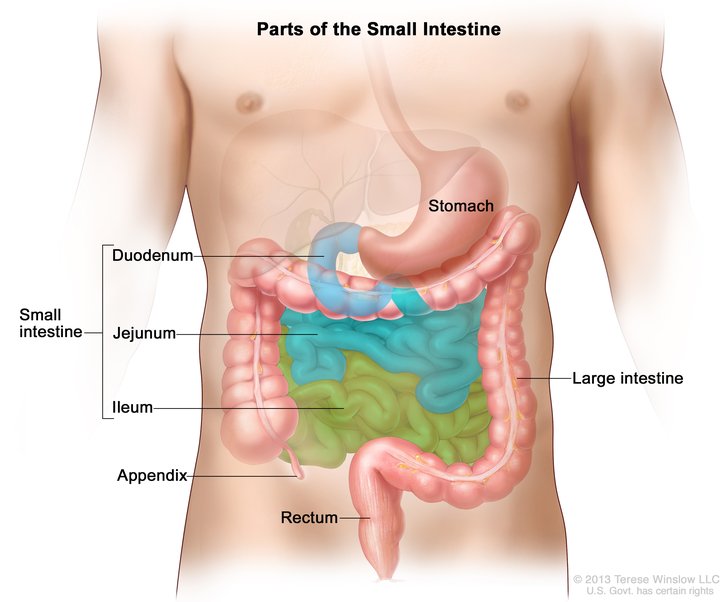
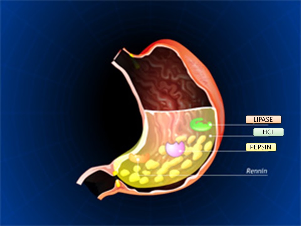
Stomach & Intestines Digestive System Anatomy: Stomach & Intestines GASTROINTESTINAL ANATOMY Stomach The stomach is a dilated, J-shaped organ of the alimentary canal, situated between the esophagus and the duodenum. Functions Storage of food: It acts as a temporary reservoir for ingested food. Mixing and mechanical digestion: It churns food with gastric juices to form chyme. Chemical digestion: Gastric juices (containing hydrochloric acid and enzymes like pepsin) initiate protein digestion. Controlled release: Regulates the slow release of chyme into the duodenum for further digestion and absorption. General Characteristics Capacity: The total capacity of the stomach is approximately 1500 ml. Shape and Position: Its position and shape vary significantly among individuals and with posture and respiration. In short and obese individuals, it tends to be high and more transverse. In tall and thin individuals, it is often elongated and vertical. It typically occupies the epigastric and umbilical regions of the abdomen and is partly covered by the costal diaphragm and the lower ribs. General Structure The stomach has two surfaces, two apertures (orifices), and two curvatures. Surfaces: Anterior surface, Posterior surface. Orifices (Apertures): Cardia: The opening from the esophagus into the stomach. Pylorus: The opening from the stomach into the duodenum. Curvatures: Lesser curvature: The shorter, concave, right border of the stomach. Greater curvature: The longer, convex, left and inferior border of the stomach. Specific Features: Cardia (Cardiac Orifice) Located at the esophagogastric junction. There is no anatomical sphincter in the traditional sense. Instead, a physiological sphincter mechanism is formed by the circular muscle layer of the distal esophagus and stomach, the acute angle of His (where the esophagus joins the stomach), and the diaphragmatic crus. This physiological sphincter relaxes during swallowing to allow food entry and closes afterward to prevent gastroesophageal reflux (regurgitation) of gastric contents back into the esophagus. Pylorus The distal opening of the stomach into the duodenum, located at the gastroduodenal junction. Lesser Curvature Located on the right margin of the stomach. Serves as the attachment site for the lesser omentum. The angular incisure (incisura angularis) is a constant notch on the lesser curvature, marking the junction between the body and the pyloric part of the stomach. Greater Curvature Located on the left margin and inferior border of the stomach. Provides attachment for the gastrosplenic ligament (connecting to the spleen) and the greater omentum (which extends inferiorly, folds back, and attaches to the transverse colon). Anterior Surface: Covered by the peritoneum. The left vagus nerve forms the anterior vagal trunk and predominantly supplies the anterior surface of the stomach. Posterior Surface: Also covered by the peritoneum. The right vagus nerve forms the posterior vagal trunk and predominantly supplies the posterior surface of the stomach. Parts of the Stomach The stomach is typically divided into four main parts: Cardia: The region immediately surrounding the cardiac orifice. Fundus: The dome-shaped part that projects superiorly and to the left of the cardia. It often contains gas. Body: The main part of the stomach, extending from the cardia/fundus to the angular incisure on the lesser curvature. Pylorus (Pyloric Part): The tubular distal part of the stomach connecting it to the duodenum. It is further divided into: Pyloric Antrum: Wider, more proximal part. Pyloric Canal: Narrower, distal part. Pyloric Sphincter: A thickened ring of circular smooth muscle at the gastroduodenal junction. It controls the discharge of chyme from the stomach into the duodenum. The pylorus typically lies on the transpyloric plane (L1). Peritoneal Attachments (Omenta): Lesser Omentum: A two-layered fold of peritoneum that extends from the porta hepatis (on the liver) to the lesser curvature of the stomach and the superior part of the duodenum. It is further divided into: Hepatogastric ligament: From the liver to the lesser curvature of the stomach. Hepatoduodenal ligament: From the liver to the superior part of the duodenum. This is clinically very important as it contains the portal triad: the common bile duct, the proper hepatic artery, and the hepatic portal vein. Greater Omentum: A large, apron-like fold of peritoneum that hangs down from the greater curvature of the stomach and covers the intestines. It contains varying amounts of fat. Gastrosplenic Omentum (Ligament): Connects the greater curvature of the stomach to the hilum of the spleen. Mucous Membrane of the Stomach: The internal lining of the stomach is thrown into numerous folds called rugae. These folds allow the stomach to expand significantly when filled with food and flatten out as it distends. Muscle Layer of the Stomach: The muscularis externa of the stomach is unique among the alimentary canal because it has three layers of smooth muscle, which contribute to its powerful churning action: Outer Longitudinal Layer: Primarily present along the curvatures (lesser and greater). Middle Circular Layer: Surrounds the entire stomach but is particularly prominent at the pylorus (forming the pyloric sphincter) and cardia. Innermost Oblique Layer: Found mainly in the body and fundus, allowing for a unique churning motion. Peritoneum of the Stomach: The stomach is almost entirely intraperitoneal, meaning it is nearly completely covered by visceral peritoneum. The peritoneum leaves the stomach to form the various omenta and ligaments (lesser omentum, greater omentum, gastrosplenic omentum). Relations of the Stomach Anteriorly Anterior abdominal wall Costal margin Left lobe of the liver Left pleura and lung (superiorly) Diaphragm Posteriorly (Stomach Bed) Separated by the lesser sac (omental bursa): Diaphragm (posteriorly, superiorly) Spleen (laterally) Splenic artery (superior border of the pancreas) Left kidney Left suprarenal gland Pancreas Transverse mesocolon and transverse colon Blood Supply of the Stomach The stomach has a rich arterial supply from branches of the celiac trunk, ensuring robust collateral circulation. Arterial Supply Lesser Curvature: Left Gastric Artery: Direct branch of the celiac trunk. Supplies the upper part of the lesser curvature and the abdominal esophagus. Right Gastric Artery: A branch of the proper hepatic artery (which comes from the common hepatic artery, a celiac trunk branch). Supplies the lower part of the lesser curvature. Greater Curvature: Left Gastro-omental (Gastroepiploic) Artery: A branch of the splenic artery. Supplies the upper part of

heart anatomy 1
Anatomy

Heart & Great Vessels

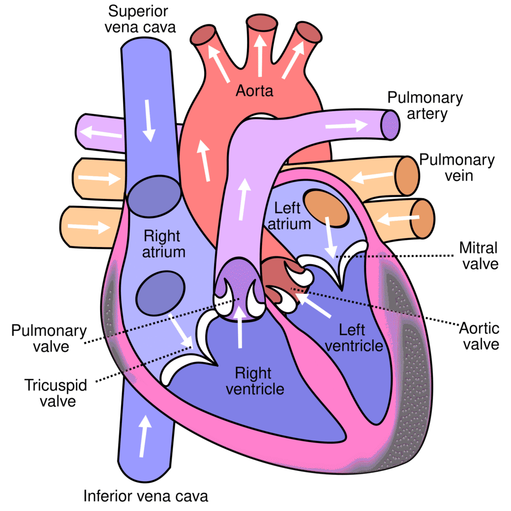
Heart & Great Vessels Anatomy: Heart, Pericardium, Great Vessels & Esophagus GROSS ANATOMY The Heart, Pericardium, and Great Vessels Pericardium The pericardium is a tough, double-layered fibroserous sac that encloses the heart and the roots of the great vessels (aorta, pulmonary trunk, venae cavae, pulmonary veins). Main Functions Lubrication: Contains a small amount of fluid that reduces friction between the moving heart and the surrounding structures. Restriction of Movement: Anchors the heart in the mediastinum, preventing excessive movement and overdistention during sudden increases in blood volume. Protection: Acts as a physical barrier against infection and external trauma. Components The pericardium is composed of two main layers: Fibrous Pericardium (Outer layer) Serous Pericardium (Inner layer) 1. Fibrous Pericardium The fibrous pericardium is the robust, outermost layer of the pericardial sac. Description: It is a thick, tough, inelastic, and conical-shaped fibrous bag that surrounds the heart. Attachments: Inferiorly (Base): It is firmly and broadly fused to the central tendon of the diaphragm. This attachment is crucial for the diaphragm’s role in cardiac stability. Anteriorly: It is loosely attached to the posterior surface of the sternum by the sternopericardial ligaments. Superiorly (Apex): It blends and is fused with the outer connective tissue coats (adventitia) of the great vessels as they enter and leave the heart (aorta, pulmonary trunk, superior vena cava, inferior vena cava, pulmonary veins). Posteriorly: It is attached to the structures in the posterior mediastinum, like the esophagus and aorta. Embryological Origin: Its primary origin is debated, but contributions are thought to come from the septum transversum (which also gives rise to the central tendon of the diaphragm) and pleuropericardial membranes. 2. Serous Pericardium The serous pericardium is a thin, delicate, two-layered membrane that lines the inner surface of the fibrous pericardium and covers the external surface of the heart. Layers: It is divided into two continuous layers: Parietal Layer of Serous Pericardium: This layer lines the inner surface of the fibrous pericardium. Visceral Layer of Serous Pericardium (Epicardium): This layer tightly adheres to the outer surface of the heart itself. It is considered the outermost layer of the heart wall. Pericardial Cavity: The potential space located between the parietal and visceral layers of the serous pericardium. This space normally contains a small amount of serous fluid. Pericardial Cavity and Sinuses A. Pericardial Cavity Description: A potential space located between the parietal and visceral layers of the serous pericardium. Contents: Normally contains a small amount (typically 15-50 mL) of thin, straw-colored serous fluid. Function of Fluid: The pericardial fluid acts as a lubricant, allowing the heart to beat smoothly and with minimal friction within the pericardial sac. Clinical Significance: a. Pericardial Effusion: An abnormal accumulation of fluid in the pericardial cavity. This can be caused by various conditions, including infections (e.g., tuberculosis), inflammation (e.g., pericarditis), autoimmune diseases, trauma, and malignancies (tumors). The term “water in the heart” is a colloquial and somewhat misleading description; it’s fluid around the heart. b. Cardiac Tamponade: A life-threatening condition where a large or rapidly accumulating pericardial effusion compresses the heart, restricting its ability to fill adequately with blood during diastole. This leads to reduced cardiac output and can be fatal if not treated promptly. c. Pericardiocentesis: A medical procedure to aspirate (remove) excess fluid from the pericardial cavity to relieve pressure in cases of pericardial effusion or cardiac tamponade. B. Pericardial Sinuses These are reflections of the serous pericardium that create cul-de-sacs or recesses. Oblique Pericardial Sinus: Location: A blind-ended, inverted U-shaped cul-de-sac located posterior to the heart’s left atrium. Boundaries: It is situated between the reflections of the serous pericardium around the four pulmonary veins and the inferior vena cava (IVC). The posterior wall of the left atrium forms its anterior boundary. Clinical Significance: Allows for surgical access to the posterior surface of the heart. Transverse Pericardial Sinus: Location: A short, transverse tunnel or passage that runs between the great arteries (aorta and pulmonary trunk) anteriorly and the great veins (superior vena cava, inferior vena cava, and pulmonary veins) posteriorly. Clinical Significance: This sinus is strategically important for cardiac surgeons. A surgical clamp can be passed through the transverse sinus to temporarily occlude the aorta and pulmonary trunk during cardiac surgery (e.g., for coronary artery bypass grafting or valve replacement), isolating the heart from the systemic circulation. Innervation and Blood Supply of Pericardium A. Blood Supply of the Pericardium The pericardium receives its arterial supply from several sources: Pericardiacophrenic Artery: The main arterial supply, a branch of the internal thoracic artery. It accompanies the phrenic nerve. Musculophrenic Artery: Another branch of the internal thoracic artery. Branches of the Thoracic Aorta: Small branches directly from the aorta (e.g., bronchial and esophageal arteries). Coronary Arteries: The visceral layer (epicardium) receives some small branches from the coronary arteries. Venous Drainage: Follows the arterial supply, draining into corresponding veins (e.g., pericardiacophrenic veins, internal thoracic veins, azygos system). B. Innervation of the Pericardium The innervation differs for the fibrous/parietal serous layers and the visceral serous layer. Fibrous Pericardium and Parietal Layer of Serous Pericardium: Innervation: Primarily supplied by the phrenic nerves (C3, C4, C5). Sensitivity: These layers are richly innervated and are sensitive to pain, temperature, pressure (touch), and stretch. Referred Pain: Because the phrenic nerves also supply sensory innervation to the C3-C5 dermatomes, pain originating from these pericardial layers is often referred to the ipsilateral (same side) shoulder, neck, and supraclavicular region. (The original mention of “left jaw” is less common for pericardial pain referral than the shoulder and neck). Visceral Layer of Serous Pericardium (Epicardium): Innervation: Supplied by the autonomic nervous system (sympathetic and parasympathetic fibers) from the cardiac plexuses. Sensitivity: This layer is generally considered insensitive to pain, temperature, and touch. It is primarily sensitive to stretch. Function: Autonomic innervation primarily modulates cardiac function rather than providing somatic sensation from the epicardium itself. The Heart The heart is a hollow, muscular organ that acts as a pump, circulating blood throughout the entire body to deliver oxygen and nutrients and remove waste products. Shape: It is

rib cage and diaphragm anatomy
Anatomy

Rib Cage & Diaphragm Anatomy

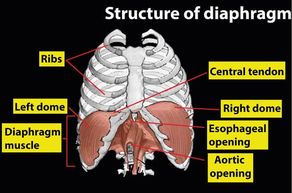
Rib Cage & Diaphragm Anatomy Anatomy: The Rib Cage and Diaphragm THORACIC ANATOMY The Rib Cage: An Overview The thoracic cage, commonly known as the rib cage, is a robust bony-cartilaginous framework that forms the skeletal wall of the chest. It serves several critical functions: Protection: Encapsulates and safeguards vital thoracic organs, including the heart, lungs, and great vessels, as well as parts of the upper abdominal organs (liver, spleen). Muscle Attachment: Provides numerous points of attachment for muscles of the neck, back, chest, and upper limbs, playing a role in posture and movement. Respiration: Its flexible, expandable structure is fundamental to the mechanics of breathing, allowing for changes in thoracic volume during inspiration and expiration. Support: Forms the central axial skeleton to which the pectoral girdle and upper limbs are attached. Shape and Location: The rib cage is situated in the thorax, the region of the trunk between the neck and the abdomen. Its overall shape is that of a truncated cone, wider inferiorly than superiorly. It is characteristically flattened anteriorly and posteriorly (dorsoventrally) but rounded laterally, creating a spacious cavity within. Boundaries of the Thoracic Cage The thoracic cage forms a well-defined compartment with distinct boundaries: Anterior Boundary: Formed by the sternum (breastbone) and the articulating costal cartilages. Posterior Boundary: Formed by the twelve thoracic vertebrae and their associated intervertebral discs. Lateral Boundaries: Consist of the twelve pairs of ribs, which extend from the thoracic vertebrae posteriorly to the sternum or costal cartilages anteriorly. Superior Boundary: Thoracic Inlet (Superior Thoracic Aperture): A relatively small, kidney-shaped opening. Formed by: The superior aspect of the first thoracic vertebra (T1) posteriorly, the medial border of the first ribs laterally, and the superior border of the manubrium anteriorly. This aperture provides passage for structures (e.g., trachea, esophagus, major vessels, nerves) between the neck and the thoracic cavity. Inferior Boundary: Thoracic Outlet (Inferior Thoracic Aperture): A much larger, irregular opening. Formed by: The 12th thoracic vertebra (T12) posteriorly, the 11th and 12th pairs of ribs laterally, and the costal margin (formed by the cartilages of ribs 7-10) and the xiphoid process anteriorly. This aperture is almost completely sealed by the diaphragm, which separates the thoracic cavity from the abdominal cavity. The Sternum (Breastbone) The sternum, or breastbone, is a flat, elongated bone positioned in the central anterior aspect of the thoracic cage. It forms the anterior articulation for the ribs via their costal cartilages. Composition: The sternum is typically divided into three fused parts: Manubrium Sterni (Handle) Location: The broadest, most superior part of the sternum. Vertebral Level: Roughly lies opposite the T3 and T4 vertebral bodies. Articulations: Superiorly: The suprasternal (jugular) notch is at its superior border. Lateral to the notch are the clavicular notches for articulation with the clavicles (forming the sternoclavicular joints). Laterally: Possesses facets for articulation with the costal cartilages of the first pair of ribs and the upper half of the second pair of ribs. These are synovial joints, permitting slight movement. Inferiorly: Articulates with the body of the sternum at the manubriosternal joint. Body of the Sternum (Gladiolus) Location: The longest, central part of the sternum. Articulations: Superiorly: Articulates with the manubrium at the manubriosternal joint. Laterally: Contains facets for articulation with the costal cartilages of the lower half of the second ribs through the seventh ribs. Inferiorly: Articulates with the xiphoid process at the xiphisternal joint. Xiphoid Process Location: The smallest, most inferior part of the sternum. Vertebral Level: The xiphisternal joint is typically at the level of the T9 vertebral body. Characteristics: It is highly variable in shape and size. It generally does not articulate with any ribs. It remains cartilaginous in young individuals and gradually undergoes ossification (hardens into bone) from its proximal end, a process that completes in adulthood and continues into old age. Joints of the Sternum: Manubriosternal Joint (Sternal Angle / Angle of Louis): A secondary cartilaginous joint (symphysis) between the manubrium and the body. It forms a palpable transverse ridge. Xiphisternal Joint: A secondary cartilaginous joint between the body and the xiphoid process. It typically fuses completely in older adults. Clinical Uses of the Sternum A. Diagnostic and Therapeutic Procedures Median Sternotomy (Median Thoracotomy): Procedure: A surgical incision made longitudinally down the center of the sternum, which is then divided using a saw. Purpose: Provides wide surgical access to the mediastinum (heart, great vessels, trachea, thymus) for procedures such as cardiac surgery (e.g., coronary artery bypass grafting, valve replacement) and lung transplantation. The sternum is typically rejoined with wires post-surgery. Bone Marrow Biopsy/Aspiration: Procedure: Due to its broad, flat, and relatively superficial nature, the sternum (particularly the manubrium) is a common site for obtaining bone marrow samples. A needle is inserted into the sternum to extract marrow for diagnostic purposes (e.g., in cases of leukemia, anemia, or other hematological disorders). This site is chosen to avoid vital organs directly below and due to its accessibility. B. Congenital Anomalies of the Sternum Congenital malformations of the sternum primarily involve abnormalities in its shape, which can affect respiratory and cardiac function in severe cases. Pectus Carinatum (Pigeon Chest): Description: A chest wall deformity characterized by an outward protrusion of the sternum and costal cartilages. Appearance: Gives the chest a prominent, “pigeon-breasted” appearance. Clinical Significance: Usually cosmetic, but severe cases can restrict lung expansion and cardiac function, especially during strenuous exercise. Pectus Excavatum (Funnel Chest): Description: The most common sternal deformity, characterized by an inward depression of the sternum and costal cartilages. Appearance: Creates a “funnel-shaped” indentation in the chest. Clinical Significance: Ranges from mild cosmetic concerns to severe cases where the sternum compresses the heart and lungs, potentially leading to respiratory and cardiac compromise (e.g., reduced exercise tolerance). (The “Cup-shaped deformity” and “Saucer-shaped” descriptions in the original text likely refer to variations or degrees of Pectus Excavatum.) Horns-of-Steer Deformity: (This term is less common in standard medical literature but may refer to a specific variant of chest wall deformity where the costal cartilages project laterally or superiorly, resembling horns, often associated

digestion
Anatomy

Digestion, Absorption & GIT Disorders

Digestion, Absorption & GIT Disorders Digestion, Absorption & GIT Disorders GASTROINTESTINAL PHYSIOLOGY Digestion, Absorption & GIT Disorders Digestion is the process of breaking down complex food molecules into simpler forms that can be absorbed by the body. Absorption is the subsequent process of transporting these digested nutrients from the lumen of the GI tract into the bloodstream or lymphatic system. I. Why Digestion? The human body relies on three main macronutrients: 1. Carbohydrates, 2. Fats, 3. Proteins. Additionally, small quantities of vitamins and minerals are essential. These macronutrients, in their natural, complex forms (e.g., starch, triglycerides, large proteins), cannot be directly absorbed through the gastrointestinal (GIT) mucosa. They are “useless as nutrients without preliminary digestion.” Digestion by Hydrolysis Digestion primarily occurs through hydrolysis, a chemical process where water molecules are added to break down larger molecules into smaller ones. This process reverses the condensation reactions that originally formed these macromolecules. Carbohydrates Exist mostly as large polysaccharides (e.g., starch) or disaccharides (e.g., sucrose, lactose). Formed by condensation (removal of a water molecule between monosaccharide units). Hydrolysis, catalyzed by specific enzymes, reverses this, yielding monosaccharides. Fats Exist as triglycerides (one glycerol molecule attached to three fatty acid molecules). Formed by condensation, with three water molecules removed. Hydrolysis, by fat-digesting enzymes, reverses this, forming fatty acids and glycerol. Proteins Formed from multiple amino acids linked by peptide bonds (a condensation reaction). Proteolytic enzymes (proteases) reverse this, breaking peptide bonds to yield smaller peptides and ultimately amino acids. II. Digestion of Specific Macronutrients A. Digestion of Carbohydrates Major Dietary Sources: Sucrose: Disaccharide, commonly known as cane sugar. Lactose: Disaccharide, found in milk. Starches: Large polysaccharides, present in almost all non-animal foods (e.g., potatoes, grains). Other Minor Sources: Amylose, glycogen, alcohol, lactic acid, pyruvic acid, pectins, dextrins, and minor carbohydrate derivatives in meats. Cellulose: Not digestible by humans as we lack the necessary enzymes. Locations of Digestion: Mouth: Salivary amylase (ptyalin) initiates starch digestion, breaking it into smaller polysaccharides (dextrins) and some maltose. Accounts for about 5% of carbohydrate digestion. Stomach: Salivary amylase continues to act in the fundus and body of the stomach until it is inactivated by the acidic gastric juice. Can digest 30-40% of starches into dextrins and maltose. Small Intestine (Final Stage): This is where the bulk of carbohydrate digestion occurs. Pancreatic Amylase: Secreted by the pancreas into the duodenum, it breaks down starches and dextrins into maltose and other small glucose polymers. Brush Border Enzymes: Located on the microvilli of enterocytes (intestinal epithelial cells). These enzymes are responsible for the final breakdown of disaccharides into monosaccharides: Lactase: Digests lactose into glucose and galactose. Sucrase: Digests sucrose into glucose and fructose. Maltase: Digests maltose and other small glucose polymers into glucose. Final Products: The final products of carbohydrate digestion are exclusively monosaccharides (glucose, galactose, fructose), which are the only forms absorbable into the bloodstream. B. Digestion of Proteins Locations of Digestion: Stomach: Pepsin: Secreted by chief cells as pepsinogen and activated by hydrochloric acid (HCl) at a pH of 2-3. Pepsin initiates protein digestion, breaking down proteins into proteoses, peptones, and large polypeptides. Accounts for 10-20% of total protein digestion. Small Intestine: Pancreatic Secretions: The majority of protein digestion occurs in the upper small intestine (duodenum and jejunum) due to powerful pancreatic proteolytic enzymes. Trypsin, Chymotrypsin, Carboxypolypeptidase, Proelastase: These enzymes (secreted as inactive zymogens and activated in the duodenum) break down proteins, proteoses, peptones, and large polypeptides into smaller polypeptides, tripeptides, dipeptides, and a few free amino acids. Brush Border Peptidases (in Enterocytes): Located on the luminal surface of enterocytes lining the intestinal villi (especially in the duodenum and jejunum). Aminopolypeptidase and Dipeptidases: These enzymes further digest the small polypeptides, tripeptides, and dipeptides into their final absorbable form: amino acids. End Products of Luminal Digestion: Dipeptides, tripeptides, and amino acids. Final Absorbable Form: Over 99% of the final protein products are absorbed as amino acids. C. Digestion of Fats Primary Location: Almost entirely occurs in the small intestine. Two Main Steps: Emulsification: Large fat globules are broken down into smaller droplets. Bile Acids and Lecithin: These components of bile, secreted by the liver, are amphipathic molecules that surround fat droplets, reducing their surface tension and preventing them from coalescing. This process increases the surface area of fat by approximately 1000-fold, making it accessible to water-soluble digestive enzymes. Enzymatic Action: Pancreatic Lipase: The most important enzyme for fat digestion, secreted by the pancreas. It hydrolyzes triglycerides into monoglycerides and free fatty acids. Enteric Lipase: Also present in the small intestine, contributing to fat digestion. Cholesterol Ester Hydrolase: Hydrolyzes cholesterol esters into cholesterol and fatty acids. Phospholipase A2: Hydrolyzes phospholipids (like lecithin) into lysophospholipids and fatty acids. Final Products: Monoglycerides, free fatty acids, cholesterol, and lysophospholipids. III. Absorption of Digested Food Absorption is the process by which digested food materials move from the lumen of the GIT into the blood or lymph. Mechanisms: Involves both passive processes (e.g., diffusion, osmosis) and active processes (e.g., active transport, co-transport). Fluid Balance: Total fluid ingested per day: ~1.5 liters. Total fluid secreted into GIT (saliva, gastric juice, bile, pancreatic juice, intestinal secretions): ~7 liters. Total fluid entering small intestine: ~8.5 liters. Total fluid absorbed per day: ~8-9 liters. Most absorption (all but ~1.5 liters) occurs in the small intestine. Only about 1.5 liters pass through the ileocecal valve into the colon each day. A. Absorptive Surface of the Small Intestine The small intestine has an enormous surface area, crucial for efficient absorption. This is achieved through multiple levels of folding: Valvulae Conniventes (Folds of Kerckring): Large circular folds of the mucosa and submucosa, particularly well-developed in the duodenum and jejunum (up to 8mm high), increasing surface area by ~3-fold. Villi: Millions of small, finger-like projections (0.5-1mm long) covering the entire surface of the small intestine. Each villus is covered by epithelial cells and contains a lacteal (lymphatic capillary) and a rich capillary network. Villi increase surface area by ~10-fold. Microvilli (Brush Border): Each epithelial cell covering the villi has thousands of microscopic, hair-like projections called

Gastrointestinal GIT Secretions 1
Anatomy

Gastrointestinal (GIT) Secretions

GIT Secretions Gastrointestinal Secretions Physiology SYSTEMS PHYSIOLOGY Gastrointestinal (GIT) Secretions The gastrointestinal tract is equipped with a diverse array of secretory glands that play two fundamental roles: Secretion of Digestive Enzymes: These enzymes are essential for breaking down complex food molecules into absorbable units. This enzymatic activity occurs from the mouth all the way to the distal end of the ileum. Provision of Mucus: Mucus serves as a lubricant and protective barrier for the entire GIT, from the mouth to the anus. Key Principle: The presence of food in the GIT is the primary stimulus for secretions. The quantity and type of secretions are precisely regulated to match the amount and type of food present, ensuring efficient digestion. Anatomical Types of Glands in the GIT The GIT houses several types of glands, each contributing to the overall secretory process: Goblet Cells/Simple Mucous Cells: These are single-celled glands interspersed among the epithelial cells. They directly extrude mucus onto the epithelial surface, providing immediate lubrication and protection. They are found throughout the GIT. Crypts of Lieberkühn: These are invaginations or pits found deep within the mucosa of the small intestine and large intestine. They contain various specialized secretory cells, including enterocytes (which secrete water and electrolytes), goblet cells, and enteroendocrine cells. Tubular Glands: These glands are typically found deeper within the mucosal layer. Stomach: Examples include the oxyntic glands (gastric glands) in the body and fundus, which secrete acid, pepsinogen, intrinsic factor, and mucus, and pyloric glands in the antrum, which secrete mucus and gastrin. Upper Duodenum: Brunner’s glands, located in the submucosa of the duodenum, secrete alkaline mucus to protect against acidic chyme from the stomach. Complex Glands (Extramural Glands): These are large, accessory glands located outside the wall of the GIT but connected to it by ducts. They provide copious secretions crucial for digestion or emulsification. Salivary Glands: Produce saliva for initial digestion and lubrication. Pancreas: Secretes pancreatic juice containing a wide array of digestive enzymes and bicarbonate. Liver: Produces bile, essential for fat emulsification. Mechanism of Secretion by Glandular Cells Glandular cells in the GIT typically secrete two main types of substances simultaneously: Organic Substances: This includes digestive enzymes (proteins), mucin (glycoproteins), and hormones. These are synthesized within the cells and packaged into vesicles before exocytosis. Water and Electrolytes: These are secreted to create a fluid environment for the organic substances and to aid in transport and hydration. The movement of water and electrolytes is often regulated by ion pumps and channels, creating osmotic gradients. Mucus: Properties and Role Mucus is a vital secretion found throughout the GIT, acting as both a lubricant and a protectant. Composition: Mucus is a thick, viscous secretion primarily composed of water, electrolytes, and a mixture of several glycoproteins. These glycoproteins are large polysaccharides with smaller quantities of protein attached. Key Properties: Adherent Qualities: Mucus readily adheres to surfaces, forming a continuous coating. Coats the Gut Wall: It has sufficient “body” or viscosity to effectively coat and protect the entire luminal surface of the GIT. Low Resistance for Slippage: Provides a slippery surface, allowing food (bolus or chyme) to move easily along the tract without causing damage. Causes Fecal Particles to Adhere: In the large intestine, mucus helps bind fecal particles together, facilitating their smooth passage. Strongly Resistant to Digestion: Its complex structure and chemical properties make it highly resistant to breakdown by digestive enzymes, ensuring its protective function. Amphoteric Glycoproteins: The glycoproteins in mucus are amphoteric, meaning they can act as both an acid and a base. This property allows mucus to buffer against both acidic and alkaline conditions, protecting the underlying mucosa. The 4 Main Secretions of the GIT 1. Saliva Saliva is the first major digestive secretion, produced by the salivary glands in the mouth. A. Salivary Glands Three Principal Glands (Major Salivary Glands): Parotid Glands: Largest salivary glands, located below and in front of the ears. They secrete entirely serous (watery, enzyme-rich) saliva. Submandibular Glands: Located under the floor of the mouth. They secrete a mixed serous and mucous saliva. Sublingual Glands: Smallest of the major glands, located under the tongue. They primarily secrete mucous saliva, with some serous component. Minor Salivary Glands: Numerous small buccal glands (and other minor glands throughout the oral cavity) secrete only mucus. Daily Secretion Volume: The total daily secretion of saliva ranges between 800 and 1500 ml, with an average of about 1000 ml. B. Composition of Saliva Saliva is a complex fluid containing two major types of protein secretions: Serous Secretion: A watery fluid containing digestive enzymes. The main enzyme is ptyalin (salivary α-amylase), which initiates carbohydrate digestion. Mucus Secretion: Contains mucin, a glycoprotein that provides lubrication. pH of Saliva: Between 6.0 and 7.0, which is slightly acidic to neutral. C. How Saliva is Secreted (Two-Stage Process) Saliva is not a simple ultrafiltrate. Its composition is modified as it passes through the ducts. A typical submandibular gland, being a compound gland, illustrates this two-stage process: Primary Secretion by Acini: The acinar cells (the secretory units) produce a “primary secretion” that is roughly isotonic with plasma. This primary secretion contains ptyalin (α-amylase) and/or mucin, along with water and ions (similar to extracellular fluid). Modification in Salivary Ducts: As the primary secretion flows through the salivary ducts, significant changes occur: Sodium (Na+) Reabsorption: Na+ is actively reabsorbed from the ductal lumen into the interstitial fluid. Potassium (K+) Secretion: K+ is actively secreted from the interstitial fluid into the ductal lumen. Chloride (Cl-) Reabsorption: Cl- is reabsorbed passively, following Na+ due to the electrical gradient. Bicarbonate (HCO3-) Secretion: Bicarbonate ions are actively secreted by the ductal epithelium into the duct lumen. Net Effect: The net reabsorption of Na+ and Cl- is greater than the secretion of K+ and HCO3-. This results in a hypotonic final saliva (more dilute than plasma), especially at lower flow rates. D. Function of Saliva in Oral Hygiene The mouth is constantly exposed to pathogenic bacteria and food particles. Saliva plays a crucial role in maintaining oral health: Washing Action: Saliva

git neuro and motility
Anatomy

GIT Neuro & Motility

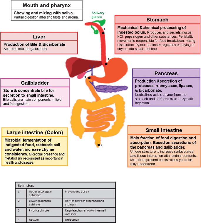
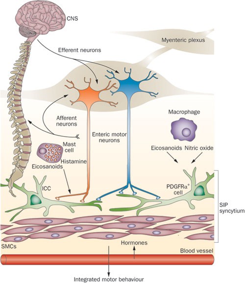
Digestive/GIT Neuro & Motility Digestive System Physiology SYSTEMS PHYSIOLOGY The Digestive System Physiology The digestive system is a vital organ system responsible for breaking down food into absorbable nutrients, water, and electrolytes, and then eliminating indigestible waste. It can be broadly divided into two main parts: the gastrointestinal tract (GIT), also known as the alimentary canal or gut, and accessory digestive organs. I. Components of the Digestive System A. The Gastrointestinal Tract (GIT) / Alimentary Canal This is a continuous, muscular tube that extends from the mouth to the anus, about 30 feet (9 meters) long in a cadaver (shorter in a living person due to muscle tone). It is the primary site where digestion and absorption occur. Mouth: The entrance, where mechanical digestion (chewing) and initial chemical digestion (salivary enzymes) begin. Pharynx: A common passageway for food and air. Esophagus: A muscular tube that transports food from the pharynx to the stomach via peristalsis. Stomach: A muscular sac for food storage, mechanical churning, and initiation of protein digestion. Small Intestine: The primary site for chemical digestion and nutrient absorption. It’s divided into the duodenum, jejunum, and ileum. Large Intestine (Colon): Primarily involved in water and electrolyte absorption, and formation/storage of feces. B. Accessory Digestive Organs These organs produce secretions that aid in digestion or help with the mechanical breakdown of food, but food does not pass directly through them. Teeth: Mechanically break down food (mastication). Tongue: Aids in tasting, chewing, and swallowing food. Salivary Glands (parotid, submandibular, sublingual): Produce saliva, containing enzymes (e.g., amylase for starch) and mucus. Liver: Produces bile (important for fat digestion), metabolizes nutrients, and detoxifies. Gallbladder: Stores and concentrates bile produced by the liver. Pancreas (exocrine part): Produces a wide range of digestive enzymes (for carbohydrates, proteins, fats) and bicarbonate to neutralize stomach acid. II. Key Roles of the GIT The primary function of the GIT is to provide the body with essential water, electrolytes, and nutrients. To achieve this, it performs six fundamental processes: Ingestion: Taking food into the digestive tract, typically through the mouth. Propulsion (Movement of Food): Moving food through the alimentary canal, which includes: Swallowing (Deglutition): Voluntary and involuntary. Peristalsis: Rhythmic waves of contraction and relaxation of smooth muscle in the organ walls, pushing food forward. Mechanical Digestion: Physical breakdown of food into smaller pieces to increase surface area for enzyme action. This includes chewing (mastication), churning in the stomach, and segmentation in the small intestine. Chemical Digestion: Enzymatic breakdown of complex food molecules into their simpler chemical building blocks (e.g., carbohydrates into monosaccharides, proteins into amino acids, fats into fatty acids and glycerol). Absorption: The passage of digested nutrients, vitamins, minerals, and water from the lumen of the GIT into the blood or lymph. Defecation: Elimination of indigestible substances and waste products from the body in the form of feces. III. Structure of the GIT Wall (The Four Tunics) The wall of the GIT from the esophagus to the anal canal has a consistent pattern of four distinct layers, or tunics, from the innermost to the outermost: Mucosa (Innermost Layer): Epithelium: Lines the lumen, specialized for secretion of mucus, digestive enzymes, and hormones, and for absorption of digested nutrients. Protects against disease. Lamina Propria: Loose connective tissue with capillaries (for absorption) and lymphoid follicles (MALT – mucosa-associated lymphoid tissue, for defense). Muscularis Mucosae: A thin layer of smooth muscle that produces local movements of the mucosa, facilitating absorption and secretion. Submucosa: Dense connective tissue containing blood and lymphatic vessels, lymphoid follicles, and nerve fibers (submucosal plexus/Meissner’s plexus). These nerves help regulate glands and smooth muscle in the mucosa. Muscularis Externa (Muscularis): Responsible for segmentation and peristalsis. Typically consists of two layers of smooth muscle: Inner Circular Layer: Fibers run around the circumference of the organ. Contraction constricts the lumen. Outer Longitudinal Layer: Fibers run parallel to the long axis of the organ. Contraction shortens the organ. An additional oblique muscle layer is found only in the stomach, aiding in its powerful churning action. Contains the myenteric plexus (Auerbach’s plexus) between the two muscle layers, which controls GIT motility. Serosa (Outermost Layer): The protective outermost layer, which is the visceral peritoneum in most parts of the alimentary canal. It is a thin layer of areolar connective tissue covered with mesothelium. In the esophagus, the outermost layer is an adventitia (fibrous connective tissue) instead of serosa. IV. Smooth Muscles of the GIT and Electrical Activity The smooth muscle of the muscularis externa is crucial for the motor functions of the GIT. A. Characteristics of GI Smooth Muscle: Individual fibers are small (200-500 µm long, 2-10 µm diameter). Arranged in bundles (up to 1000 fibers) separated by loose connective tissue. Functional Syncytium: Muscle fibers within a layer (and between layers) are electrically connected by gap junctions. This allows action potentials to spread rapidly from one fiber to the next, causing the entire muscle layer or bundle to contract as a single unit. B. Electrical Activity: The resting membrane potential (RMP) of GI smooth muscle is unstable and fluctuates, typically averaging around -56 mV. Two basic types of electrical waves characterize its activity: 1. Slow Waves (Basic Electrical Rhythm – BER) Not true action potentials. They are undulating, rhythmic fluctuations in the RMP, oscillating between -50 and -60 mV. Caused by pacemaker cells called Interstitial Cells of Cajal (ICCs), which act as electrical pacemakers. They set the maximum frequency of contraction. Slow waves themselves usually do not cause muscle contraction, except in some areas like the stomach where they might be strong enough. Their primary role is to set the stage for action potentials. 2. Spike Potentials (True Action Potentials) These are true action potentials that occur when the RMP of a slow wave depolarizes sufficiently (typically becoming less negative than -40 mV, reaching the threshold for excitation). The higher the peak of the slow wave rises above the threshold, the greater the frequency of spike potentials (1 to 10 spikes/second), leading to stronger and more prolonged muscle contraction. Ionic Basis: These action

Scroll to Top