Doctors Revision

Author name: doctorsrevision@gmail.com

Parathyroid Gland & Calcium Metabolism
Anatomy

Parathyroid Gland & Calcium Metabolism

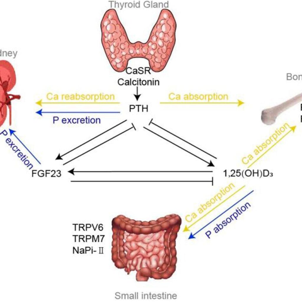
Parathyroid Gland & Calcium Metabolism Physiology: Calcium, Phosphate, and Bone Metabolism MINERAL METABOLISM INTRODUCTION TO CALCIUM METABOLISM Calcium (Ca²⁺) is the most abundant mineral in the human body, playing a pivotal role far beyond its primary association with bone health. It is an indispensable second messenger in virtually every cell, a key player in nerve impulse transmission, muscle contraction, and blood coagulation. Similarly, phosphate (PO₄³⁻) is a crucial component of bones, cell membranes (phospholipids), genetic material (DNA, RNA), and energy currency (ATP). The body maintains extremely tight control over the levels of these ions, particularly calcium, in the extracellular fluid (ECF) and plasma. Deviations, even slight ones, can have profound and immediate physiological consequences. This section will explore the regulation of calcium and phosphate, their distribution in the body, and the critical physiological roles they play. CALCIUM REGULATION IN ECF AND PLASMA The concentration of calcium ions in the extracellular fluid (ECF) and plasma is precisely and tightly regulated. It rarely deviates significantly from normal levels, highlighting its critical importance for life. Normal Value: The normal value of total calcium in the ECF is approximately 9.4 mg/dL (or 2.4 mEq/L). This represents a very small fraction, about 0.1%, of the total calcium in the body. Vital Physiological Processes: Calcium ions are absolutely vital to numerous physiological processes, including: Contraction of muscles: Essential for the excitation-contraction coupling in skeletal, cardiac, and smooth muscles. Blood clotting: A critical cofactor in several steps of the coagulation cascade, facilitating the formation of a stable blood clot. Transmission of nerve impulses: Involved in the release of neurotransmitters from presynaptic terminals and influencing neuronal excitability. Enzyme activation, hormone secretion, and cell signaling. Impact of Deviations: Any significant deviations from the normal ECF calcium levels have immediate and direct effects: Low Ca²⁺ (Hypocalcemia): Directly excites neuromuscular systems, leading to increased neuronal excitability, tetany, and muscle spasms. High Ca²⁺ (Hypercalcemia): Directly depresses neuromuscular and cardiac systems, leading to muscle weakness, lethargy, and cardiac arrhythmias. Distribution of Total Body Calcium: Approximately 99% of total body calcium is stored in the bones, serving as a large and readily available reservoir. About 1% of total calcium is found in cells, where it functions as a crucial intracellular messenger. The remaining very small fraction is in the ECF and plasma. CALCIUM IN PLASMA AND INTERSTITIAL FLUID In plasma and interstitial fluid, calcium exists in three distinct forms, contributing to the total calcium level: 41% Combined with Plasma Proteins: This fraction is primarily bound to albumin and, to a lesser extent, globulins. This protein-bound calcium is non-diffusible through capillary membranes and therefore not physiologically active in terms of directly influencing cell excitability. 9% Diffusible, Combined with Anionic Substances: This portion is bound to various anionic substances present in plasma and interstitial fluid, such as citrates and phosphates. This calcium is diffusible across capillary membranes but is not ionized, meaning it is not biologically active in the same way as free calcium ions. 50% Diffusible and Ionized (Free Ca²⁺): This is the most crucial form of calcium. It is diffusible across capillary membranes and, most importantly, exists as free calcium ions (Ca²⁺). This ionized calcium is the physiologically active form that participates in muscle contraction, nerve impulse transmission, blood clotting, and other vital cellular processes. Its normal level in plasma is approximately 1.2 mmol/L (or 2.4 mEq/L), which corresponds to roughly 4.7 mg/dL. The ionized calcium fraction is the one that is tightly regulated by hormones like parathyroid hormone (PTH), vitamin D, and calcitonin. PHOSPHATE REGULATION IN ECF AND PLASMA Phosphate is also a vital mineral, but its regulation in the ECF is generally less precise and less tightly controlled than calcium. Distribution of Total Body Phosphate: Approximately 85% of the body’s phosphate is found in bones, predominantly as hydroxyapatite crystals. 14-15% is located within cells, where it is integral to intracellular processes (e.g., ATP, DNA, RNA, phospholipids). Less than 1% is in the ECF, indicating its relatively minor extracellular presence compared to its intracellular and bone stores. ECF Concentration: The concentration of inorganic phosphate in the ECF is typically around 4 mg/dL. This level can vary slightly: Adults: Generally 3 to 4 mg/dL. Children: Tend to have slightly higher levels, typically 4 to 5 mg/dL, due to higher growth rates. Forms in ECF: Inorganic phosphate exists in the ECF in two primary forms: HPO₄²⁻ (Divalent Phosphate Ion): Approximately 1.05 mmol/L. H₂PO₄⁻ (Monovalent Phosphate Ion): Approximately 0.26 mmol/L. Relationship with pH: An increase in total ECF phosphate will generally increase the concentrations of both forms. A low pH (acidosis) increases the concentration of H₂PO₄⁻ and decreases HPO₄²⁻. A high pH (alkalosis) has the reverse effect, increasing HPO₄²⁻ and decreasing H₂PO₄⁻. Regulation: Although less tightly regulated than calcium, many of the same factors that regulate ECF calcium concentration (e.g., PTH, Vitamin D) also influence phosphate levels, mainly by affecting its renal excretion and intestinal absorption. NON-BONE EFFECTS OF ALTERED CA AND PHOSPHATE CONCENTRATIONS IN THE BODY FLUIDS The immediate physiological impact of altered calcium and phosphate levels differs significantly: Phosphate: Changing the level of phosphate in the ECF from far below normal to two to three times normal does not cause major immediate effects on the body. While chronic alterations can have serious consequences, acute changes are often well-tolerated because most phosphate is intracellular or in bone. Calcium: In stark contrast, even slight increases or decreases of ionized calcium in the ECF can cause extreme immediate physiological effects. This underscores the body’s meticulous regulatory mechanisms for calcium. Hypocalcemia: (Low ECF ionized calcium) leads to increased neuromuscular excitability, manifesting as tetany, muscle cramps, tingling, and potentially seizures. Hypercalcemia: (High ECF ionized calcium) leads to depressed neuromuscular activity, manifesting as muscle weakness, lethargy, constipation, confusion, and cardiac arrhythmias. Hence, clinical conditions are primarily discussed in terms of: Hypocalcemia vs. Hypercalcemia (which are acutely life-threatening due to effects on excitable tissues) Hypophosphatemia vs. Hyperphosphatemia (which tend to have more chronic and metabolic implications, rather than immediate severe effects on excitability). ALTERED CALCIUM LEVELS HYPOCALCEMIA Hypocalcemia occurs when
Endocrine System Physiology
Anatomy

Hypothalamus & Pituitary Physiology

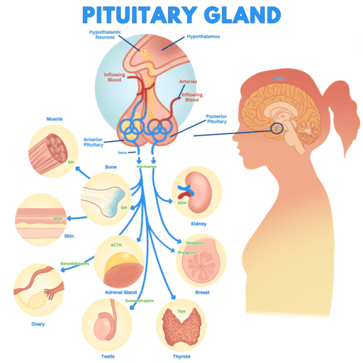
Hypothalamus & Pituitary Gland Physiology Endocrine Physiology: Hypothalamus & Pituitary Gland NEUROENDOCRINOLOGY INTRODUCTION TO THE HYPOTHALAMUS & PITUITARY GLAND The hypothalamus and pituitary gland form a crucial functional unit at the base of the brain, acting as the primary link between the nervous system and the endocrine system. Together, they regulate virtually all hormonal functions of the body, maintaining homeostasis, governing growth, metabolism, reproduction, and stress responses. The hypothalamus, a small but immensely powerful region of the diencephalon, serves as the command center, integrating neural signals from the brain and translating them into hormonal signals. The pituitary gland (also known as the hypophysis), often dubbed the “master gland,” receives these signals from the hypothalamus and, in turn, secretes hormones that control other endocrine glands throughout the body. FUNCTIONS OF THE HYPOTHALAMUS The hypothalamus is a highly specialized region of the brain responsible for maintaining various homeostatic functions and integrating responses to internal and external stimuli. Its diverse functions include: 1. Autonomic Nervous System Regulation The hypothalamus is a major control center for the autonomic nervous system (ANS), influencing both its sympathetic and parasympathetic divisions. It regulates involuntary functions such as heart rate, blood pressure, digestion, respiration, and pupil dilation, adapting the body’s internal environment to changing conditions. 2. Hormone Production The hypothalamus itself produces several hormones. These include: Releasing hormones and inhibiting hormones that control the secretion of hormones from the anterior pituitary. Antidiuretic hormone (ADH, or vasopressin) and oxytocin, which are synthesized in the hypothalamic nuclei and then transported to the posterior pituitary for storage and release. 3. Endocrine Regulation This is a primary function. Through its production of releasing and inhibiting hormones, the hypothalamus controls the secretion of nearly all anterior pituitary hormones, thereby indirectly regulating many other endocrine glands (e.g., thyroid, adrenal cortex, gonads). 4. Circadian Rhythm Regulation The suprachiasmatic nucleus (SCN) within the hypothalamus acts as the body’s primary biological clock, regulating circadian rhythms such as the sleep-wake cycle, body temperature, and hormone secretion patterns in response to light-dark cues. 5. Limbic System Interaction The hypothalamus is intimately connected with the limbic system, the part of the brain involved in emotion, motivation, and memory. This connection allows the hypothalamus to integrate emotional responses with physiological functions, influencing behaviors like feeding, aggression, and sexual drive. 6. Integration of Basic Drives It is involved in regulating fundamental physiological drives and behaviors, such as thirst, hunger, satiety, sexual behavior, and defensive reactions. 7. Temperature Regulation The hypothalamus contains specialized thermoreceptors and serves as the body’s thermostat. It initiates physiological responses (e.g., sweating, shivering, vasodilation/vasoconstriction) to maintain a stable core body temperature within a narrow range. 8. Feeding Specific nuclei within the hypothalamus (e.g., ventromedial nucleus for satiety, lateral hypothalamus for hunger) play critical roles in regulating food intake and energy balance. THE PITUITARY GLAND (HYPOPHYSIS, MASTER GLAND) The pituitary gland is a small, pea-sized endocrine gland, approximately 1 cm in diameter and weighing about 0.5 to 1 gram. It is strategically located within the sella turcica, a bony cavity at the base of the brain, protecting it from injury. The pituitary gland is functionally and anatomically connected to the hypothalamus by the pituitary stalk (or hypophysial stalk/infundibulum), a slender structure containing blood vessels and nerve fibers. Structurally and functionally, the pituitary gland is divided into two distinct lobes: Anterior Pituitary Lobe (Adenohypophysis): Constitutes about two-thirds of the gland. Posterior Pituitary Lobe (Neurohypophysis): Constitutes about one-third of the gland. A. THE ANTERIOR PITUITARY LOBE (ADENOHYPOPHYSIS) The anterior pituitary is an endocrine gland in its own right, synthesizing and secreting a variety of vital hormones. Pars Intermedia: In the fetus, there is a small, avascular tissue called the pars intermedia located between the anterior and posterior lobes. It is much more functional in some lower animals (fish, amphibians, reptiles) but is largely vestigial and no longer present as a distinct functional unit in adult humans, though some of its cells may be dispersed within the anterior lobe. Adult Structure: In adults, the anterior pituitary consists of two main parts: Pars Distalis: This is the rounded, major endocrine part of the gland, responsible for secreting most of the anterior pituitary hormones. This is what is commonly referred to as the “anterior pituitary.” Pars Tuberalis: A thin, upward extension that wraps around the infundibulum (pituitary stalk). Its precise function in humans is less understood but is believed to contribute to seasonal and circadian rhythms. The Anterior Pituitary Gland Cells: Histologically, the anterior pituitary contains various types of secretory cells, traditionally classified by their staining properties as chromophils (acidophils and basophils) or chromophobes. Each cell type is typically responsible for producing a specific hormone or hormones: Chromophils: Acidophils Somatotropes: Constitute approximately 30-40% of anterior pituitary cells and secrete human Growth Hormone (hGH). Lactotropes (or Mammotropes): Constitute approximately 3-5% of anterior pituitary cells and secrete Prolactin (PRL). Chromophils: Basophils Corticotropes: Constitute approximately 20% of anterior pituitary cells and secrete Adrenocorticotropic Hormone (ACTH). Thyrotropes: Constitute approximately 3-5% of anterior pituitary cells and secrete Thyroid-Stimulating Hormone (TSH). Gonadotropes: Constitute approximately 3-5% of anterior pituitary cells and secrete the gonadotropic hormones: Luteinizing Hormone (LH) and Follicle-Stimulating Hormone (FSH). Embryological Origin: The anterior pituitary is embryologically derived from Rathke’s pouch, an upward invagination of epithelial tissue from the roof of the primitive pharynx (mouth). This epithelial origin distinguishes it from the posterior pituitary, which has a neural origin. B. THE POSTERIOR PITUITARY LOBE (NEUROHYPOPHYSIS) The posterior pituitary is functionally an extension of the hypothalamus, serving as a storage and release site for hormones produced by hypothalamic neurons. Embryological Origin: It is embryologically derived from a downward outgrowth of nervous tissue from the hypothalamus. This neural origin explains its structural and functional connection to the brain. Structure: It is in direct contact with the infundibulum (pituitary stalk) and physically associated with the adenohypophysis. It consists mainly of the Pars Nervosa, which is essentially a collection of axons and nerve terminals originating from the hypothalamus, along with specialized glial cells called pituicytes. Neural Part: The posterior pituitary is distinctly the

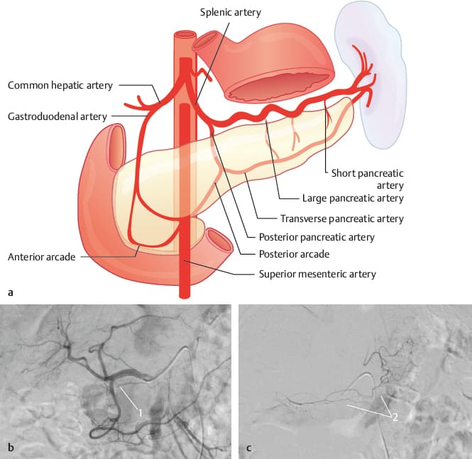
Blood Supply Pancreas 1
Anatomy

Pancreas Physiology

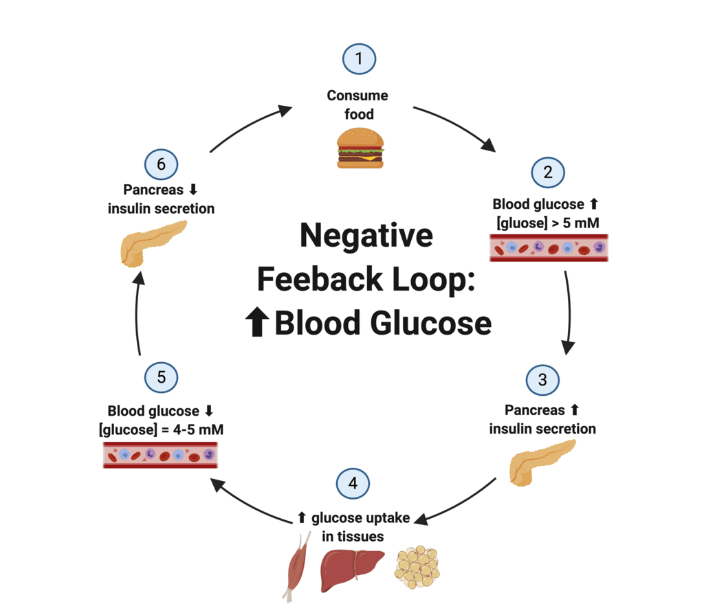
Pancreas Physiology Endocrine Physiology: The Pancreas ENDOCRINE PHYSIOLOGY THE PANCREAS The pancreas is an elongated, flattened gland located deep in the abdomen, nestled between the stomach and the spine. It is a unique organ because it serves two vital functions: Exocrine Function: The vast majority (about 98-99%) of the pancreas is dedicated to its exocrine role. It produces digestive enzymes (e.g., amylase, lipase, proteases) that are secreted into the duodenum via a system of ducts to aid in the digestion of carbohydrates, fats, and proteins in the small intestine. Endocrine Function: The remaining 1-2% of the pancreas is composed of specialized clusters of cells called the islets of Langerhans. These islets are hormone-producing factories that secrete critical hormones directly into the bloodstream to regulate blood glucose levels. I. ENDOCRINE FUNCTION OF THE PANCREAS: THE ISLETS OF LANGERHANS The endocrine pancreas is composed of approximately 1 to 2 million islets of Langerhans, which collectively constitute only about 1-2% of the total pancreatic mass. Despite their small proportion, these islets are vital for maintaining metabolic homeostasis, especially blood glucose regulation. Structure of the Islets: Each islet is a microscopic cluster of endocrine cells, typically 0.3 mm in diameter. They are highly vascularized and organized around small capillaries into which their cells directly secrete their hormones, allowing for rapid systemic distribution. The cells within each islet are in close proximity to each other, facilitating paracrine communication (local hormone action between neighboring cells). Major Cell Types within the Islets: The islets contain several distinct cell types, each producing specific hormones. These cells are distinguished by their morphological and staining characteristics. 1. Alpha (α) cells Constitute about 25% of the islet cells. Typically located at the periphery of the islets. Their primary secretion is glucagon, often referred to as the “fasting hormone.” 2. Beta (β) cells Are the most abundant, making up about 60% of the islet cells. Predominantly located in the center of the islets. Their primary secretions are insulin (the “feasting hormone”) and amylin. Amylin is co-secreted with insulin and helps regulate glucose homeostasis by slowing gastric emptying and promoting satiety. 3. Delta (δ) cells Comprise about 10% of the islet cells. Distributed around the periphery of the islets. They secrete somatostatin, a hormone that acts locally (paracrine effect) to inhibit the secretion of both insulin and glucagon, thus modulating their release. It also has systemic effects, inhibiting growth hormone and TSH from the pituitary. 4. PP cells (or Gamma (γ) cells) Present in small numbers (less than 1%). They secrete pancreatic polypeptide, whose exact physiological role is still not entirely clear but is thought to be involved in regulating pancreatic secretion and satiety. II. BLOOD GLUCOSE HOMEOSTASIS: The primary role of the endocrine pancreas is to maintain blood glucose homoeostasis (or homeokinesis), ensuring that blood glucose levels remain within a narrow, healthy range despite variations in food intake and energy expenditure. This delicate balance is achieved through the coordinated actions of insulin and glucagon. Outline of Glucose Regulation: The “Feasting” Hormone – Insulin: Primarily secreted after meals, when blood glucose is high, to promote glucose uptake and storage. The “Fasting” Hormone – Glucagon: Primarily secreted during fasting or between meals, when blood glucose is low, to raise blood glucose levels. Integrated Hormonal Control of Blood Glucose Concentration: The interplay between insulin, glucagon, and other hormones (like somatostatin, amylin, and gut hormones) orchestrates the precise regulation of glucose. Diabetes, Pathophysiological Changes: Disruptions in this hormonal control, particularly insulin function, lead to diabetes mellitus, a chronic metabolic disorder. III. INSULIN AND ITS METABOLIC EFFECTS Insulin is the most important anabolic hormone in the body, primarily responsible for lowering blood glucose levels after a meal and promoting energy storage. Structure and Synthesis: Insulin is a small protein hormone (peptide hormone) with a molecular weight of 5808. It consists of two peptide chains (A and B chains) linked together by two disulfide bonds. This specific structure is crucial for its biological activity. It is secreted by the beta (β) cells of the pancreatic islets via exocytosis, primarily in response to elevated blood glucose levels (a signal of “energy abundance”). Half-Life and Degradation: The “half-life” of insulin in circulation is remarkably short, about 6 minutes, meaning half of the secreted insulin is cleared from the bloodstream within this time. Consequently, it is cleared completely from the blood in approximately 10-15 minutes. This rapid turnover allows for precise and dynamic control of blood glucose levels. Insulin is primarily degraded by the enzyme insulinase, which is found abundantly in the liver and kidneys, and to a lesser extent in muscles and most other tissues. Role in Blood Glucose Maintenance: Insulin maintains blood glucose levels close to 5 mmol/L (90 mg/dL) under fasting conditions. After a meal, blood glucose levels may temporarily rise as high as 8 mmol/L (144 mg/dL), but insulin rapidly brings these levels back down to the normal range. Overweight and obese individuals often exhibit higher basal levels of insulin (hyperinsulinemia) as a compensatory mechanism to overcome insulin resistance, where their cells do not respond as effectively to insulin. IV. SYNTHESIS OF INSULIN: Insulin synthesis is a classic example of protein synthesis for secretion, involving several steps within the β-cells: Preproinsulin Formation: Insulin RNA, transcribed from the insulin gene, is translated by ribosomes attached to the endoplasmic reticulum (ER). The initial product is a larger precursor molecule called preproinsulin (molecular weight = 11,500). This molecule contains a signal peptide that directs it into the ER lumen. Proinsulin Formation: Inside the ER, the signal peptide is cleaved off, converting preproinsulin into proinsulin (molecular weight = 9,000). Proinsulin consists of the A and B chains linked by a C-peptide. Insulin and C-peptide Cleavage: Proinsulin then travels to the Golgi apparatus. Here, specific enzymes (prohormone convertases) cleave the proinsulin molecule, removing the C-peptide and leaving behind the mature, active insulin molecule (A and B chains linked by disulfide bonds) and the peptide fragments (C-peptide). Packaging and Secretion: Both insulin and C-peptide are then packaged together into secretory granules. These

Adrenal Gland Physiology
Anatomy

Adrenal Gland Physiology

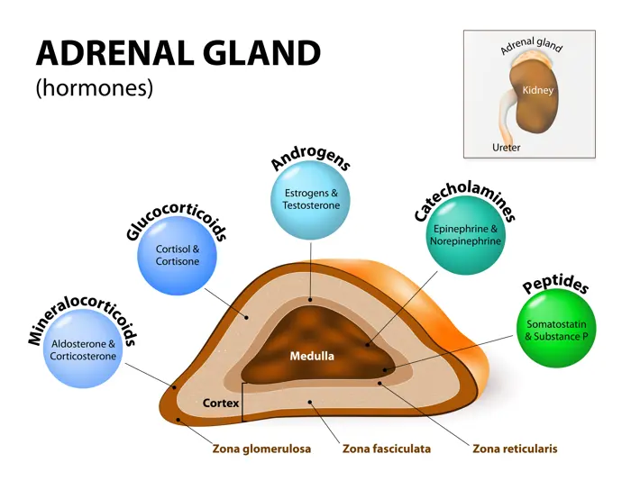
Adrenal Gland Physiology Endocrine Physiology: The Adrenal Gland ENDOCRINE PHYSIOLOGY ADRENAL GLAND FUNCTIONS AND REGULATION The adrenal glands, also known as suprarenal glands, are a pair of small, triangular-shaped endocrine glands located on top of each kidney (ad-renal, meaning “near the kidney”). Despite their small size, these glands are absolutely vital for life, playing a central role in managing stress, regulating metabolism, blood pressure, fluid balance, and even influencing immune function and sexual development. “The Stress Glands”: Often referred to as the “stress glands,” the adrenals orchestrate the body’s response to both acute (e.g., immediate danger) and chronic (e.g., prolonged illness, psychological strain) stressors through the rapid release of powerful hormones. Their proper functioning is critical for maintaining homeostasis, and dysfunction can lead to a wide array of serious health problems. The adrenal gland is not a single, homogenous organ but rather a composite of two distinct endocrine glands, each with unique origins, structures, and hormonal secretions. These two compartments are: The Adrenal Cortex (Outer Layer) The Adrenal Medulla (Inner Layer) These two regions, though anatomically juxtaposed, function almost as separate organs, producing different classes of hormones that contribute synergistically to the body’s complex physiological responses. I. THE ADRENAL GLAND: TWO DISTINCT COMPARTMENTS A. The Adrenal Cortex (Outer Layer) The adrenal cortex is the outer, yellowish layer of the adrenal gland, accounting for approximately 80-90% of the gland’s total mass. It is derived from embryonic mesoderm and is responsible for synthesizing and secreting a class of steroid hormones known as corticosteroids. These hormones are all synthesized from cholesterol and are lipid-soluble, allowing them to easily pass through cell membranes to exert their effects. The adrenal cortex is further subdivided into three distinct layers or zones, each characterized by its unique cellular structure, enzymatic machinery, and primary hormonal products. These layers are arranged concentrically, starting from the outermost layer just beneath the capsule, and moving inward towards the medulla: 1. Zona Glomerulosa (15% of the Cortex) Location: Outermost layer, directly beneath the adrenal capsule. Cells arranged in small, spherical or arched clusters (glomeruli). Primary Hormones: Sole site of mineralocorticoid synthesis, with aldosterone being the most important. Function of Aldosterone: Regulates mineral balance (sodium and potassium). Main actions on kidneys: Sodium (Na+) reabsorption: Leads to water retention, maintaining blood volume/pressure. Potassium (K+) excretion: Essential for maintaining normal potassium levels. Hydrogen (H+) excretion: Contributing to acid-base balance. Regulation: Primarily regulated by the Renin-Angiotensin-Aldosterone System (RAAS) and plasma potassium concentration. ACTH has a permissive role. 2. Zona Fasciculata (75% of the Cortex) Location: Middle and largest layer. Cells arranged in long, straight cords or fascicles. Cells are often called “spongiocytes” due to vacuolated appearance from abundant lipid droplets (cholesterol esters). Primary Hormones: Primary site of glucocorticoid synthesis, with cortisol (hydrocortisone) being the most important. Small amounts of adrenal androgens are also produced here. Function of Cortisol: Crucial “stress hormone”. Glucose Metabolism: Promotes gluconeogenesis; decreases peripheral glucose utilization. Protein Metabolism: Increases protein catabolism (breakdown). Fat Metabolism: Promotes lipolysis. Anti-inflammatory/Immunosuppressive: Suppresses immune responses. Regulation: Primarily regulated by ACTH (from anterior pituitary), which is regulated by CRH (from hypothalamus). Forms the HPA axis. 3. Zona Reticularis (10% of the Cortex) Location: Innermost layer, adjacent to the medulla. Cells arranged in an irregular, anastomosing network (reticular). Primary Hormones: Primary site of adrenal androgen synthesis. Includes dehydroepiandrosterone (DHEA) and androstenedione (weak androgens). Some glucocorticoids produced here. Function of Androgens: Contribute to secondary sexual characteristics (pubic/axillary hair, libido). In males, masked by testicular androgens. In females, DHEA is a significant estrogen precursor. Regulation: Primarily regulated by ACTH. B. The Adrenal Medulla (Inner Layer) The adrenal medulla is the central, reddish-brown core of the adrenal gland, completely surrounded by the adrenal cortex. It comprises about 10-20% of the gland’s total mass. Nature: Embryologically derived from neuroectoderm (neural crest cells), making it essentially a specialized sympathetic ganglion. Cells are called chromaffin cells (modified postganglionic sympathetic neurons). Innervation: Directly innervated by preganglionic sympathetic fibers (cholinergic). Allows rapid release of hormones in response to acute stress. Primary Hormones: Synthesizes and secretes catecholamines: Epinephrine (Adrenaline): ~80% of secretion. Norepinephrine (Noradrenaline): ~20% of secretion. Dopamine: Smaller amounts. Function: Central to the “fight-or-flight” response. Cardiovascular: Increased heart rate, contraction force, and blood pressure. Metabolic: Increased blood glucose (glycogenolysis/gluconeogenesis), lipolysis. Respiratory: Bronchodilation. Blood Flow: Shunting blood to muscles and brain. Regulation: Regulated by direct neural stimulation from the sympathetic nervous system in response to stress (pain, fear, hypoglycemia, etc.). III. GLUCOCORTICOIDS: CORTISOL AND ITS ACTIONS Glucocorticoids, with cortisol being the prime example, are powerful steroid hormones essential for life. Produced by the zona fasciculata and zona reticularis. Regulation of Cortisol Secretion (HPA Axis): CRH (Corticotropin-Releasing Hormone): Secreted by the hypothalamus. Major regulator of ACTH secretion. ACTH (Adrenocorticotropic Hormone): Released from anterior pituitary. Stimulates cortisol synthesis/secretion. ADH (Vasopressin): A potent ACTH secretagogue, especially during stress. Pulsatile Secretion: Leads to diurnal (24-hour) variations. Peak activity is in the early morning (6-8 AM), diminishing to a nadir around midnight. Negative Feedback: Cortisol exerts negative feedback on both the hypothalamus (inhibiting CRH) and anterior pituitary (inhibiting ACTH). Actions of Glucocorticoids (Cortisol): 1. Metabolic Response to Fasting (Anti-insulin Effects) Cortisol promotes processes ensuring glucose availability during stress or fasting. Gluconeogenesis: Increases enzymes in the liver; mobilizes amino acids from muscle. Mobilization of Stored Fat: Promotes lipolysis, releasing free fatty acids (FFA) for energy and glycerol for gluconeogenesis. Diagrammatic Representation of Metabolic Effects: Muscle Cell: Promotes proteolysis (protein breakdown) releasing amino acids. Reduces protein synthesis. Fat Cell: Promotes lipolysis (releasing FFA and glycerol). Can promote lipogenesis in central areas. Liver Cell: Takes up amino acids/glycerol. Stimulates gluconeogenesis and glycogen synthesis. Overall Effect: Increase in circulating glucose (hyperglycemia) and free fatty acids (FFA). 2. Anti-inflammatory and Immunosuppressive Effects Cortisol suppresses immune responses, inhibits pro-inflammatory cytokines, stabilizes lysosomal membranes, and decreases capillary permeability. Synthetic glucocorticoids are widely used as anti-inflammatory medications. 3. Other Important Actions Cardiovascular: Increases cardiac output; enhances vasoconstrictive effects of catecholamines (permissive action). Bone Metabolism: Chronic high levels inhibit bone formation, accelerate resorption (osteoporosis). Connective Tissue: Decreases collagen synthesis. CNS: Affects mood/behavior.

Thyroid Physiology
Anatomy

Thyroid Physiology

Thyroid Gland Physiology Endocrine Physiology: The Thyroid Gland ENDOCRINE PHYSIOLOGY THE THYROID GLAND The endocrine system is a complex network of ductless glands that play a crucial role in maintaining homeostasis. Unlike exocrine glands that secrete substances through ducts, endocrine glands release their chemical messengers, known as hormones, directly into the surrounding extracellular fluid. These hormones then pass into the bloodstream for general circulation, traveling throughout the body to reach distant target organs or tissues. Upon arrival, hormones excite or inhibit the activity of these target cells, coordinating a vast array of physiological processes, including growth, metabolism, reproduction, and mood. The thyroid gland is a vital component of this system, holding the distinction of being one of the largest endocrine glands in the human body. Its primary function is to regulate metabolism, energy production, and body temperature. II. HORMONES SECRETED BY THE THYROID GLAND The thyroid gland produces two main types of hormones: 1. Thyroid Hormones (from Follicular Cells) Thyroxine (T4): Also known as tetraiodothyronine. This is the more abundant hormone secreted by the thyroid. While less biologically active at the cellular level compared to T3, it serves as a prohormone and a major reservoir for T3. Triiodothyronine (T3): This is the more potent and biologically active form of thyroid hormone. Most of the T3 in the body is generated from the deiodination (removal of an iodine atom) of T4 in peripheral tissues, rather than directly secreted by the thyroid gland. Origin: Both T4 and T3 are synthesized and secreted by the follicular cells of the thyroid gland. 2. Calcitonin (from Parafollicular Cells) Calcitonin: This hormone plays a role in calcium homeostasis. Origin: It is secreted by specialized cells located interspersed between the thyroid follicles, known as parafollicular cells (also called C cells). Calcitonin’s primary function is to lower blood calcium levels, acting in opposition to parathyroid hormone. III. FUNCTIONAL ANATOMY OF THE THYROID GLAND Understanding the structure of the thyroid gland is key to comprehending its function. Location: The thyroid gland is strategically located in the neck, immediately below the larynx (voice box) and anterior (in front of) to the upper part of the trachea (windpipe). Its superficial position makes it palpable during physical examination. Weight: In a healthy adult, it typically weighs about 15-20 grams. Its size can fluctuate with age, sex, and physiological states (e.g., pregnancy). Gross Structure: The gland consists of two distinct, fleshy lateral lobes, one on each side of the trachea. These lobes are connected by a narrow band of thyroid tissue called the isthmus. The isthmus usually overlies the region from the 2nd to the 4th tracheal cartilage, anchoring the gland in place. Sometimes, a third lobe, the pyramidal lobe, may extend upwards from the isthmus. IV. PHYSIOLOGICAL ANATOMY: THE FUNCTIONAL UNIT The microscopic structure of the thyroid gland reveals its unique functional organization: Thyroid Follicles: The lobes of the thyroid gland are composed of numerous, tiny, hollow, spherical structures called follicles. These follicles are the functional units of the thyroid gland. Each follicle is lined by a single layer of cuboidal epithelial cells – the follicular cells. Colloid: Each follicle is filled with a thick, sticky, protein-rich substance known as colloid. The major constituent of colloid is a large glycoprotein called thyroglobulin (Tg). This thyroglobulin serves as the scaffold for thyroid hormone synthesis and, crucially, as the storage site for thyroid hormones. Storage Uniqueness: Unlike most other endocrine glands, which store minimal amounts of pre-formed hormones and secrete them rapidly upon synthesis, the thyroid gland has a remarkable capacity to store considerable amounts of thyroid hormones within the colloid (bound within thyroglobulin). This stored reserve can last for several weeks or even months, providing a buffer against fluctuations in iodine intake or acute demand. C Cells (Parafollicular Cells): Interspersed between the follicles are the C cells (or parafollicular cells), which, as mentioned, are responsible for secreting calcitonin. V. SYNTHESIS, STORAGE, AND SECRETION OF THYROID HORMONES The production of thyroid hormones is a multi-step process that requires specific raw materials and intricate cellular machinery. Essential Precursors: Tyrosine: An amino acid that forms the backbone of thyroid hormones. It is synthesized by the body and is a crucial component of the thyroglobulin protein. Iodine: A trace element that is dietary essential. The body cannot synthesize iodine; it must be consumed through diet (e.g., iodized salt, seafood). Iodine is absolutely critical for thyroid hormone synthesis, as it is incorporated directly into the tyrosine residues of thyroglobulin. Location of Synthesis: Hormone synthesis primarily occurs on the thyroglobulin molecule within the colloid. Steps Involved in Synthesis, Storage, and Secretion: 1. Thyroglobulin Production and Release (Into Colloid) Process: Follicular cells synthesize the large glycoprotein thyroglobulin (Tg) in their endoplasmic reticulum and Golgi apparatus. Delivery: Tg is then packaged into vesicles and released into the follicular lumen (colloid space) by exocytosis. 2. Iodine Uptake (Iodide Trapping) Process: Follicular cells actively transport iodide (I-) from the bloodstream into their cytoplasm. This is an energy-dependent process mediated by the Sodium-Iodide Symporter (NIS), which co-transports two Na+ ions with one I- ion. Concentration: This mechanism allows the thyroid gland to concentrate iodide to levels 20-40 times higher than in the blood (and even higher under TSH stimulation). Transfer to Colloid: Iodide is then transported from the follicular cell cytoplasm into the colloid space via a chloride-iodide counter-transporter called pendrin. 3. Oxidation and Iodination of Tyrosine (Organification) Process: Once in the colloid, iodide is rapidly oxidized to iodine (I0 or I+) by the enzyme thyroid peroxidase (TPO). Attachment to Tyrosine: This highly reactive iodine then attaches to specific tyrosine residues within the thyroglobulin molecule. One iodine atom attached to a tyrosine forms Monoiodotyrosine (MIT). Two iodine atoms attached to a tyrosine form Diiodotyrosine (DIT). 4. Coupling Processes (Formation of T4 and T3) Process: Still within the thyroglobulin molecule, two iodinated tyrosine molecules are coupled together by TPO: DIT + DIT → T4 (Thyroxine): This is the primary coupling reaction, forming the most abundant thyroid hormone. MIT + DIT → T3 (Triiodothyronine): This

Hormone Regulation, Disorders & Transport
Anatomy

Hormone Regulation, Disorders & Transport

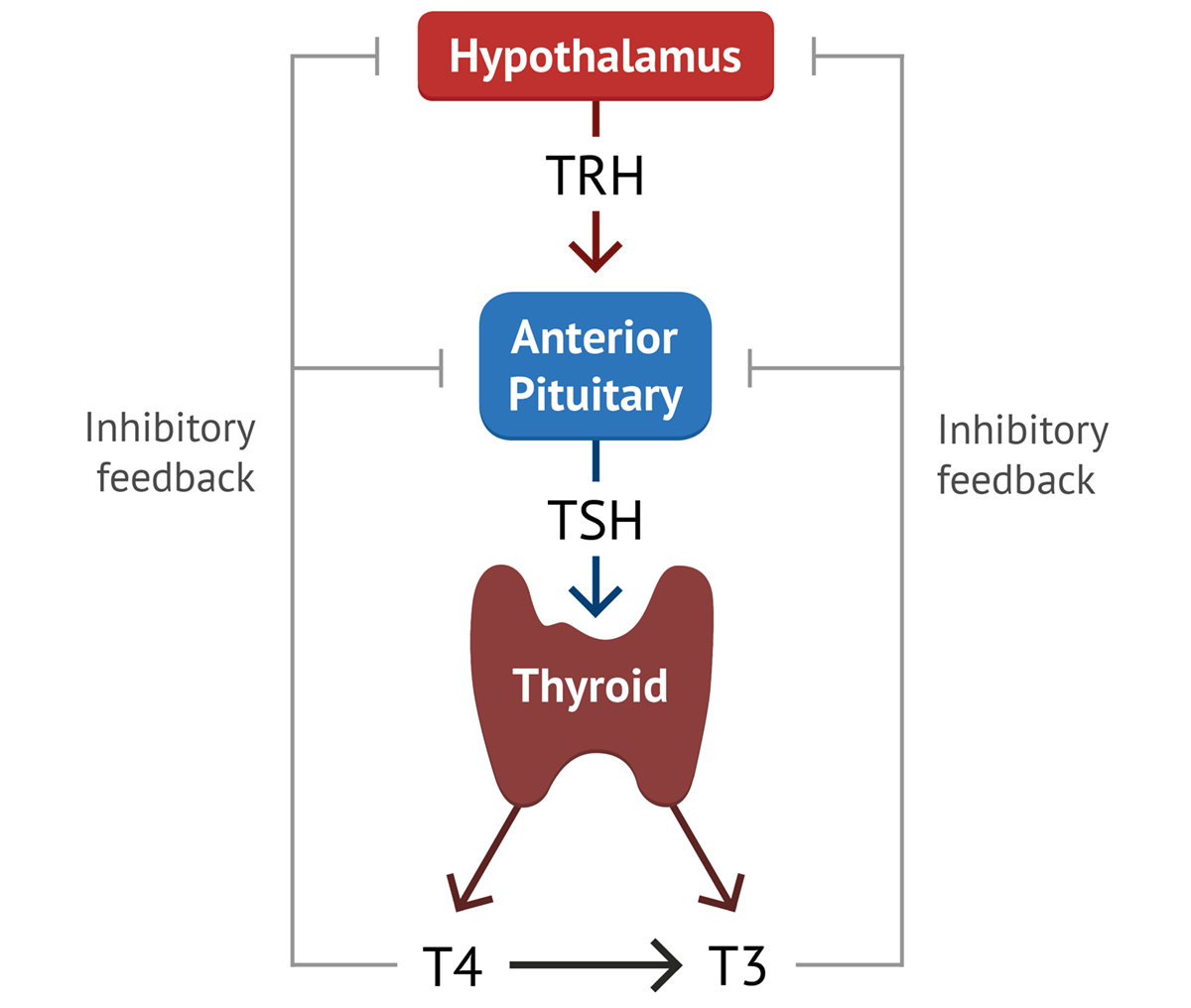
Endocrine System Physiology Reference: Regulation of Hormone Secretion ENDOCRINOLOGY Regulation of Hormone Secretion The endocrine system meticulously controls hormone secretion to ensure physiological homeostasis and appropriate responses to environmental cues. This regulation involves complex feedback loops, neural reflexes, and intrinsic biological rhythms. I. Characteristics of Hormone Secretion and Action Before delving into the control mechanisms, it’s important to understand some general characteristics of hormone dynamics: Variable Latency and Duration of Action Rapid-Acting Hormones: Some hormones, typically those involved in immediate stress responses or neural signaling, are secreted within seconds after stimulation. Examples include epinephrine and norepinephrine from the adrenal medulla. Their full physiological actions can develop within another few seconds to minutes (e.g., increased heart rate, blood pressure, glucose mobilization). Slower-Acting Hormones: Others, like thyroxine (thyroid hormone) or growth hormone, may take hours, days, or even months to exert their full physiological effects. Thyroxine, for instance, influences metabolic rate over an extended period, and its full impact on growth and development is gradual. Growth hormone’s effects on skeletal growth accumulate over years. Extremely Low Serum Concentrations Hormones are incredibly potent, meaning they can exert significant effects even at very low concentrations. Their serum concentrations are typically measured in picograms to micrograms per 100 ml of blood. Consequently, they are secreted in equally small amounts, often in micrograms or milligrams per day. This underscores the efficiency and sensitivity of the endocrine signaling system, where a tiny amount of chemical messenger can orchestrate widespread physiological changes. II. Mechanisms of Control of Hormonal Secretion Hormone secretion is not a constant process but is dynamically regulated by several intricate mechanisms to maintain precise control. 1. Negative-Feedback Control This is the most common and fundamental regulatory mechanism in the endocrine system, ensuring stability and preventing overactivity. Definition: Negative feedback exists when the output of a system counteracts a change in input, thereby maintaining a controlled variable within a narrow, desired range (around a “set point”). Mechanism: A stimulus (e.g., deviation from a set point) triggers an endocrine gland to release a hormone. This hormone then acts on target cells to produce a specific physiological response. Crucially, the physiological response itself, or the hormone’s direct effect on the gland, then inhibits the further release of the initial hormone. This “turns off” the stimulatory signal once the desired level or effect is achieved. Example: Thyroid Hormone Regulation (HPT Axis) Initial Stimulus: If the plasma concentration of free circulating thyroid hormone (T₃ and T₄) falls below a given set point. Hypothalamic Response: The hypothalamus senses this drop and secretes Thyrotropin-Releasing Hormone (TRH). Anterior Pituitary Response: TRH stimulates the anterior pituitary to secrete Thyroid-Stimulating Hormone (TSH). Thyroid Gland Response: TSH stimulates the thyroid gland to increase its secretion of thyroid hormones (T₃ and T₄). Negative Feedback Loop: As the levels of T₃ and T₄ in the blood rise back to the set point (or above), they exert negative feedback at two levels: On the Anterior Pituitary: T₃ and T₄ directly inhibit the secretion of TSH. On the Hypothalamus: T₃ and T₄ also inhibit the secretion of TRH. Outcome: This inhibition ensures that once thyroid gland secretion has been “turned on” by TSH, it will not continue unabated. Instead, it will be “turned off” when the appropriate level of free circulating thyroid hormone has been achieved, thus preventing excessive thyroid hormone levels. Significance: Prevents Overactivity: Negative feedback is critical for preventing the overproduction or underproduction of hormones, maintaining stable internal conditions (homeostasis). Maintains Set Point: It ensures that hormone levels and their physiological effects remain within a relatively narrow, healthy range. 2. Positive-Feedback Mechanisms While less common for maintaining ongoing homeostasis, positive feedback plays a vital role in specific, self-amplifying processes. Definition: Positive feedback mechanisms control events that are often “out of control” or self-amplifying, and do not require continuous adjustment to a set point; instead, they push the system further in the same direction. Nature: Rarely used to maintain homeostasis because they tend to drive physiological variables away from a stable state. Mechanism: A stimulus triggers an endocrine response. This response then amplifies the original stimulus, leading to an even greater response. This cycle continues until a specific event or external factor breaks the loop. Example: Childbirth Initial Stimulus: Pressure of the baby’s head against the cervix during labor. Neural Signal: Nerve impulses from the cervix are sent to the hypothalamus. Hormone Release: The hypothalamus stimulates the posterior pituitary to release oxytocin. Target Response: Oxytocin stimulates and enhances the contractions of the uterine smooth muscle. Positive Feedback Loop: The stronger uterine contractions push the baby further down, increasing the pressure on the cervix. This increased cervical stretching sends more nerve impulses to the hypothalamus, leading to the release of more oxytocin, which in turn causes even stronger contractions. Outcome: This intensifies contractions until the baby is delivered from the birth canal. Once the baby is born, the cervical stretching stops, the oxytocin production significantly decreases, and the labor contractions cease, thus breaking the positive feedback loop. 3. Neuroendocrine Reflexes These mechanisms highlight the intimate connection between the nervous and endocrine systems, often allowing for rapid, precise hormonal responses to external stimuli. Definition: Many endocrine control systems involve neuroendocrine reflexes, which include both neural and hormonal components. They typically involve nerve impulses triggering hormone release. Mechanism: Neural input to an endocrine gland directly stimulates or inhibits hormone secretion. This often produces a sudden increase in hormone secretion (like “turning up the thermostat setting”) in response to a specific stimulus, which is frequently an external one. Pure Neural Control Example: Adrenal Medulla Secretion Mechanism: In some instances, neural input to the endocrine gland is the only factor regulating the secretion of a hormone. Example: The secretion of epinephrine and norepinephrine by the adrenal medulla is solely controlled by the sympathetic nervous system. When faced with a perceived threat (fight-or-flight response), sympathetic preganglionic neurons directly stimulate chromaffin cells in the adrenal medulla to release these catecholamines into the bloodstream. There is no hormonal intermediate from a higher endocrine gland. Combined Feedback

Endocrine System Physiology
Anatomy

Endocrine System Physiology

Endocrine System Physiology Reference: Endocrine System ENDOCRINOLOGY Introduction to the Endocrine System The endocrine system is a crucial communication network within the body, working in conjunction with the nervous system to maintain homeostasis and regulate various physiological processes. Unlike the nervous system, which primarily uses electrical signals for rapid, short-duration responses, the endocrine system employs chemical messengers called hormones that travel through the bloodstream, eliciting slower, more sustained responses. Overall Functions of the Endocrine System The endocrine system is responsible for a wide array of vital functions, ensuring the body’s proper growth, development, and adaptation. Its key roles include: Regulating Nutrient Metabolism and Water & Electrolyte Balance: This is fundamental for maintaining a constant internal environment (homeostasis). Hormones control how the body processes carbohydrates, fats, and proteins for energy, storage, or conversion. They also play a critical role in balancing water and crucial ions like sodium, potassium, and calcium, essential for cell function and overall fluid balance. Inducing Adaptive Changes to Help the Body Cope with Stressful Situations: The endocrine system orchestrates the body’s “fight or flight” response and longer-term stress adaptation. Hormones like cortisol and adrenaline prepare the body for perceived threats by mobilizing energy stores and altering physiological functions. Promoting Smooth, Sequential Growth and Development: From fetal development through childhood and adolescence, hormones meticulously control the growth of tissues, organs, and the entire body. They also dictate the timing and progression of puberty and the maturation of reproductive organs. Controlling Reproduction: Hormones are central to all aspects of reproduction, including the development of sex characteristics, the regulation of menstrual cycles in females, sperm production in males, pregnancy, childbirth, and lactation. Regulating Red Blood Cell Production: Erythropoietin, a hormone primarily produced by the kidneys, stimulates the bone marrow to produce red blood cells, which are vital for oxygen transport. Controlling and Integrating Activities of the Circulatory and Digestive Systems (along with the Autonomic Nervous System): Hormones influence heart rate, blood pressure, blood vessel tone, and blood volume (circulatory system). In the digestive system, they regulate appetite, enzyme secretion, motility, and nutrient absorption. This integration ensures that these systems function efficiently and are coordinated with the body’s overall needs. Definitions Hormone: A hormone is a chemical messenger synthesized by specialized cells, typically within endocrine glands. These messengers are transported by the blood to target organs or tissues (their “site of action”), where they bind to specific receptors and cause an alteration in the function of the cell. An example is Growth Hormone, which promotes overall body growth. Endocrine Glands: Endocrine glands are specialized organs or tissues that synthesize and release classical hormones directly into the blood. They are often referred to as ductless glands because, unlike exocrine glands (which release secretions through ducts), their hormones are secreted directly into the bloodstream without the need for ducts. Examples include the thyroid gland, adrenal glands, pituitary gland, and pancreas (for its endocrine function). Tropic Hormones Definition: Tropic hormones are a special class of hormones that have as their primary function the regulation of hormone secretion by another endocrine gland. The term “tropic” means “nourishing,” which reflects their role in stimulating and maintaining their endocrine target tissues. Function: Tropic hormones not only stimulate the target gland to produce and secrete its own hormones but also maintain the structural integrity (trophic effect) of that gland. Without their tropic hormone, target glands can atrophy (shrink) and become underactive. Example: Thyroid-Stimulating Hormone (TSH): TSH is a tropic hormone secreted by the anterior pituitary gland. It stimulates the thyroid gland to secrete thyroid hormones (T3 and T4). Additionally, TSH maintains the normal size and health of the thyroid gland. In the absence of TSH, the thyroid gland would atrophy (shrink) and produce very low levels of its hormones, leading to hypothyroidism. Complexity of Endocrine Function The endocrine system, while seemingly straightforward in its definition, exhibits remarkable complexity in its operations: A Single Endocrine Gland May Produce Multiple Hormones: Many glands are multi-functional. For instance, the anterior pituitary gland is a master gland that secretes at least six different hormones, each with distinct target tissues and functions (e.g., TSH, Growth Hormone, ACTH, FSH, LH, Prolactin). A Single Hormone May Be Secreted by More Than One Endocrine Gland: Some hormones are produced by different endocrine sources, sometimes serving different roles depending on their origin. For example, somatostatin is secreted by both the hypothalamus (where it inhibits Growth Hormone release) and the pancreas (where it acts as a paracrine to inhibit insulin and glucagon secretion). In the stomach, somatostatin also acts as a paracrine to inhibit gastric acid secretion. A Single Hormone May Have More Than One Type of Target Cell and Induce Multiple Effects: Hormones often exert diverse effects by binding to different subtypes of receptors on various target cells. Example: Vasopressin (Antidiuretic Hormone – ADH): Water Reabsorption: Vasopressin promotes water reabsorption by the kidney tubules by binding with V2 receptors on the distal and collecting tubular cells. This helps conserve body water. Vasoconstriction: It also causes vasoconstriction of arterioles throughout the body by binding with V1 receptors on arteriolar smooth muscle. This helps increase blood pressure. Hormones with Multiple Target-Cell Types Can Coordinate Activities Toward a Common End: Sometimes, a single hormone’s actions on different tissues converge to achieve a unified physiological goal. Example: Insulin: After a meal, insulin acts on muscle cells, liver cells, and fat cells. In muscle and fat, it promotes glucose uptake and storage. In the liver, it promotes glucose conversion to glycogen and fat synthesis. All these actions work in concert to store nutrients after absorption of a meal, thus lowering blood glucose. The Rate of Secretion of Some Hormones Varies Considerably in a Cyclic Pattern (Temporal Coordination): Endocrine systems often provide temporal (time) coordination of function, with hormone levels fluctuating in predictable cycles. This is particularly evident in: Reproductive cycles: Such as the menstrual cycle, where highly specific patterns of hormone secretion (e.g., estrogens, progesterone, LH, FSH) are required for normal ovarian and uterine function. Circadian rhythms: Many hormones follow a

Transport Across Nephron Segments 1
Anatomy

Urinary System

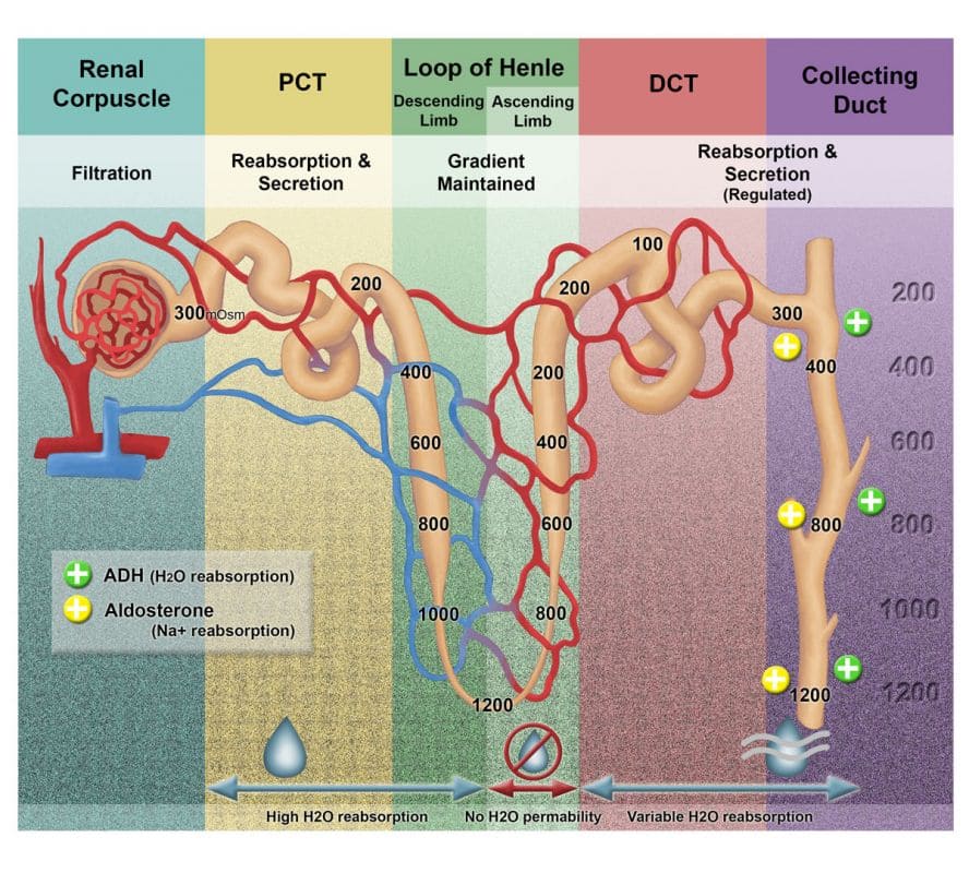
Urinary System Anatomy: The Urinary System RENAL ANATOMY The Urinary System The urinary system, also known as the renal system, is a vital organ system responsible for filtering waste products from the blood, maintaining fluid and electrolyte balance, regulating blood pressure, and producing hormones. It is composed of the following main organs: Kidneys: The primary organs of filtration and urine formation. Ureters: Tubes that transport urine from the kidneys to the urinary bladder. Urinary Bladder: A muscular sac that stores urine. Urethra: A tube that excretes urine from the bladder to the outside of the body. Main Functions of the Urinary System Maintenance of Water and Electrolyte Balance: Regulates the body’s total water volume and the concentration of various electrolytes (e.g., sodium, potassium, calcium, phosphate). Regulation of Osmotic Pressure: By controlling water and solute excretion, the kidneys help maintain the appropriate osmolarity of body fluids. Excretion of Metabolic Wastes: Removes nitrogenous wastes such as urea (from protein metabolism), creatinine (from muscle metabolism), and uric acid (from nucleic acid metabolism). Also excretes drugs and toxins. Regulation of Blood pH: By adjusting the excretion of hydrogen ions (H+) and bicarbonate ions (HCO3-), the kidneys play a crucial role in maintaining acid-base balance. Regulation of Blood Pressure: Produces the enzyme renin, which initiates the renin-angiotensin-aldosterone system (RAAS), a key mechanism for regulating blood pressure. Production of Erythropoietin: Secretes the hormone erythropoietin (EPO), which stimulates red blood cell production in the bone marrow. Activation of Vitamin D: Converts inactive vitamin D to its active form (calcitriol), which is essential for calcium absorption and bone health. Gluconeogenesis: During prolonged fasting, the kidneys can synthesize glucose from non-carbohydrate precursors. Parts of the Urinary System: Upper and Lower Tracts For anatomical and clinical convenience, the urinary system is often divided into two main parts: 1. Upper Urinary Tract Location: Primarily located in the abdominal cavity. Components: Two Kidneys Upper two-thirds of the Ureters 2. Lower Urinary Tract Location: Primarily located in the pelvis and perineum. Components: Lower one-third of the Ureters Urinary Bladder Urethra Upper Urinary Tract Details Kidneys Description: Two reddish-brown, bean-shaped organs, each about the size of a fist. They are the primary blood-filtering organs of the urinary system. Location: Located on the posterior abdominal wall, one on each side of the vertebral column, retroperitoneally (behind the peritoneum). Specific Positioning: Typically extend from the level of the T12 vertebra to the L3 vertebra. The right kidney is usually positioned slightly lower than the left kidney (approximately half an inch) due to the large size and position of the liver on the right side of the abdominal cavity. Mobility: Kidneys are not rigidly fixed. They exhibit some mobility and move vertically with respiration (descending during inspiration and ascending during expiration) and with changes in body position. Hilum: The medial concave border of each kidney features a vertical cleft called the renal hilum. This is the entry and exit point for the renal artery and vein, nerves, and the renal pelvis (which narrows to become the ureter). The hilum is typically located around the level of the L1 vertebra. Function: Urine Formation: Filters blood to produce urine, removing metabolic wastes and excess water/electrolytes. Erythropoietin Production: Produces the hormone erythropoietin, essential for red blood cell synthesis. Renin Production: Produces renin, vital for blood pressure regulation. Vitamin D Activation: Activates vitamin D. Coverings of the Kidney (from innermost to outermost) The kidneys are protected and held in place by four layers of tissue: Renal Capsule (Fibrous Capsule): Description: The innermost layer. A thin, tough, transparent, fibrous connective tissue capsule that directly surrounds the kidney parenchyma. Attachment: It is closely and firmly attached to the kidney surface. Function: Provides a barrier against infection and trauma. Perirenal Fat (Adipose Capsule): Description: A layer of adipose tissue (fat) that surrounds the renal capsule. Function: Cushions the kidney against physical trauma and helps hold it in position. Its presence provides insulation. Renal Fascia (Gerota’s Fascia): Description: A condensation of connective tissue that encloses both the kidney and the adrenal gland (which sits atop the kidney). It has an anterior and a posterior layer. Function: Anchors the kidney to the posterior abdominal wall and surrounding structures, helping to maintain its position. It also separates the kidney from the surrounding pararenal fat. Pararenal Fat (Paranephric Fat): Description: The outermost layer, consisting of a variable amount of fat located external to the renal fascia. It is part of the broader retroperitoneal fat. Function: Provides additional cushioning and support for the kidney. Internal Structure of Kidneys When sectioned, the kidney reveals two distinct regions: an outer cortex and an inner medulla. Renal Cortex Description: The outer, lighter reddish-brown region of the kidney. It is granular in appearance. Extension into Medulla: The cortex extends inwards, forming structures called renal columns (of Bertin). These columns separate the renal pyramids within the medulla. Contents: Contains the renal corpuscles (glomeruli and Bowman’s capsules), convoluted tubules (proximal and distal), and cortical collecting ducts. It’s the primary site of blood filtration. Renal Medulla Description: The inner, darker reddish-brown region, appearing striated. Renal Pyramids: The medulla is organized into 8-18 cone-shaped masses called renal pyramids. Base: The broad base of each pyramid faces the renal cortex. Apex: The pointed apex of each pyramid, known as the renal papilla, projects medially into a minor calyx. Contents: Primarily contains the loops of Henle, collecting ducts, and vasa recta (blood vessels that parallel the loops of Henle). It’s crucial for establishing the osmotic gradient necessary for concentrating urine. Renal Lobes: Each renal pyramid, along with its overlying cap of cortical tissue and half of each adjacent renal column, constitutes a renal lobe. There are typically 8-18 renal lobes per kidney. Collecting System within the Kidney Hilum: As mentioned, the hilum is the indentation on the medial aspect of the kidney. It serves as the point of entry for the renal artery, nerves, and lymphatic vessels, and the point of exit for the renal vein and the ureter. The arrangement of these structures from anterior to posterior

Female Reproductive System
Anatomy

Genitourinary System: Female Reproductive System

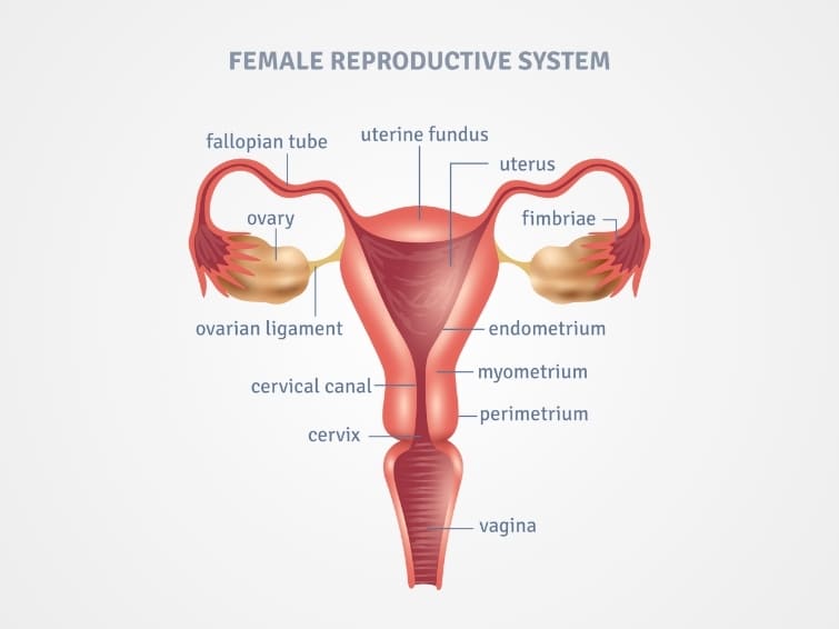
Genitourinary System Female Reproductive System Anatomy: Female Reproductive System REPRODUCTIVE ANATOMY Female External Genitalia (Vulva) The female external genitalia, collectively known as the vulva (or pudendum), encompass all the visible structures that extend from the pubic region to the perineum. These structures protect the internal reproductive organs, play a role in sexual arousal and function, and contain the openings of the urethra and vagina. The vulva is composed of the following main structures: Mons Pubis (Mons Veneris) Labia Majora Labia Minora Clitoris Vestibule Urethral Orifice Vaginal Orifice Greater Vestibular Glands (Bartholin’s Glands) (and Lesser Vestibular Glands) 1. Mons Pubis (Mons Veneris) Description: A rounded, fleshy prominence composed of adipose tissue (fat pad) overlying the pubic symphysis (the joint between the two pubic bones). Location: Anterior to the labia majora and superior to the clitoris. Covering: After puberty, it becomes covered with pubic hair. Hair Pattern: Female Pattern: Typically forms an inverted triangle, with a horizontal upper border (concave or convex downwards). Male Pattern: Tends to be diamond-shaped, extending upwards towards the umbilicus. Clinical Relevance: A change in female hair pattern towards a masculine distribution (hirsutism) can indicate underlying hormonal imbalances (e.g., polycystic ovary syndrome, adrenal disorders) and may be associated with infertility or other endocrine issues. Function: Acts as a protective cushion over the pubic bone during sexual intercourse. 2. Labia Majora Description: Two prominent, longitudinal, fleshy folds of skin that extend downwards and backwards from the mons pubis. They are homologous to the scrotum in males. Composition: Primarily composed of fat, elastic tissue, and some smooth muscle. Their outer surface is covered with pubic hair after puberty, while the inner surface is hairless and smooth. Extension: They unite anteriorly at the mons pubis and posteriorly, they tend to merge into the perineum, often forming a posterior commissure anterior to the anal canal. Erectile Tissue: The tissue within the labia majora is non-erectile. Function: Act as a protective barrier, enclosing and safeguarding the more delicate internal structures of the vulva (labia minora, clitoris, and vestibule). 3. Labia Minora (Nymphae) Description: Two smaller, hairless folds of highly vascularized, reddish-pink skin located medial to the labia majora. They are homologous to the ventral aspect of the penis in males. Composition: Rich in erectile tissue, blood vessels, nerve endings, and sebaceous glands, giving them a rich sensory innervation. Anatomical Configuration: Anteriorly: Each labium minus splits into two folds: Medial Fold: Passes over the clitoris to form the frenulum of the clitoris on its undersurface. Lateral Fold: Passes over the clitoris to form the prepuce (hood) of the clitoris on its dorsal surface. Posteriorly: In nulliparous (never given birth) women, the labia minora may fuse to form a transverse fold called the fourchette anterior to the vaginal opening. This often disappears after childbirth. Erectile Tissue: The labia minora contain erectile tissue, and they engorge with blood during sexual arousal. Function: Sexual Arousal: Highly sensitive due to rich nerve supply, contributing significantly to sexual pleasure. They swell and darken during arousal. Protection: Further protect the urethral and vaginal orifices. Lubrication: Contain numerous sebaceous glands for lubrication. Surgical Use: The tissue’s elasticity and rich blood supply make it suitable for grafting, particularly in reconstructive surgery (e.g., for facial wounds, though this is a specialized application and not its primary function). 4. Clitoris Description: A highly sensitive organ composed of erectile tissue, homologous to the penis in males. It is the primary organ for sexual pleasure in females. Location: Located anteriorly, where the labia minora converge. Structure: Fixed Root: Attached to the pubic arch. Composed of three masses of erectile tissue: Crura (Corpora Cavernosa): Two crura, continuous with the corpora cavernosa of the body of the clitoris, attach to the pubic rami and are covered by the ischiocavernosus muscles. Bulbs of the Vestibule (Corpus Spongiosum): Two masses of erectile tissue, located on either side of the vaginal orifice (split in females due to the presence of the vagina), covered by the bulbospongiosus muscles. These merge anteriorly to form the body of the clitoris. Free Body: Protrudes externally, composed of two corpora cavernosa and a small amount of corpus spongiosum (which forms the glans). Glans Clitoris: The most external, highly sensitive, pea-sized tip of the clitoris. Coverings: Covered by the prepuce (hood) and frenulum, formed by the labia minora. Erection Mechanism (Similar to penis): Psychic and Tactile Stimulation: Pleasurable sights, sounds, smells, and touch (nipples, inner thighs, clitoris itself) trigger a parasympathetic discharge from the sacral spinal cord segments (S2, S3, S4). Vasodilation: This leads to the release of nitric oxide, causing vasodilation of arteries supplying the erectile tissue (corpora cavernosa and bulbs of vestibule). Engorgement: Increased blood flow rapidly fills the vascular spaces within the erectile tissue. Venous Occlusion: The engorged erectile tissue compresses the veins draining the clitoris against the surrounding tough fibrous capsule (tunica albuginea) and pelvic bones, retarding venous outflow. Erection: The combination of increased arterial inflow and decreased venous outflow leads to engorgement and rigidity of the clitoris. Function: Solely dedicated to sexual sensation and arousal. 5. Vestibule Description: The almond-shaped space or cleft enclosed by the labia minora. Boundaries: Laterally: Labia minora. Anteriorly: Clitoris. Posteriorly: Fourchette (if present) or posterior commissure. Contents/Openings: It contains the openings of several important structures: Urethral Orifice Vaginal Orifice Ducts of the Greater Vestibular Glands (Bartholin’s Glands) Ducts of the Lesser Vestibular Glands (Skene’s Glands/Paraurethral Glands): These small mucus-secreting glands are located around the urethral orifice. 6. Urethral Orifice (External Urethral Meatus) Description: The opening of the female urethra. Location: Located posterior to the clitoris and anterior to the vaginal orifice, typically about 2.5 cm below the clitoris. It often appears as a small, slit-like or star-shaped opening. Function: Serves as the sole passage for urine in females. Clinical Relevance: Infections: Its short length (about 4 cm, much shorter than in males) and close proximity to the anal canal and vaginal opening make females more susceptible to recurrent urinary tract infections (UTIs), as bacteria can easily ascend into the bladder. Trauma: Can be traumatized during sexual

Male Reproductive System
Anatomy

Genitourinary System: Male Reproductive System

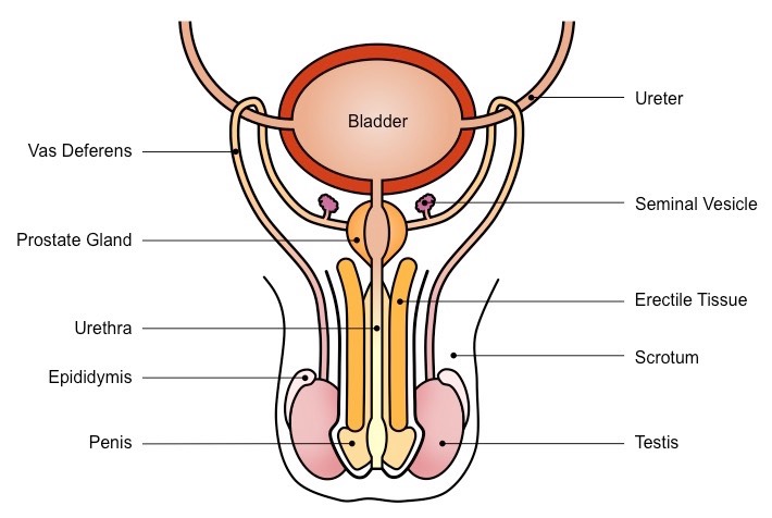
Genitourinary System Male Reproductive System The Genitourinary System: Male Reproductive System GENITOURINARY SYSTEM The Genitourinary System: An Introduction The genitourinary system encompasses the reproductive organs and the urinary system. While functionally distinct in many ways, they share developmental origins and anatomical proximity, particularly in males, hence their common classification. This section will focus on the reproductive system. The Reproductive System: Core Principles The reproductive system is a unique and fundamental biological system with several distinguishing characteristics: Sexual Dimorphism: It exhibits marked differences between males and females in terms of anatomy, physiology, and hormonal regulation. This is the most obvious difference from other organ systems. Latency in Development: Unlike most other body systems which are functional from birth, the reproductive system undergoes a prolonged period of dormancy (childhood) before maturing and becoming fully functional at puberty. Not Essential for Individual Survival: While crucial for the propagation of the species, an individual can survive perfectly well without a functional reproductive system. This contrasts with systems like the cardiovascular, respiratory, or nervous systems, which are indispensable for individual life. Functional Classification of the Reproductive System: 1. Primary Reproductive Organs (Gonads) Males: Testes (produce sperm and male hormones). Females: Ovaries (produce ova/eggs and female hormones). These organs are responsible for gamete (sex cell) production and hormone synthesis. 2. Accessory Reproductive Organs (Ducts and Glands) These structures are responsible for the transport, maturation, and nourishment of gametes, and facilitate fertilization. Males: Epididymis, vas deferens, ejaculatory ducts, seminal vesicles, prostate gland, bulbourethral glands. Females: Uterine tubes (fallopian tubes), uterus, vagina. 3. Secondary Sexual Characteristics These are physical traits that distinguish males from females but are not directly involved in reproduction. They develop under the influence of sex hormones at puberty. Males: Deepening voice, facial and body hair growth, increased muscle mass, broader shoulders. Females: Breast development, widening of hips, growth of pubic and axillary hair. These characteristics also serve as “sexual attractants” or signals for reproductive fitness. Anatomical Classification of the Reproductive System: External Genitalia (Perineal Structures): Males: Penis and Scrotum (containing the testes). Females: Vulva (comprising the labia majora, labia minora, clitoris, vestibule, and associated glands). Internal Genitalia: Males: Testes (though partially in scrotum, functionally internal), epididymis, vas deferens, ejaculatory ducts, seminal vesicles, prostate gland, bulbourethral glands. Females: Ovaries, uterine tubes, uterus, vagina. Male External Genitalia The male external genitalia are located in the perineum and consist of the penis and the scrotum. The Penis The penis is the male organ of copulation and urination. Analogue to the Female Clitoris: Both develop from the same embryonic structure (genital tubercle), hence they are homologous organs. However, their primary functions in adulthood differ significantly. Primary Function: To deliver sperm into the female reproductive tract during sexual intercourse and to serve as the exit for urine. Structure of the Penis: The penis consists of a fixed root and a free, pendulous body (shaft), terminating in the glans penis. 1. Root of the Penis The internal, attached portion of the penis. Composed of three erectile tissue structures: Bulb of the Penis: A single, ventrally placed, rounded mass of erectile tissue. It is the proximal continuation of the corpus spongiosum and is attached to the perineal membrane. It is covered externally by the bulbospongiosus muscle. Crura of the Penis (Crus, singular): Two diverging masses of erectile tissue (right and left) that are the proximal continuations of the corpora cavernosa. They are firmly attached to the inferior rami of the pubis and ischium. Each crus is covered externally by an ischiocavernosus muscle. 2. Body (Shaft) of the Penis The free, movable portion of the penis, covered by skin. Composed of three cylindrical masses of erectile tissue arranged longitudinally: Corpus Spongiosum (Corpus Cavernosum Urethrae): A single, ventrally placed column of erectile tissue. It is the distal continuation of the bulb of the penis. Crucially, it is traversed throughout its length by the penile (spongy) urethra. Distally, it expands to form the glans penis. Corpora Cavernosa (Corpus Cavernosum, singular): Two dorsally placed (relative to the corpus spongiosum and urethra), parallel columns of erectile tissue. They are the distal continuations of the crura of the penis. Their distal ends are covered by the expanded glans penis. These are the primary erectile bodies responsible for penile rigidity during erection. 3. Glans Penis: The expanded, conical distal end of the corpus spongiosum. Covers the distal ends of the corpora cavernosa. Highly sensitive due to abundant nerve endings. Corona: The prominent rim at the base of the glans. Prepuce (Foreskin): A retractable fold of skin that covers the glans penis in uncircumcised males. Coverings of the Penis: Skin: Thin, loose, and usually hairless, allowing for movement during erection. Superficial (Dartos) Fascia: A layer of loose connective tissue and smooth muscle (dartos muscle) just beneath the skin. Deep (Buck’s) Fascia of the Penis: A strong, inelastic, fibrous sheath that binds the three corpora together and surrounds the neurovascular structures. This fascia plays a crucial role in maintaining erection by compressing emissary veins. Neurovascular Supply: Arterial Supply: Primarily from the internal pudendal arteries, which give rise to the: Dorsal arteries of the penis: Supply the skin, fascia, and glans. Deep arteries of the penis (Cavernosal arteries): Run within the corpora cavernosa and are essential for erection. Bulbourethral arteries: Supply the bulb of the penis and corpus spongiosum. Venous Drainage: Deep veins (e.g., deep dorsal vein of the penis) drain the erectile tissue, and superficial veins drain the skin. Innervation: Somatic (Pudendal Nerve): Provides sensory innervation to the skin of the penis and glans. Autonomic (Pelvic Splanchnic Nerves – Parasympathetic; Hypogastric Plexus – Sympathetic): Essential for the erectile and ejaculatory reflexes. Parasympathetic stimulation causes vasodilation leading to erection, while sympathetic stimulation leads to ejaculation and detumescence. The Scrotum The scrotum is a cutaneous fibromuscular sac that encloses and protects the testes, epididymides, and the inferior parts of the spermatic cords. Analogue to the Female Labia Majora: Both develop from the same embryonic structures (labioscrotal folds), hence they are homologous. Location: An outpouching of the anterior abdominal wall, located in

Scroll to Top