Male Reproduction
Male reproductive functions include:
- Spermatogenesis – production of sperm cells.
- Male sexual act (coitus) – processes involved in sexual intercourse.
- Hormonal regulation – control of puberty, sexual function, spermatogenesis, and fertility.
Male Reproductive Organs
1. Primary sex organs
- Two testes – produce sperm and testosterone.
2. Accessory sex organs
- Epididymis
- Vas deferens
- Ejaculatory ducts
- Seminal vesicles
- Prostate gland
- Bulbourethral (Cowper’s) glands, Urethra and Penis
Physiological Anatomy of the Male Sexual Organs
- Each testis contains about 900 coiled seminiferous tubules, where sperm are produced.
- Sperm pass into the epididymis, a highly coiled tube where they mature and are stored.
- Epididymis continues into the vas deferens, which enlarges to form the ampulla near the prostate.
- Seminal vesicles empty their secretions into the vas deferens near the ampulla.
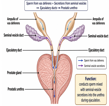
- The vas deferens and seminal vesicle duct join to form the ejaculatory duct.
- Ejaculatory ducts pass through the prostate gland and empty into the urethra.
Prostate and Urethra
- Prostatic ducts empty prostatic secretions into the prostatic urethra.
- The urethra is the final passage that carries semen to the exterior.
- Mucus is secreted by numerous urethral glands and the paired bulbourethral (Cowper’s) glands.
- These mucus secretions help lubricate and protect the urethra during ejaculation.
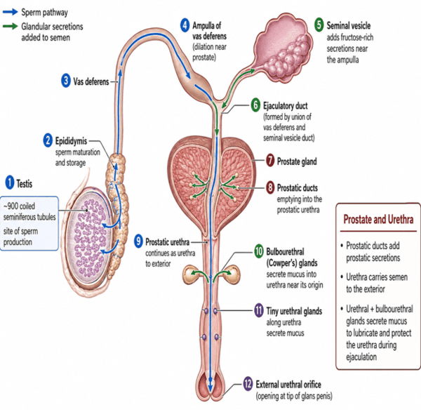
Physiological Anatomy of the Male Sexual Organs
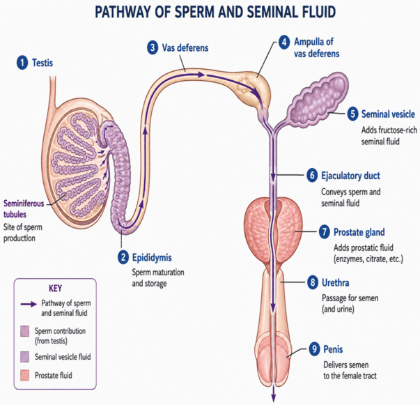
Sequential Transit of Spermatozoa
Seminiferous tubules → Tubuli recti → Rete testis → Efferent ductules → Epididymis (Caput → Corpus → Cauda) → Vas deferens → Ampulla → Ejaculatory duct → Prostatic urethra → Membranous urethra → Spongy (penile) urethra → External urethral orifice.
Functional Anatomy of the Testes
- Testes are the male gonads responsible for producing sperm and androgens, mainly testosterone.
- They are oval-shaped organs located within the scrotum.
- Each testis is covered by:
- Tunica vaginalis – an outer serous covering with parietal and visceral layers.
- Tunica albuginea – a tough, dense connective tissue capsule beneath the tunica vaginalis.
- Tunica albuginea extends inward to form septa, dividing the testis into about 250–300 lobules.
- Each lobule contains seminiferous tubules, where sperm are produced.
Seminiferous Tubules and Sperm Pathway
- Each testicular lobule contains tightly coiled seminiferous tubules (main site of sperm production).
- Sperm move from the seminiferous tubules into the straight tubules (tubuli recti).
- From there they then enter the rete testis (network of interconnected tubules).
- From the rete testis, sperm leave the testis through about 15–20 efferent ductules & enter the epididymis.
- Seminiferous tubules contain:
- Sertoli (sustentacular) cells – support and nourish developing sperm.
- Germ cells – which develop through successive stages to form mature spermatozoa.
Summary: Seminiferous tubules → Tubuli recti → Rete testis → Efferent ductules → Epididymis.
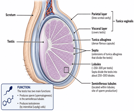
Functional Anatomy of the Testis
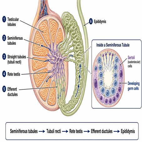
Seminiferous Tubules and Sperm Pathway
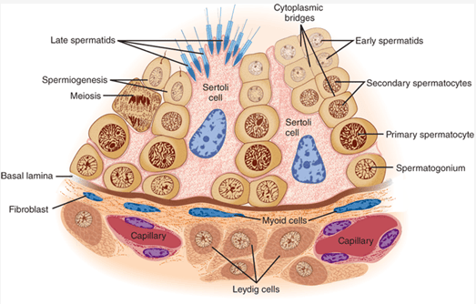
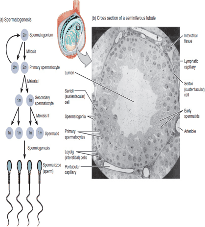
Section of the human testis. Cross section shows the diversity of cellular anatomy within the testis. Seminiferous epithelium.: Note that maturing germ cells remain connected by cytoplasmic bridges through the early spermatid stage and that these cells are closely invested by Sertoli cell cytoplasm as they move from the basal lamina to the lumen
Blood Supply to the Testes
- Arterial supply: mainly through the testicular arteries.
- Venous drainage: through the pampiniform plexus, a network of veins surrounding the testicular artery.
Counter-Current Exchange System
- Testicular arteries and veins run closely together with blood flowing in opposite directions.
- This arrangement helps:
- Cool arterial blood before it reaches the testes, maintaining the lower temperature required for spermatogenesis.
- Facilitate some local exchange of substances, including testosterone, between venous and arterial blood.
- Heat exchange: Warm arterial blood transfers heat to the cooler venous blood returning from the testes.
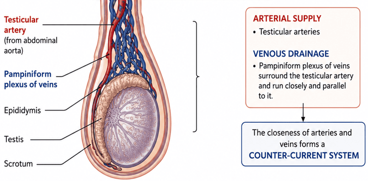
BLOOD SUPPLY TO THE TESTES
Thermoregulation via Counter-Current Exchange
Arterial blood at core body temperature (~37°C) is cooled to ~34°C before entering the testicular parenchyma as heat dissipates into the cooler (~32°C → 35°C) venous blood of the pampiniform plexus. This maintains the essential 2–3°C gradient below core body temperature required for viable spermatogenesis.
Functions of the Testes
- Endocrine function – secretion of male sex hormones, mainly testosterone.
- Gametogenic function – production of sperm through spermatogenesis.
1. Endocrine Functions of the Testes
- Testosterone is the principal androgen produced by the adult testes.
- Testosterone and its more potent metabolite, dihydrotestosterone (DHT), promote the development and maintenance of male secondary sexual characteristics.
- Testosterone stimulate protein synthesis, muscle growth, bone growth, and the typical male body physique.
- At puberty, testosterone promotes growth and function of the accessory reproductive organs, especially the prostate and seminal vesicles.
- Testosterone is important for:
- Libido and sexual function
- Maintenance of spermatogenesis
- Male sexual differentiation
- Development and maintenance of male secondary sexual characteristics
- Influence on some aspects of male sexual and behavioral development
2. Gametogenic Function (Spermatogenesis)
- Spermatogenesis is the process by which spermatozoa (sperm cells) are produced from primitive germ cells called spermatogonia located in the seminiferous tubules of the testes.
- Spermatogenesis begins at puberty and continues throughout adult life, although efficiency may decline with age.
- In humans, development from spermatogonia to mature spermatozoa takes approximately 64–74 days.
Spermatogenesis
Learning Objectives
By the end of this section, students should be able to:
- Describe the stages of spermatogenesis by which diploid germ cells develop into haploid spermatozoa.
- Explain the roles of mitosis, meiosis, and spermiogenesis in sperm formation.
- Identify the role of testosterone in the initiation and maintenance of spermatogenesis.
- Relate testosterone to normal male reproductive function, including sexual development and fertility.
Overview of Spermatogenesis
- During embryonic development, primordial germ cells migrate to the testes and develop into spermatogonia located near the basement membrane of the seminiferous tubules.
- At puberty, spermatogonia begin continuous mitotic division, producing cells that differentiate into spermatozoa.
- As development progresses, germ cells move from the basal region toward the lumen of the seminiferous tubules.
- Sertoli cells surround, nourish, protect, and support the developing germ cells throughout spermatogenesis.
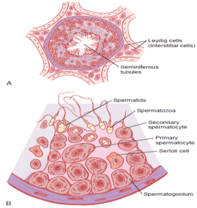
A, Cross section of a seminiferous tubule. B, Stages in the development of sperm from spermatogonia
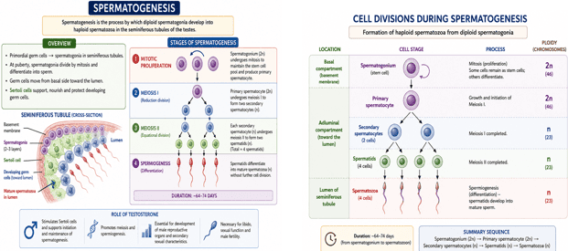
SPERMATOGENESIS: CELL DIVISIONS DURING SPERMATOGENESIS
Cell Divisions During Spermatogenesis
- During embryonic development, primordial germ cells migrate to the testes and develop into spermatogonia.
- At puberty, spermatogonia multiply by mitosis; some differentiate into primary spermatocytes.
- Primary spermatocytes undergo meiosis I to form secondary spermatocytes.
- Secondary spermatocytes undergo meiosis II to form haploid spermatids.
- Spermatids then differentiate by spermiogenesis into spermatozoa.
Sequence:
Spermatogonium (2n) → Primary spermatocyte (2n) → Secondary spermatocytes (n) → Spermatids (n) → Spermatozoa (n)
Process of Spermatogenesis
Stage 1: Spermatogoniogenesis
a. Spermatogonia
- Spermatogenesis begins with mitotic proliferation of spermatogonia in the seminiferous tubules.
- Spermatogonia are diploid (2n) cells containing 46 chromosomes (23 pairs).
b. Formation of primary spermatocytes
- Other spermatogonia differentiate and enlarge to become primary spermatocytes.
- Primary spermatocytes subsequently enter meiosis I.
Stage 2: Spermatocytogenesis
a. Primary spermatocytes undergo meiosis I
- Each primary spermatocyte (2n) undergoes meiosis I.
- Homologous chromosome pairs separate, producing two secondary spermatocytes.
- Each secondary spermatocyte is haploid (n) with 23 chromosomes, although each chromosome still has two sister chromatids.
b. Secondary spermatocytes undergo meiosis II
- Each secondary spermatocyte undergoes meiosis II.
- Sister chromatids separate, producing two spermatids from each secondary spermatocyte.
- Therefore, one primary spermatocyte ultimately produces four haploid spermatids (n).
Process of Spermatogenesis
| Cell Stage | Anatomical Location | Division / Process Type | Ploidy & Chromosome Number |
|---|---|---|---|
| Spermatogonium | Basal compartment (basement membrane) | Mitosis (proliferation & self-renewal) | Diploid (2n) = 46 |
| Primary Spermatocyte | Moves toward adluminal compartment | Undergoes Meiosis I (reduction division) | Diploid (2n) = 46 (4C DNA) |
| Secondary Spermatocyte | Adluminal compartment | Undergoes Meiosis II (equational division) | Haploid (n) = 23 (2C DNA) |
| Spermatid | Adluminal compartment near lumen | Spermiogenesis (differentiation without division) | Haploid (n) = 23 (1C DNA) |
| Spermatozoon | Tubular lumen → efferent ducts | Released via spermiation | Haploid (n) = 23 (1C DNA) |
Stage 3: Spermiogenesis (Spermatid Maturation)
- Transformation of haploid spermatids into spermatozoa without further cell division.
- Major changes include:
- Acrosome formation – develops an enzyme-containing cap needed for fertilization.
- Flagellum formation – produces the sperm tail for motility.
- Nuclear condensation – compacts the genetic material.
- Loss of excess cytoplasm – produces the streamlined shape of mature sperm.
- Mature spermatozoa are released into the lumen of the seminiferous tubules by spermiation and then transported toward the epididymis for further maturation and storage.
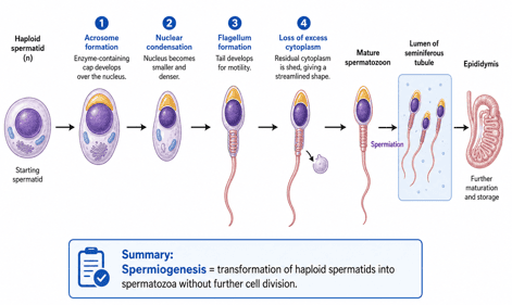
Stage 3: Spermiogenesis (Spermatid Maturation)
Spermatocytogenesis vs. Spermiogenesis vs. Spermiation
Spermatocytogenesis: Meiotic phase where primary spermatocytes form haploid spermatids.
Spermiogenesis: Morphological packaging (no cell division) — round spermatid transforms into a streamlined flagellated spermatozoon.
Spermiation: Physical release of mature spermatozoa from Sertoli cell cytoplasmic crypts into the seminiferous tubule lumen.
Structure of a Mature Sperm Cell
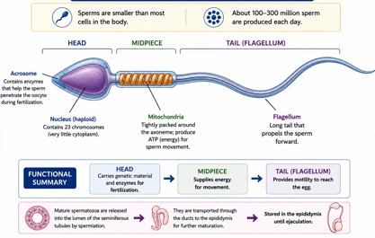
Spermatozoa are small, highly specialized male gametes. About 100–300 million sperm may be produced daily in adult males.
a. Head
- Contains a haploid nucleus (23 chromosomes).
- Covered anteriorly by the acrosome, which contains enzymes that help the sperm penetrate the oocyte during fertilization.
b. Midpiece
- Contains numerous mitochondria. Produces ATP required for sperm movement.
c. Tail (Flagellum)
- Extends from the neck and midpiece.
- Generates the propulsive movement that allows the sperm to swim.
Physiology of Mature Sperm
- Mature sperm are motile and move by flagellar action at about 1–4 mm/min.
- Sperm motility is greatest in a neutral to slightly alkaline medium, such as semen.
- Acidic conditions reduce sperm motility, while strongly acidic environments may rapidly kill sperm.
- Increasing temperature may increase sperm motility and metabolism, but excessive heat reduces sperm survival and function.
- Sperm can remain viable for prolonged periods while stored in the male reproductive tract, especially the epididymis.
- After ejaculation, sperm may survive in the female reproductive tract for about 3–5 days under favorable conditions.
Sperm Maturation and Capacitation
- After leaving the seminiferous tubules, sperm pass through the epididymis, a highly coiled duct about 6 meters long when uncoiled.
- Sperm entering the epididymis are immotile and functionally immature and are initially unable to fertilize an ovum.
- After 18–24 hours in the epididymis, sperm develop the capacity for motility, although their movement remains largely suppressed by inhibitory factors in the epididymal fluid.
- After ejaculation, these inhibitory influences are reduced and sperm become actively motile, however, the sperms are not yet fully capable of fertilization.
- Epididymal epithelium secretes substances that support sperm maturation, including proteins, enzymes, ions, and nutrients.
- Epididymal environment is also influenced by androgens (testosterone) and dihydrotestosterone (DHT) which are important for normal epididymal function and sperm maturation.
- Sertoli cells also secrete testicular fluid that supports the developing sperm and facilitates their transport from the seminiferous tubules.
- Final fertilizing capacity is achieved through capacitation in the female reproductive tract, NOT in the epididymis, which enables the sperm to undergo the acrosome reaction and fertilize the ovum.
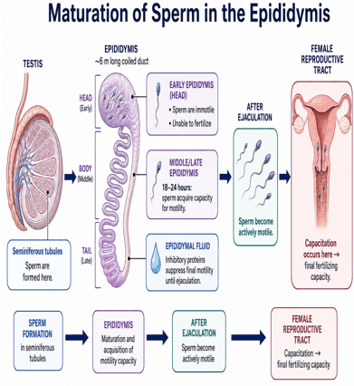
Epididymal Maturation vs. Female Tract Capacitation
Epididymal Maturation (18–24 hours): Sperm acquire forward motility potential and glycoprotein surface coats, but stay held in check by local decapacitation factors.
Capacitation (Female Reproductive Tract, ~7 hours): Removal of cholesterol and inhibitory glycoprotein coat from the acrosome cap by female fluids → Calcium influx → hyperactivated motility and readiness for the acrosome reaction upon binding ZP3 on the zona pellucida.
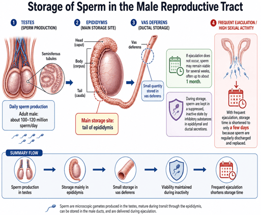
Storage of Sperm in the Male Reproductive Tract
- Two testes of an adult male produce approximately 100–120 million sperm per day.
- After maturation, most sperm are stored in the epididymis, especially in its tail, while a smaller number may be stored in the vas deferens.
- Stored sperm can remain viable and retain their fertilizing capacity for several weeks, often up to about one month, if ejaculation does not occur.
- During storage, sperm are maintained in a relatively inactive and suppressed state by inhibitory substances present in the secretions of the epididymis and other reproductive ducts.
- With frequent sexual activity and repeated ejaculation, sperm storage time is shortened to only a few days because stored sperm are regularly discharged and replaced.
Note: Mature sperm are stored mainly in the epididymis, not within the testes.
Factors Affecting Spermatogenesis
1. Hormonal Factors
a. Gonadotropin-Releasing Hormone (GnRH)
- Secreted by the hypothalamus in a pulsatile manner.
- It stimulates the anterior pituitary gland to release FSH & LH.
- Reduced GnRH secretion leads to reduced gonadotropin secretion and impaired spermatogenesis.
b. Follicle-Stimulating Hormone (FSH)
- Acts mainly on Sertoli cells in the seminiferous tubules.
- It stimulates Sertoli cells to:
- Support developing germ cells.
- Produce androgen-binding protein (ABP).
- Maintain a high concentration of testosterone within the seminiferous tubules.
- Produce nutrients and growth factors required for germ-cell development.
- Thus, FSH is particularly important for the initiation and maintenance of normal sperm production.
c. Luteinizing Hormone (LH)
- Acts on the Leydig cells of the testes.
- Stimulates the production of testosterone.
- Testosterone then supports spermatogenesis within the seminiferous tubules.
d. Testosterone
- Essential for normal spermatogenesis. It:
- Promotes growth and division of germ cells.
- Supports meiosis and spermiogenesis.
- Maintains the structure and function of the seminiferous epithelium.
- Works together with FSH to support Sertoli-cell function.
- A high intratesticular concentration of testosterone is required for normal sperm production.
e. Inhibin B
- Secreted by Sertoli cells and provides negative feedback primarily on FSH secretion.
- Helps regulate the rate of sperm production.
| Hormone | Source Gland / Cell | Primary Target | Physiological Action on Spermatogenesis |
|---|---|---|---|
| GnRH | Hypothalamus (Arcuate nucleus) | Anterior Pituitary Gonadotropes | Pulsatile release drives LH and FSH secretion |
| FSH | Anterior Pituitary | Sertoli Cells | Stimulates ABP production, supports germ cell nourishment |
| LH | Anterior Pituitary | Leydig (Interstitial) Cells | Triggers steroidogenesis → Testosterone production |
| Testosterone | Leydig Cells | Seminiferous Epithelium / Sertoli | Maintains blood-testis barrier, drives meiosis and spermiogenesis |
| Inhibin B | Sertoli Cells | Anterior Pituitary | Selective negative feedback on FSH |
2. Temperature
- Normal spermatogenesis requires the testes to remain approximately 2–3°C below core body temperature.
- Testes are therefore located in the scrotum outside the abdominal cavity.
- Mechanisms regulating testicular temperature include:
- Pampiniform plexus – provides counter-current heat exchange.
- Dartos muscle – alters wrinkling and surface area of the scrotum.
- Cremaster muscle – raises or lowers the testes according to temperature.
- Scrotal sweating and thin scrotal skin also facilitate heat loss.
- Increased testicular temperature can impair spermatogenesis.
- Causes include: Prolonged high fever, Cryptorchidism, Varicocele, Prolonged exposure to excessive environmental heat, Certain occupational heat exposures.
- Excessive heat may decrease sperm concentration, sperm motility & normal sperm morphology.
3. Cryptorchidism
- Failure of one or both testes to descend normally into the scrotum.
- The abdominal temperature is higher than the scrotal temperature.
- Persistent exposure to higher temperatures damages the seminiferous epithelium.
- This may markedly impair or stop spermatogenesis.
- Early correction is important to reduce the risk of:
- Infertility.
- Testicular degeneration.
- Testicular malignancy.
4. Nutrition
- Adequate nutrition is required for continuous production of sperm.
- Important nutrients include:
- Proteins and amino acids: Required for cell division and synthesis of structural proteins.
- Zinc: Important for: Testosterone synthesis, Sperm development, Sperm motility and membrane stability.
- Selenium: Important for antioxidant defense and normal sperm structure and motility.
- Folate: Required for DNA synthesis and cell division.
- Vitamins:
- Vitamin A: important for differentiation of germ cells.
- Vitamin C and vitamin E: act as antioxidants and protect sperm from oxidative damage.
- Vitamin D: may contribute to normal reproductive function.
- Severe malnutrition may suppress the hypothalamic–pituitary–gonadal axis and reduce sperm production.
5. Age
- Spermatogenesis begins at puberty.
- It continues throughout adult life.
- However, with increasing age there may be gradual reductions in:
- Testosterone production.
- Sperm concentration.
- Motility.
- Normal morphology.
- Overall reproductive efficiency.
- Advanced paternal age may also be associated with increased sperm DNA damage and some genetic risks in offspring.
6. General Health and Systemic Disease
- Normal sperm production requires good general health.
- Conditions that may impair spermatogenesis include: severe chronic illness, prolonged fever, chronic kidney disease, chronic liver disease, poorly controlled diabetes mellitus, severe systemic infections, significant hormonal disorders.
- Illness can interfere with the hypothalamic–pituitary–gonadal axis and may also directly affect testicular function.
7. Endocrine Disorders
- Hormonal abnormalities can markedly affect sperm production. Examples include:
- Hypogonadotropic hypogonadism: Reduced GnRH, LH, or FSH results in reduced testicular stimulation.
- Hyperprolactinemia: Excess prolactin can suppress GnRH secretion, resulting in reduced LH, FSH, testosterone, and sperm production.
- Thyroid disorders: Both severe hypothyroidism and hyperthyroidism may adversely affect testicular and sexual function.
- Pituitary disorders: Damage to the pituitary gland may reduce FSH and LH secretion and consequently impair spermatogenesis.
8. Varicocele
- Abnormal dilatation of the veins of the pampiniform plexus.
- It may impair spermatogenesis through:
- Increased testicular temperature.
- Venous congestion.
- Oxidative stress.
- Altered testicular blood flow.
- Varicocele is an important potentially reversible cause of impaired male fertility.
9. Drugs and Hormonal Agents
- Several drugs may reduce sperm production. Examples include:
- Anabolic steroids and exogenous testosterone
- Some chemotherapeutic agents
- Certain antiandrogen drugs
- Some medications that interfere with the hypothalamic–pituitary–gonadal axis
10. Radiation
- The rapidly dividing germ cells are particularly sensitive to ionizing radiation.
- Radiation exposure may cause:
- Reduced sperm production.
- Temporary or permanent azoospermia.
- DNA damage in germ cells.
- The severity depends on the dose and duration of exposure.
11. Chemicals and Environmental Toxins
- Certain toxic substances may damage germ cells, Sertoli cells, Leydig cells, or interfere with reproductive hormones.
- Examples include:
- Heavy metals such as lead and cadmium.
- Some pesticides.
- Industrial chemicals.
- Organic solvents.
- Endocrine-disrupting chemicals.
13. Smoking
- Cigarette smoke contains numerous toxic and oxidizing substances. Smoking may be associated with:
- Reduced sperm concentration.
- Reduced motility.
- Increased abnormal morphology.
- Increased oxidative stress.
- Increased sperm DNA damage.
14. Alcohol and Recreational Drugs
- Heavy or chronic alcohol consumption may:
- Suppress testosterone production.
- Alter LH and FSH secretion.
- Damage testicular cells.
- Reduce sperm production and quality.
- Some recreational drugs may also disturb reproductive hormone secretion or directly impair testicular function.
15. Testicular Infections and Inflammation
- Inflammation of the testes (orchitis) can damage the seminiferous tubules. Examples include:
- Viral orchitis, particularly following mumps.
- Severe bacterial infections.
- Sexually transmitted infections affecting the reproductive tract.
- Severe bilateral testicular damage may result in infertility.
16. Psychological and Physical Stress
- Severe or prolonged stress may disturb the hypothalamic–pituitary–gonadal axis.
- Stress-related elevation of glucocorticoids may suppress:
- GnRH secretion.
- LH and FSH secretion.
- Testosterone production.
- This can indirectly reduce spermatogenesis.
Male Accessory Organs of Reproduction
Structures that assist in the maturation, storage, transport, nourishment, protection, and delivery of spermatozoa.
- Epididymis
- Vas deferens
- Ejaculatory ducts
- Urethra
- Seminal vesicles
- Prostate gland
- Bulbourethral glands
- Penis
- Scrotum
1. Epididymis
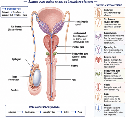
- Long, highly coiled duct located along the posterior surface of each testis.
- When uncoiled, it measures approximately 5–6 meters in length.
- Parts of the Epididymis is divided into: Head (caput), Body (corpus), Tail (cauda).
- The head receives sperm from the testis through the efferent ductules, while the tail continues as the vas deferens.
Functions of the Epididymis
- a. Sperm maturation: Sperm leaving the testes are initially immotile and functionally immature. During passage through the epididymis, they acquire the capacity for progressive motility and fertilization.
- b. Sperm storage: Most mature sperm are stored in the tail of the epididymis.
- c. Concentration of sperm: Excess fluid from the testis is absorbed by the epididymal epithelium.
- d. Transport of sperm: Smooth-muscle contractions move sperm toward the vas deferens.
- e. Secretion of substances: Epididymal epithelium secretes proteins, enzymes, nutrients, ions, and other factors that support sperm maturation.
2. Vas Deferens
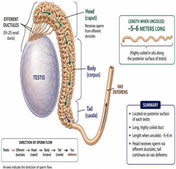
- Also called the ductus deferens, is a thick-walled muscular tube that transports sperm from the epididymis toward the ejaculatory duct.
- Begins at the tail of the epididymis and ascends within the spermatic cord.
- It passes through the inguinal canal and enters the pelvic cavity. It enlarges near its terminal portion to form the ampulla.
- It Joins the duct of the seminal vesicle to form the ejaculatory duct.
Functions
- Transports sperm during ejaculation.
- Provides temporary storage for some sperm.
- Produces strong peristaltic contractions during emission. The thick smooth-muscle wall of the vas deferens is especially important for rapid propulsion of sperm.
3. Ejaculatory Ducts
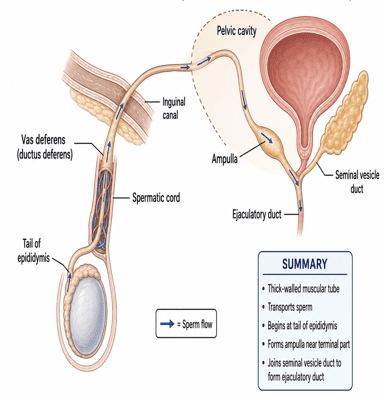
- Short paired ducts formed by the union of:
- The ampulla of the vas deferens.
- The duct of the seminal vesicle.
- Each ejaculatory duct passes through the prostate gland and opens into the prostatic urethra.
Function
- Conducts sperm mixed with seminal-vesicle secretions into the urethra during ejaculation.
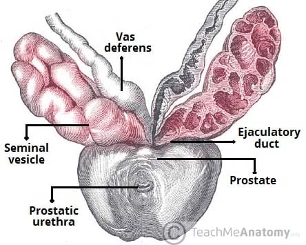
4. Seminal Vesicles
- Paired elongated glands located posterior to the urinary bladder.
- Contribute approximately 60–70% of the volume of semen.
Composition of Seminal Vesicle Secretion
- Secretion is rich in: Fructose, Prostaglandins, Citrate, Proteins, Semen-coagulating proteins, Bicarbonate and other alkaline substances.
Functions
- a. Fructose: Provides an important energy source for sperm.
- b. Alkaline secretion: Helps neutralize the acidic environment of the male urethra and female vagina. It promotes sperm survival and motility.
- c. Prostaglandins: May assist sperm transport by:
- Stimulating contractions in the female reproductive tract.
- Influencing cervical mucus.
- d. Semen coagulation: Seminal-vesicle proteins contribute to the temporary coagulation of semen shortly after ejaculation.
Seminal Vesicle Function
The seminal vesicles do not store sperm. Their main role is to produce nutrient-rich seminal fluid that nourishes and supports sperm function and survival.
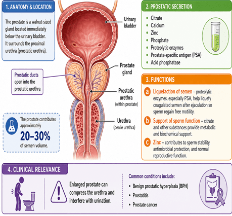
5. Prostate Gland
- Single gland located immediately below the urinary bladder and surrounds the proximal urethra.
- Its ducts open into the prostatic urethra.
- The prostate contributes approximately 20–30% of the volume of semen.
Prostatic Secretion
- Prostatic fluid contains: Citrate, Calcium, Zinc, Phosphate, Proteolytic enzymes, Prostate-specific antigen (PSA), Acid phosphatase.
Functions
- a. Liquefaction of semen: Proteolytic enzymes (prostate-specific antigen), produced mainly by the prostate gland and released into seminal fluid help break down the coagulated semen after ejaculation. This liquefaction allows sperm to regain free motility.
- b. Support of sperm function: Citrate and other substances provide metabolic and biochemical support.
- c. Zinc: contributes to sperm stability, Antimicrobial protection. Normal reproductive function.
Clinical Relevance
- Enlargement of the prostate can compress the urethra and interfere with urination.
- Common conditions include: Benign prostatic hyperplasia (BPH), Prostatitis, Prostate cancer.
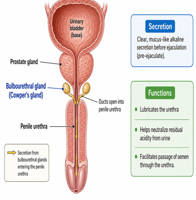
6. Bulbourethral glands
- Also called Cowper’s glands, are small paired glands located below the prostate.
- Their ducts open into the penile urethra.
Secretion
- They produce a clear, mucus-like alkaline secretion before ejaculation.
- This fluid is commonly called pre-ejaculate.
Functions
- Lubricates the urethra.
- Helps neutralize residual acidity from urine.
- Facilitates passage of semen through the urethra.
7. Penis
- Male copulatory organ responsible for depositing semen in the female reproductive tract.
Major Structures
- Penis contains three erectile bodies: Two corpora cavernosa and One corpus spongiosum (surrounds the penile urethra and expands distally to form the glans) penis.
Functions
- Sexual intercourse
- Delivery of semen
- Passage of urine
Erection
- Occurs when parasympathetic stimulation causes vasodilation of penile blood vessels.
- This leads to: Increased arterial blood flow, Filling of erectile tissues. Compression of venous outflow, Enlargement and rigidity of the penis.
- Nitric oxide is a major mediator of penile vasodilation.
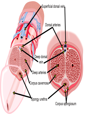
Cross Section and Vascular Architecture of the Penis
8. Scrotum
- Skin-covered sac that contains the testes and part of the spermatic cords.
Functions
- Main function of the scrotum is temperature regulation.
- Normal spermatogenesis requires a temperature approximately 2–3°C below core body temperature.
Mechanisms of Temperature Regulation
a. Dartos muscle
- Smooth muscle within the scrotal wall that contracts in cold conditions, producing wrinkling of the skin and reducing heat loss.
- Relaxes in warm conditions, increasing surface area for heat loss.
b. Cremaster muscle
- Elevates the testes closer to the body in cold conditions and relaxes in warm conditions, allowing the testes to descend.
c. Pampiniform plexus
- Network of veins surrounding the testicular artery that functions as a counter-current heat exchanger. It also cools arterial blood before it enters the testes.
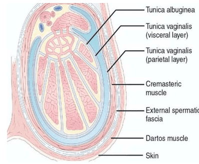
Anatomical Layers and Thermoregulatory Mechanisms of the Scrotum
9. Urethra
- Male urethra serves both the:
- Urinary system
- Reproductive system
It carries:
- Urine from the bladder.
- Semen during ejaculation.
Parts of the Male Urethra
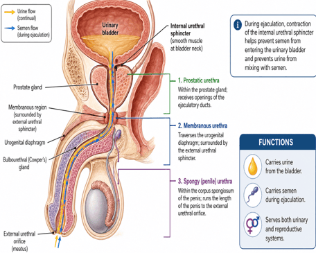
- Prostatic urethra
- Membranous urethra
- Spongy or penile urethra
During ejaculation, contraction of the internal urethral sphincter helps prevent semen from entering the urinary bladder and prevents urine from mixing with semen.
| Gland | % Volume of Semen | Primary Constituents | Key Physiological Role |
|---|---|---|---|
| Seminal Vesicles | 60–70% | Fructose, prostaglandins, coagulating proteins, bicarbonate | Sperm nutrition, alkaline buffering, female smooth muscle contraction |
| Prostate Gland | 20–30% | PSA, citrate, zinc, acid phosphatase, calcium | Liquefaction of coagulated semen, sperm motility & stability |
| Bulbourethral (Cowper's) | <1–5% | Clear alkaline mucus (pre-ejaculate) | Urethral neutralization of acidic urine, lubrication |
| Testes / Epididymis | ~2–5% | Spermatozoa, epididymal transport fluid | Fertilizing gamete payload |
Semen
- Whitish fluid ejaculated from the male reproductive tract during orgasm.
- It consists of spermatozoa suspended in seminal plasma, which is produced mainly by the accessory reproductive glands.
Composition of Semen
- Spermatozoa – produced in the testes and stored mainly in the epididymis.
- Seminal vesicle fluid – contributes about 60–70% of semen volume.
- Prostatic fluid – contributes about 20–30%.
- Small amounts of secretions from:
- Epididymis
- Vas deferens
- Bulbourethral glands
Major Constituents of Seminal Plasma
Seminal plasma contains:
- Fructose – provides energy for sperm.
- Citrate – supports sperm metabolism.
- Prostaglandins – may facilitate sperm transport in the female reproductive tract.
- Bicarbonate – helps maintain an alkaline environment.
- Zinc – supports sperm stability and antimicrobial defense.
- Calcium and phosphate
- Proteins and enzymes
- Prostate-specific antigen (PSA) – helps liquefy semen after ejaculation.
Physical Characteristics
Normal semen is usually:
- Whitish-gray and opalescent
- Slightly viscous immediately after ejaculation
- Slightly alkaline, with a pH of approximately 7.2–8.0
- Usually ejaculated in a volume of about 1.5–6 mL, although normal values vary between individuals.
Coagulation and Liquefaction
- Immediately after ejaculation, semen temporarily coagulates because of proteins mainly from the seminal vesicles.
- Within about 15–30 minutes, proteolytic enzymes from the prostate, particularly PSA, liquefy the semen.
Functions of Semen
- Transports sperm into the female reproductive tract.
- Provides nutrients, especially fructose, for sperm metabolism.
- Buffers acidity in the male urethra and female reproductive tract.
- Supports sperm motility and survival.
- Provides a suitable fluid medium for sperm movement.
- Contains substances that may facilitate sperm transport within the female reproductive tract.
Sperm Concentration
- A semen sample normally contains millions of spermatozoa per milliliter.
- Male fertility depends not only on sperm number but also on:
- Sperm motility
- Normal morphology
- Semen volume
- Sperm viability
- Integrity of sperm DNA
| Parameter | Lower Reference Limit | Clinical Significance |
|---|---|---|
| Semen Volume | ≥ 1.4 mL | Low volume (<1.4 mL) indicates ejaculatory duct obstruction or retrograde ejaculation |
| Sperm Concentration | ≥ 16 million sperm/mL | <16 million/mL = Oligozoospermia |
| Total Sperm Number | ≥ 39 million per ejaculate | Absolute minimum threshold for natural reproductive potential |
| Total Motility (PR + NP) | ≥ 42% motile | <42% = Asthenozoospermia |
| Normal Morphology | ≥ 4% normal forms | <4% = Teratozoospermia (strict Kruger criteria) |
| pH | 7.2 – 8.0 | Acidic pH (<7.0) with low fructose suggests seminal vesicle agenesis or obstruction |
Cryptorchidism
- Failure of one or both testes to descend completely into the scrotum.
- During fetal development, the testes originate from the genital ridges on the posterior abdominal wall.
- They normally descend through the inguinal canals and reach the scrotum during late fetal life, usually by about the 7th–9th month of gestation.
- In cryptorchidism, one or both testes may remain:
- In the abdominal cavity
- Within the inguinal canal
- At another point along the normal pathway of descent
Why Cryptorchidism Affects Spermatogenesis
- Normal spermatogenesis requires the testes to remain approximately 2–3°C below core body temperature.
- An undescended testis is exposed to the higher temperature of the abdomen or inguinal region.
- Prolonged heat exposure damages the seminiferous tubules and germinal epithelium, reducing or preventing sperm production.
- Early treatment, usually orchiopexy during infancy, helps preserve fertility potential and facilitates later examination of the testis.
Consequences of Cryptorchidism
- Impaired spermatogenesis: untreated abdominal testis may produce few or no sperm.
- Degeneration of seminiferous tubules: Germ cells and seminiferous epithelium progressively deteriorate with prolonged exposure to higher temperatures.
- Reduced fertility or infertility: Risk is greater in bilateral cryptorchidism than in unilateral disease.
- Leydig-cell function may be relatively preserved: Testosterone secretion may continue even when sperm production is severely impaired.
- Increased risk of testicular cancer: Cryptorchidism is associated with an increased lifetime risk of testicular malignancy.
- Other complications: Increased risk of testicular torsion, inguinal hernia, and trauma.
Treatment of Cryptorchidism
1. Observation During Early Infancy
- Some undescended testes descend spontaneously during the first few months after birth.
- If spontaneous descent has not occurred by 6 months of age (corrected for prematurity), further spontaneous descent is unlikely.
- The child should therefore be referred for definitive treatment.
2. Hormonal Therapy
- Testosterone is not recommended as routine treatment for cryptorchidism.
- Hormonal agents such as human chorionic gonadotropin (hCG) or GnRH analogues have historically been used to stimulate testicular descent.
- However, their success rate is relatively low, and the testis may subsequently re-ascend.
- Therefore, hormonal therapy is not routinely recommended for inducing testicular descent.
3. Surgical Treatment – Orchiopexy
- Orchiopexy is the standard treatment for persistent cryptorchidism.
- It involves:
- Locating the undescended testis.
- Mobilizing the testis and spermatic cord.
- Bringing the testis into the scrotum.
- Fixing it in the scrotum to prevent re-ascent.
Timing of Surgery
- If the testis has not descended by 6 months, surgery should generally be performed within the following year.
- Ideally, orchiopexy should be completed by 12 months, and no later than 18 months of age.
Early treatment helps:
- Preserve fertility potential.
- Reduce progressive damage to germ cells.
- Facilitate examination of the testis later in life.
- Reduce, although not completely eliminate, the risk of testicular malignancy.
Surgical Approach
- Palpable undescended testis: usually treated by inguinal or scrotal orchiopexy.
- Non-palpable/intra-abdominal testis: diagnostic laparoscopy may be required, followed by appropriate surgical management.
Male Sexual Act
Introduction: Male Sexual Act — Neuronal Stimuli
- Male sexual act is coordinated by sensory, spinal, autonomic, and higher brain mechanisms.
- The glans penis and penile skin are major sources of sensory input, responding to touch, pressure, and friction.
- These impulses are transmitted mainly through the dorsal nerve of the penis and pudendal nerve to the sacral spinal cord (S2–S4), where they activate reflex pathways involved in erection and ejaculation.
- Signals also ascend to the brain to contribute to sexual arousal, motivation, and pleasure, while sensory input from the scrotum, perineum, and perianal region may further enhance the response.
Stages of the Male Sexual Act
1. Penile Erection — Role of Parasympathetic Nerves
- Erection is the first major response to sexual stimulation.
- Sexual stimulation may be psychogenic (erotic) or physical (mechanical).
- Parasympathetic impulses arise from the sacral spinal cord (S2–S4) and travel through the pelvic nerves to the penis.
- These impulses cause vasodilation and increased blood flow into the erectile tissues, making the penis longer, wider, rigid, and warm.
- The degree of erection generally increases with the intensity of sexual stimulation.
Summary pathway: Sexual stimulation → S2–S4 parasympathetic activation → pelvic nerves → increased penile blood flow → erection.
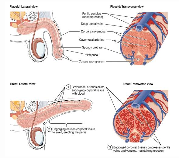
Stages of Penile Erection and Vascular Engorgement
Mechanism of Penile Erection
- Parasympathetic stimulation promotes release of nitric oxide (NO), with contributions from ACh and VIP.
- NO activates guanylyl cyclase, increasing cyclic GMP (cGMP).
- cGMP relaxes penile arterial and trabecular smooth muscle increasing blood flow into the corpora cavernosa and corpus spongiosum, producing erection.
- Increased blood flow also stimulates endothelial NO release, further enhancing vasodilation.
Summary: Parasympathetic stimulation → NO → ↑ cGMP → smooth muscle relaxation → ↑ penile blood flow → erection.
Erectile Tissue During Erection
- Corpora cavernosa contain large vascular sinusoids that are relatively empty when the penis is flaccid.
- During erection, arterial inflow increases while venous outflow is reduced, causing the sinusoids to fill with blood.
- The surrounding strong fibrous tissue limits expansion, raising intracavernosal pressure.
- This makes the penis enlarged, elongated, and rigid.
Summary: ↑ Arterial inflow + ↓ venous outflow → sinusoidal filling → ↑ pressure → penile rigidity and erection.
Role of nitric oxide in erection of the penis:
- Nitric oxide is released by parasympathetic axons in the penis and the endothelial cells in blood vessels of the penis.
- Nitric oxide activates guanylate cyclase, causing the production of cGMP from GTP.
- cGMP causes Ca2+ ion channels to close in the plasma membrane of the vascular smooth muscle cells decreasing the cytoplasmic Ca2+ ions concentration.
- This produces relaxation of the smooth muscle cells causing vasodilation in the penis and engorgement of the erectile tissue.
- Viagra and related drugs inhibit the phosphodiesterase (PDE-5) that catalyzes the breakdown of cGMP, thereby increasing the availability of cGMP to promote an erection.
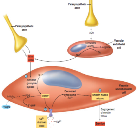
Role of Nitric Oxide and cGMP in Penile Erection
2. Lubrication — A Parasympathetic Function
- During sexual stimulation, parasympathetic impulses (S2–S4) stimulate the bulbourethral and urethral glands to secrete mucus.
- This mucus enters the urethra and helps lubricate the urethral passage and glans penis during sexual activity.
- Male lubrication is relatively small; most lubrication during coitus is provided by secretions from the female genital tract.
- Adequate lubrication reduces friction and discomfort, thereby facilitating sexual activity.
- Inadequate lubrication may cause painful friction, which can inhibit sexual arousal and interfere with the sexual response.
Summary: Parasympathetic stimulation → bulbourethral/urethral mucus secretion → reduced friction → facilitates coitus.
3. Emission and Ejaculation
- Emission and ejaculation represent the final phases of the male sexual act.
- As sexual stimulation intensifies, sympathetic impulses from T11–L2 travel mainly through the hypogastric nerves and pelvic sympathetic plexus.
Emission
- Sympathetic stimulation causes contraction of the:
- vas deferens → moves sperm into the posterior urethra,
- seminal vesicles → add seminal fluid,
- prostate gland → adds prostatic secretions.
- These secretions mix with sperm and bulbourethral mucus to form semen in the urethra.
Summary pathway: Intense sexual stimulation → sympathetic activation (T11–L2) → contraction of vas deferens + accessory glands → semen enters posterior urethra = emission.
Note: Emission is mainly sympathetic, whereas the forceful expulsion of semen during ejaculation also involves somatic motor activity through the pudendal nerve.
4. Ejaculation, Orgasm, and Resolution
- Accumulation of semen in the posterior urethra activates sensory receptors, sending afferent impulses to the sacral spinal cord and triggering the ejaculatory reflex.
- Somatic impulses through the pudendal nerve produce rhythmic contractions of the bulbospongiosus and ischiocavernosus muscles.
- These contractions, together with contractions of the reproductive ducts and urethra, generate pressure that expels semen through the urethra to the exterior. This is ejaculation.
- Rhythmic contractions of the pelvic floor muscles may accompany ejaculation and contribute to pelvic thrusting.
- Orgasm is the intense pleasurable sensation that usually accompanies emission and ejaculation.
- After ejaculation, sexual arousal decreases and the penis returns to the flaccid state; this phase is called resolution and is usually followed by a refractory period.
Summary pathway: Semen enters posterior urethra → spinal ejaculatory reflex → pudendal nerve activation → rhythmic pelvic muscle contractions → semen expulsion → orgasm → resolution.
Male Orgasm and Phases of the Male Sexual Response
Intense pleasurable sensation that usually accompanies emission and ejaculation, resulting from coordinated autonomic, somatic, spinal, and brain activity during the male sexual response.
- Arousal/Erection Phase: Sexual stimulation activates parasympathetic pathways (S2–S4). Increased blood flow fills the erectile tissues, producing penile erection.
- Lubrication Phase: The bulbourethral (Cowper’s) and urethral glands secrete mucus. This secretion lubricates the urethra and glans penis and reduces friction.
- Emission Phase: Mainly mediated by sympathetic nerves (T11–L2). Sperm are propelled from the vas deferens into the posterior urethra. The seminal vesicles and prostate contract and add their secretions to form semen. The internal urethral sphincter/bladder neck contracts, helping prevent semen from entering the urinary bladder.
- Ejaculation and Orgasm: Filling of the posterior urethra triggers the ejaculatory reflex. Rhythmic contractions of the bulbospongiosus, ischiocavernosus, pelvic floor muscles, and reproductive ducts propel semen through the urethra. These contractions may be accompanied by pelvic thrusting. There are general autonomic responses which include increased heart rate, blood pressure, and respiratory rate.
- Resolution and Refractory Period: After ejaculation, sympathetic tone and sexual arousal decline. Blood leaves the erectile tissues, causing the return of the penis to the flaccid state. Physiological responses gradually return to baseline; relaxation or sleepiness may occur. A refractory period usually follows, during which another erection and ejaculation are temporarily difficult or impossible.
Summary Pathway: Arousal/erection → lubrication → emission → ejaculation/orgasm → resolution → refractory period.
| Sexual Response Phase | Predominant Innervation | Primary Neurotransmitters / Signals | Primary Effector Organs & Action |
|---|---|---|---|
| Erection | Parasympathetic (S2–S4 via Pelvic nerves) | Nitric Oxide (NO), ACh, VIP, cGMP | Cavernous helicine arteries dilate; venous compression |
| Lubrication | Parasympathetic (S2–S4) | Acetylcholine | Bulbourethral and urethral glands release clear mucus |
| Emission | Sympathetic (T11–L2 via Hypogastric nerves) | Norepinephrine | Vas deferens, seminal vesicles, prostate, internal sphincter |
| Ejaculation | Somatic motor (S2–S4 via Pudendal nerve) | Acetylcholine (nicotinic) | Bulbospongiosus and ischiocavernosus rhythmic spasm |
| Resolution | Sympathetic tone elevation | Norepinephrine (alpha-1 vasoconstriction) | Arterial vasoconstriction; blood exits cavernous spaces |
Basic Sexual Functions of the Male: Testosterone
- Principal male sex hormone (androgen).
- Steroid hormone secreted mainly by the interstitial cells of Leydig in the testes.
- Secretion is stimulated primarily by luteinizing hormone (LH) from the anterior pituitary.
- Testosterone is essential for the development and maintenance of the male reproductive organs, secondary sexual characteristics, spermatogenesis, sexual behavior, and anabolic functions.
Synthesis of Testosterone
- Synthesized from cholesterol in the Leydig cells.
- LH stimulates testosterone synthesis by activating steroidogenic enzymes within Leydig cells.
Transport of Testosterone in Blood
- Because testosterone is poorly soluble in plasma, most of it circulates bound to plasma proteins:
- Approximately 98% is protein-bound:
- About 65% is strongly bound to sex hormone-binding globulin (SHBG), previously called gonadal steroid-binding globulin.
- About 33% is loosely bound to albumin.
- Approximately 2% circulates as free testosterone.
Physiological Significance
- Free testosterone is readily available to enter target cells and exert biological effects.
- Albumin-bound testosterone is weakly bound and can readily dissociate; therefore, free plus albumin-bound testosterone is often referred to as bioavailable testosterone.
- SHBG-bound testosterone acts mainly as a circulating reservoir and is less immediately available to tissues.
Roles of Testosterone in Sexual Development and Function
1. Male Sexual Differentiation and Spermatogenesis
- Testosterone does not initiate formation of the testes; testicular differentiation is primarily determined by the SRY gene/testis-determining factor.
- During fetal life, testosterone produced by the Leydig cells promotes development of the Wolffian ducts into the: Epididymis, Vas deferens, Seminal vesicles and Ejaculatory ducts.
- Testosterone is converted by 5α-reductase to dihydrotestosterone (DHT), which promotes development of the: Penis, Scrotum, Prostate, Male urethra.
2. Growth and Maintenance of Male Reproductive Organs
- At puberty, testosterone and DHT stimulate the growth, maturation, function, and maintenance of male reproductive structures, including: Penis and scrotum, Epididymis, Vas deferens, Seminal vesicles, Prostate gland, Ejaculatory ducts and Bulbourethral glands.
3. Development of Male Secondary Sexual Characteristics
Testosterone is responsible for the development and maintenance of many characteristic male features after puberty.
a. Hair Growth and Male-Pattern Baldness
- Androgens stimulate growth of facial, pubic, axillary, chest, and other body hair.
- In genetically susceptible individuals, DHT acts on scalp hair follicles, particularly in the frontal and vertex regions, causing progressive follicular miniaturization and male-pattern baldness (androgenetic alopecia).
b. Libido and Sexual Function
- Testosterone is important for the development and maintenance of sexual desire (libido).
- Adequate testosterone also supports normal male sexual function and contributes to erectile responsiveness, although erection itself depends mainly on neural and vascular mechanisms.
c. Voice Changes
- Testosterone causes enlargement of the larynx and thickening of the vocal cords during puberty.
- This produces the characteristic deep male voice.
d. Muscle and Skeletal Development
- Testosterone increases protein synthesis and skeletal muscle mass, producing greater muscular development.
- It promotes bone growth, increases bone density, and contributes to the broader shoulders and characteristic male body build.
e. Behavioral Effects
- Testosterone influences sexual motivation, energy, mood, competitiveness, and certain aspects of behavior.
- It may contribute to increased assertiveness or aggression, but aggressive behavior is also strongly influenced by psychological, social, and environmental factors.
f. Testosterone increases the basal metabolic rate
- Testosterone can increase the basal metabolic rate. This increased rate of metabolism is possibly an indirect result of the effect of testosterone on protein anabolism, with the increased quantity of proteins—the enzymes especially— increasing the activities of all cells.
g. Testosterone increases red blood cells
- There is a strong association of testosterone and increased hematocrit (PCV).
- Testosterone does not appear to directly increase erythropoietin levels or red blood cell production, instead, the effect of testosterone to increase red blood cell production may be at least partly indirect because of the increased metabolic rate that occurs after testosterone administration.
Target Divergence: Testosterone vs. DHT
Testosterone: Mainly mediates internal male genital development (Wolffian derivatives), spermatogenesis, libido, muscle/skeletal mass, metabolic rate, and secondary sexual characteristics.
DHT (Dihydrotestosterone): Mediates external male genitalia virilization, prostate growth, facial and body hair growth, acne, and androgen-dependent scalp hair loss (androgenetic alopecia).
Male Infertility
Definition: Refers to infertility caused wholly or partly by male reproductive factors, resulting in failure to achieve pregnancy after 12 months or more of regular unprotected sexual intercourse.
Causes of Male Infertility
1. Low Sperm Count (Oligozoospermia)
- Refers to a reduced concentration of sperm in the ejaculate.
- Possible contributing factors include:
- Excessive testicular heat exposure
- Certain medications and chemotherapy
- Environmental or occupational toxins such as lead and arsenic
- Recreational drugs, including cannabis and cocaine
- Anabolic steroid use
- Hormonal disorders, infections, varicocele, and genetic abnormalities
Effect of Sperm Count on Fertility
- Normal fertility depends partly on an adequate semen volume and sperm number.
- Semen volume: lower reference limit ≈ 1.4 mL
- Sperm concentration: lower reference limit ≈ 16 million sperm/mL
- Total sperm number: lower reference limit ≈ 39 million sperm per ejaculate
Effect on Fertility
- Sperm concentration below about 16 million/mL may reduce the probability of conception.
- A low sperm count does not necessarily mean infertility; pregnancy may still occur.
- Fertility depends not only on sperm number but also on:
- Motility – ability of sperm to move effectively
- Morphology – normal sperm structure
- Vitality – proportion of living sperm
- Semen quality and reproductive tract function
- Female reproductive factors
Why Are Millions of Sperm Needed?
Although only one sperm fertilizes the ovum, millions are released because most sperm are lost, destroyed, or fail to reach the ampulla of the uterine tube, where fertilization usually occurs.
2. Poor Sperm Motility (Asthenozoospermia)
- Reduced ability of sperm to move effectively, particularly to move progressively forward.
- Normal sperm motility is essential for sperm to travel through the female reproductive tract and reach the ovum.
- Poor motility reduces the probability that sufficient sperm will reach and fertilize the ovum.
- Causes may include:
- abnormal semen viscosity,
- genital tract infections,
- varicocele,
- oxidative stress,
- excessive heat,
- toxins and certain drugs, and
- structural abnormalities of the sperm flagellum.
That is: ↓ Progressive sperm motility → ↓ sperm transport to the ovum → ↓ probability of fertilization.
3. Abnormal Spermatogenesis and Male Infertility
- Refers to impaired production or maturation of sperm within the seminiferous tubules of the testes.
Major causes include:
- Testicular inflammation (orchitis): severe or bilateral orchitis, including that associated with mumps, may damage testicular tissue and impair sperm production.
- Excessive testicular temperature: prolonged heat exposure can temporarily suppress spermatogenesis.
- Cryptorchidism: failure of testicular descent exposes the testes to higher abdominal temperatures and may impair sperm production.
- Genetic abnormalities, hormonal disorders, toxins, radiation, chemotherapy, and some drugs may also damage the seminiferous epithelium.
- Obstruction of the reproductive ducts can prevent normally produced sperm from reaching the ejaculate, causing obstructive infertility rather than defective spermatogenesis.
Thus: Damage to seminiferous tubules → impaired spermatogenesis → ↓ sperm number/quality → reduced male fertility.
4. Abnormal Sperm Morphology (Teratozoospermia)
- Refers to an abnormally high proportion of sperm with structural abnormalities. Normal sperm morphology is important for effective movement, penetration of the ovum, and fertilization.
- Abnormalities may involve the:
- Head – abnormal size or shape, double head
- Midpiece – thick, bent, or irregular
- Tail – short, coiled, bent, or multiple tails
- A man may have a normal sperm concentration but reduced fertility if a large proportion of sperm have abnormal morphology or poor motility.
- Abnormal sperm structure, particularly defects of the tail or midpiece, may also impair sperm movement through the female reproductive tract.
Thus: Abnormal morphology ± poor motility → impaired sperm transport and fertilizing ability → reduced male fertility.
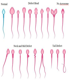
Abnormal infertile sperm, compared with a normal sperm on the right. Morphology of Sperm
Other Causes of Male Infertility
5. Hormonal Imbalance – Hypogonadism
- Deficiency of testosterone and/or gonadotropins (LH and FSH) may impair spermatogenesis.
- Causes include disorders of the hypothalamus, pituitary gland, or testes.
- Reduced FSH and intratesticular testosterone can result in low sperm production and infertility.
6. Varicocele
- Abnormal dilatation of the veins of the pampiniform plexus within the scrotum may increase testicular temperature and oxidative stress, thereby impairing sperm production, motility, and quality. It is a common potentially correctable cause of male infertility.
7. Cryptorchidism
- Failure of one or both testes to descend into the scrotum.
- Prolonged exposure of the testes to the higher abdominal or inguinal temperature may damage the seminiferous epithelium and impair spermatogenesis.
- Bilateral cryptorchidism carries a greater risk of infertility.
8. Testicular Trauma or Injury
- Direct injury to the testes may damage the seminiferous tubules, Leydig cells, or testicular blood supply.
- Severe or bilateral damage can reduce sperm production and testosterone secretion.
9. Cancer Treatment
- Chemotherapy and radiotherapy can damage rapidly dividing germ cells within the testes.
- Depending on the treatment type and dose, this may cause temporary or permanent oligospermia or azoospermia.
10. Genetic Disorders – Klinefelter Syndrome
- Chromosomal disorder in males, most commonly characterized by an additional X chromosome (47,XXY).
- It causes primary testicular failure, usually associated with:
- Small, firm testes
- Reduced testosterone
- Increased FSH and LH
- Markedly reduced sperm production or azoospermia and Infertility
11. Seminal Fructose
- Fructose is produced mainly by the seminal vesicles and provides an important energy source for spermatozoa.
- Its secretion is androgen-dependent.
- Low or absent seminal fructose may suggest:
- Seminal vesicle dysfunction
- Ejaculatory duct obstruction
- Congenital abnormalities of the seminal vesicles or vas deferens
- Severe androgen deficiency
12. Vitamin B12 and Vitamin E Deficiencies
a. Vitamin B12
- Essential for DNA synthesis, cell division, and normal sperm production.
- Deficiency may impair spermatogenesis and may be associated with reduced sperm count, motility, and overall sperm quality.
- Correcting a true vitamin B12 deficiency may therefore support normal reproductive function.
b. Vitamin E
- Fat-soluble antioxidant that protects sperm cell membranes from oxidative damage.
- Adequate vitamin E may help maintain:
- Sperm membrane integrity
- Sperm motility
- Sperm viability
- Vitamin E deficiency may increase susceptibility of sperm to oxidative stress and lipid peroxidation.
c. Vitamin C
- Antioxidant and can help regenerate oxidized vitamin E.
- Together, vitamins C and E contribute to protection of sperm from reactive oxygen species (ROS).
Fertility Is Reduced When:
- Semen abnormalities are present, such as:
- Reduced semen volume
- Low seminal fructose
- Increased semen viscosity
- Sperm abnormalities are present, including:
- Reduced sperm count
- Reduced sperm motility
- Increased proportion of abnormal sperm forms (abnormal morphology)
Management Strategy in Male Infertility
Male fertility depends on both semen quality and sperm quality. Abnormalities in volume, seminal composition, sperm number, motility, or morphology can reduce the likelihood of successful fertilization.
Male infertility is very resistant to treatment. The best approach is to treat the woman, so as to maximize her fertility, hence create room for the couple to have children.
Treatment of Male Infertility
Treatment is cause-specific and should be based on clinical evaluation, semen analysis, hormonal assessment, and other investigations when indicated.
1. Treatment of Genital Tract Infections
- Identify and treat confirmed reproductive or urinary tract infections.
- Culture and sensitivity testing or other microbiological tests may be performed when infection is suspected to identify the organism and guide appropriate antibiotic therapy.
- Leukocytes in semen alone do not necessarily indicate bacterial infection and should not automatically lead to antibiotic treatment.
2. Treatment of Hormonal Abnormalities
- Hormonal therapy depends on the underlying endocrine disorder.
- For hypogonadotropic hypogonadism, fertility may be restored using:
- Human chorionic gonadotropin (hCG) – stimulates Leydig cells to produce testosterone.
- FSH or human menopausal gonadotropin (hMG) – may be added to stimulate Sertoli cells and spermatogenesis.
- Pulsatile GnRH may be used in selected patients with hypothalamic GnRH deficiency.
3. Avoid Exogenous Testosterone in Men Seeking Fertility
- Testosterone preparations should NOT be used to treat low sperm count in men who wish to father children.
- Exogenous testosterone causes negative feedback on the hypothalamic–pituitary–testicular axis:
Mechanism: Testosterone administration → ↓ GnRH → ↓ LH and FSH → ↓ intratesticular testosterone → ↓ spermatogenesis. This may result in oligospermia or complete azoospermia.
5. Nutritional Treatment
- Correct documented nutritional deficiencies, including deficiencies of vitamin B12, folate, zinc, or other essential nutrients.
- Antioxidants such as vitamins C and E have been studied because they may reduce oxidative stress in sperm.
- However, routine antioxidant or vitamin supplementation has not consistently been shown to improve pregnancy or live-birth rates and should not replace treatment of an identifiable cause.
6. Treatment of Varicocele
- In infertile men with a clinical varicocele and abnormal semen parameters, treatment may improve semen quality and fertility.
- Options include:
- Varicocelectomy, particularly microsurgical repair
- Radiological embolization in selected patients, i.e blocking the abnormal scrotal vein from inside the blood vessel without open surgery.
7. Treatment of Reproductive Tract Obstruction
- Obstruction of the epididymis, vas deferens, or ejaculatory ducts may be managed by:
- Surgical reconstruction, where appropriate
- Surgical sperm retrieval for use in assisted reproduction
8. Lifestyle Modification
Patients should be advised to:
- Stop smoking and recreational drug use
- Avoid anabolic steroids and exogenous testosterone
- Limit excessive alcohol consumption
- Avoid excessive testicular heat exposure
- Maintain a healthy body weight
- Manage chronic diseases appropriately
9. Assisted Reproductive Techniques
When natural conception remains difficult, assisted reproductive techniques may be considered:
- Intrauterine insemination (IUI)
- In vitro fertilization (IVF)
- Intracytoplasmic sperm injection (ICSI)
- Surgical sperm retrieval combined with IVF/ICSI in selected cases of azoospermia
10. Fertility Preservation
- Before treatments that may damage spermatogenesis, particularly chemotherapy or radiotherapy, sperm should preferably be collected and cryopreserved whenever possible.
Cryopreservation
- Process of preserving biological cells, tissues, or reproductive material at very low temperatures so they can be stored and used later.
- In reproductive medicine, it commonly refers to freezing:
- Sperm
- Eggs (oocytes)
- Embryos
- Sometimes testicular or ovarian tissue
- The material is usually stored in liquid nitrogen at about −196°C, which greatly slows or stops cellular metabolism and preserves viability.
Physiology of the Female Sexual Act
Female sexual act involves coordinated psychological, sensory, autonomic, vascular, and muscular responses that prepare the reproductive tract for sexual intercourse and may culminate in orgasm.
1. Sexual Arousal
Female sexual arousal may be initiated by:
- Psychological stimuli – thoughts, emotions, visual or auditory stimuli
- Tactile stimulation of the:
- Clitoris
- Labia
- Vagina
- Breasts and nipples
- Other sensitive areas
- Sensory impulses are transmitted mainly through the pudendal nerve to the sacral spinal cord.
2. Parasympathetic Response
- Parasympathetic nerves arising from S2–S4 play an important role in genital arousal.
- They cause:
- Dilatation of genital blood vessels
- Increased blood flow to the clitoris and vulva
- Engorgement of the erectile tissues
- Vaginal lubrication
3. Clitoral and Vulval Engorgement
During sexual stimulation:
- Clitoris becomes enlarged and erect because of increased blood flow.
- Labia minora and other vulval tissues become congested.
- These changes increase sensitivity and facilitate sexual intercourse.
4. Vaginal Lubrication
- Lubrication is essential for reducing friction during intercourse.
- It is produced mainly by:
- Transudation of fluid through the vaginal wall due to increased blood flow.
- Secretions from the greater vestibular (Bartholin) glands, which help lubricate the vaginal opening.
- Small amounts of cervical and other genital tract secretions may also contribute.
5. Vaginal and Uterine Changes
During arousal:
- The vagina becomes longer and wider, especially in its upper part.
- The vaginal walls become more congested.
- The uterus may elevate slightly within the pelvis, producing vaginal tenting.
- These changes facilitate penile penetration and semen deposition.
6. Female Orgasm
- With sufficient sexual stimulation, orgasm may occur.
- It is characterized by:
- Intense pleasurable sensation
- Rhythmic contractions of the:
- Pelvic floor muscles
- Vaginal muscles
- Uterus in some women
- Increased heart rate, respiratory rate, and blood pressure
- Increased generalized muscle tension
- Orgasm involves coordinated activity of the autonomic and somatic nervous systems.
7. Role of the Sympathetic Nervous System
- Sympathetic activity contributes particularly during orgasm and produces rhythmic contractions of the reproductive tract and associated pelvic muscles.
8. Resolution
After sexual stimulation ends:
- Genital vasocongestion gradually decreases.
- The clitoris and vulval tissues return to their resting state.
- Vaginal and uterine changes subside.
- Heart rate, respiration, and muscle tension return toward normal.
Causes and Treatment of Female Infertility
Female infertility may result from disorders affecting ovulation, the ovaries, fallopian tubes, uterus, cervix, or hormonal regulation.
1. Ovulatory Disorders
Failure or irregularity of ovulation is one of the commonest causes. Examples include:
- Polycystic ovary syndrome (PCOS): common hormonal disorder in women of reproductive age characterized by irregular or absent ovulation, excess androgen activity, and/or polycystic-appearing ovaries.
- Hypothalamic dysfunction due to severe stress, excessive exercise, or marked weight loss: Excessive exercise and marked weight loss can reduce female fertility by suppressing the hypothalamic–pituitary–ovarian (HPO) axis.
That is: ↓ GnRH → ↓ FSH and LH → impaired follicular development → ↓ estrogen → anovulation or amenorrhea → reduced fertility.
- Hyperprolactinemia: ↑ Prolactin → ↓ GnRH → ↓ FSH and LH → impaired follicular development and ovulation → infertility.
- Thyroid disorders: Thyroid dysfunction → altered GnRH/FSH/LH and prolactin balance → disturbed follicular development and ovulation → menstrual abnormalities → reduced fertility.
- Premature ovarian insufficiency: ↓ Ovarian follicular function → ↓ estrogen and inhibin → loss of negative feedback → ↑ FSH and LH → irregular or absent ovulation → reduced fertility.
These conditions may disturb GnRH, FSH, and LH secretion and interfere with normal follicular development and ovulation.
2. Increasing Age and Reduced Ovarian Reserve
- Female fertility declines progressively with age, particularly after the mid-30s.
- This occurs because of a reduction in both the number and quality of oocytes.
3. Fallopian Tube Damage or Obstruction
- Tubal blockage prevents sperm from reaching the ovum or prevents the fertilized ovum from reaching the uterus.
- Causes include:
- Pelvic inflammatory disease (PID)
- Previous sexually transmitted infections, especially chlamydia or gonorrhea
- Previous pelvic surgery
- Pelvic adhesions
- Genital tuberculosis in some populations
4. Endometriosis
- Endometrial-like tissue grows outside the uterus.
- It may cause pelvic inflammation, adhesions, tubal distortion, and impaired ovarian function, thereby reducing fertility.
5. Uterine Abnormalities
Conditions that may interfere with implantation or maintenance of pregnancy include:
- Uterine fibroids, particularly those distorting the uterine cavity
- Endometrial polyps
- Intrauterine adhesions
- Congenital uterine abnormalities
6. Cervical Factors
Abnormalities of the cervix or cervical mucus may interfere with sperm passage. Examples include:
- Cervical stenosis
- Previous cervical surgery and Inadequate or abnormal cervical mucus
7. Hyperandrogenism
Excess androgen production, commonly associated with PCOS, may cause:
- Irregular or absent menstruation
- Anovulation
- Acne
- Hirsutism
- Reduced fertility
8. Psychological and Lifestyle Factors
- Severe or chronic stress may alter hypothalamic function and contribute to menstrual or ovulatory disturbances.
- Other factors include:
- Very low or excessive body weight
- Excessive strenuous exercise
- Smoking
- Heavy alcohol consumption and Poorly controlled chronic diseases
9. Timing of Intercourse
- Failure to have intercourse during the fertile window around ovulation can reduce the chance of conception.
- However:
- Sexual position has not been shown to determine fertility.
- Standing up after intercourse does not cause infertility.
- Leakage of some semen from the vagina after intercourse is normal and does not mean that all sperm have been lost.
10. Unexplained Infertility
- In some couples, routine investigations show normal ovulation, patent fallopian tubes, normal reproductive anatomy, and satisfactory semen parameters, yet conception does not occur.
- This is termed unexplained infertility.
Treatment of Female Infertility
Treatment depends on the underlying cause, age of the woman, duration of infertility, ovarian reserve, and partner factors.
1. Treat Ovulatory Disorders
- PCOS: lifestyle modification and ovulation induction, commonly with letrozole.
- Hyperprolactinemia: dopamine agonists such as cabergoline or bromocriptine.
- Thyroid disorders: correct hypo- or hyperthyroidism.
- Hypogonadotropic hypogonadism: may require gonadotropins or pulsatile GnRH.
2. Correct Tubal or Pelvic Disease
- Treat pelvic infections appropriately.
- Selected cases of tubal blockage or adhesions may require laparoscopic surgery.
- Severe bilateral tubal disease is often managed with IVF.
3. Treat Endometriosis
- Mild disease may be managed surgically when fertility is desired.
- Moderate or severe disease may require laparoscopic treatment or assisted reproduction.
4. Treat Uterine Abnormalities
Conditions that distort the uterine cavity may be corrected, for example:
- Submucosal fibroids
- Endometrial polyps
- Intrauterine adhesions
- Selected congenital uterine abnormalities
5. Lifestyle and General Health
Encourage:
- Healthy body weight
- Adequate nutrition
- Smoking cessation
- Limiting excessive alcohol
- Moderate physical activity
- Good control of chronic illnesses
- Intercourse during the fertile window
6. Assisted Reproductive Techniques
When simpler treatment fails or is unsuitable:
- Intrauterine insemination (IUI)
- In vitro fertilization (IVF)
- Intracytoplasmic sperm injection (ICSI) when male-factor infertility is also present
- Donor oocytes may be considered in selected cases of severe ovarian insufficiency.
7. Fertility Preservation
Before chemotherapy, radiotherapy, or other treatments that may damage ovarian function, fertility may be preserved by:
- Oocyte cryopreservation
- Embryo cryopreservation
- Ovarian tissue cryopreservation in selected patients
Puberty and Adolescence
- During childhood, the hypothalamic–pituitary–gonadal (HPG) axis is relatively inactive, so levels of GnRH, FSH, LH, and gonadal sex hormones remain low.
Puberty
- Stage when the gonads become functionally mature, leading to the onset of gamete production, reproductive capability, and secondary sexual characteristics.
- It begins with increased secretion of GnRH from the hypothalamus.
- GnRH stimulates the anterior pituitary to release FSH and LH, which act on the gonads to produce:
- Testosterone in males &
- Estrogens and progesterone in females
- These hormones cause maturation of the reproductive organs, development of secondary sexual characteristics, and establishment of reproductive function.
Adolescence
Broader transitional period from childhood to adulthood that includes the physical, psychological, emotional, and social changes associated with puberty.
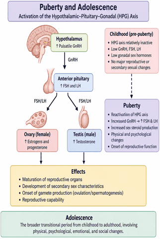
Puberty and Adolescence: Activation of the Hypothalamic–Pituitary–Gonadal (HPG) Axis
Age of Puberty
- Puberty is the period during which the reproductive system matures and an individual develops secondary sexual characteristics and reproductive capability.
- It results from reactivation of the hypothalamic–pituitary–gonadal (HPG) axis:
Pathway: ↑ Pulsatile GnRH → ↑ FSH and LH → activation of gonads → ↑ sex hormones → pubertal changes.
- Puberty normally begins earlier in girls than in boys.
Normal Age of Onset
- Girls: approximately 8–13 years, with breast development commonly beginning around 10 years.
- Boys: approximately 9–14 years, with testicular enlargement commonly beginning around 11–12 years.
- These ages vary among individuals and populations.
Factors Affecting the Age of Puberty
1. Genetic Factors
- Genetics is one of the strongest determinants of pubertal timing.
- Children often begin puberty at approximately the same age as their parents did.
- Genetic factors influence the timing of activation of the GnRH pulse generator.
2. Nutrition and General Health
- Adequate nutrition is necessary for normal reproductive maturation. Undernutrition and chronic illness may delay puberty.
- Improved nutrition and general health may be associated with earlier pubertal development.
- Severe nutritional deficiency reduces the energy available for growth and reproductive function.
3. Body Fat and Energy Availability
- Adequate energy stores are important for the onset and maintenance of puberty.
- Adipose tissue produces leptin, which provides information to the hypothalamus about the body's energy reserves.
Pathway: Adequate body fat → ↑ leptin signalling → supports GnRH secretion → puberty.
- Very low body fat or negative energy balance may:
- Reduce GnRH pulsatility
- Reduce FSH and LH secretion
- Delay ovarian maturation
- Delay menarche
- This may occur in individuals with:
- Severe undernutrition
- Eating disorders
- Intense athletic training
- Very low body weight
4. Environmental Factors
- Environmental influences may modify pubertal timing, including:
- Exposure to endocrine-disrupting chemicals
- Living conditions
- Chronic illness
- Geographic and population-related factors
5. Socioeconomic Factors
- Socioeconomic conditions affect puberty mainly through their influence on:
- Nutrition
- Healthcare
- Chronic disease
- Physical activity
- Living environment
6. Physical Activity
- Moderate physical activity normally does not delay puberty.
- However, very intense exercise combined with inadequate energy intake may suppress the HPG axis and delay puberty.
- This is particularly observed in some:
- Competitive gymnasts
- Dancers
- Long-distance runners
- High-intensity athletes
7. Psychological and Chronic Stress
- Severe or prolonged stress may alter hypothalamic function and interfere with GnRH secretion.
- Chronic stress may therefore contribute to abnormal or delayed pubertal development, although its effects vary between individuals.
8. Chronic Diseases
- Chronic systemic illnesses may delay puberty, particularly when associated with poor nutrition or reduced body weight.
- Examples include:
- Chronic kidney disease
- Chronic gastrointestinal diseases
- Severe endocrine disorders
- Chronic inflammatory diseases
First Pubertal Changes in Boys
The first sign of puberty in boys is usually testicular enlargement, not growth of facial hair or deepening of the voice.
Typical Sequence
- Testicular enlargement – usually the first sign
- Enlargement of the scrotum (Hyperpigmentation of the scrotum and presence of rugae folds) and penis
- Pubic and axillary hair growth
- Growth spurt
- Voice deepening
- Facial and body hair development including temporal hair recession due to DHT (Dihydrotestosterone)
- Increasing muscle mass
- Attainment of adult height (Linear and lateral broadening of the chest)
- Spermatogenesis commences (sperm production) and first ejaculation occurs
- Growth of accessory reproductive organs
Pubertal Changes in Girls
First visible sign of puberty in most girls is breast development.
1. Breast Development – Thelarche
- Usually the first sign of female puberty.
- Caused mainly by rising estrogen levels.
- Breast tissue and the nipple–areolar complex begin to enlarge.
2. Pubic and Axillary Hair – Pubarche
- Pubic hair appears, followed later by axillary hair. Pubic hair appears in the pubic region in a characteristic flat-topped pattern.
- Females develop less body hair than males, but more scalp hair is present.
- Increased sexual drive (libido) in females due to stimulation by estrogen.
- High pitched and thin voice.
- Ovulation commences and changes in the cervical vaginal mucus.
3. Growth Spurt and Body Development
- Rapid increase in height occurs during early to mid-puberty. Estrogen, growth hormone, and IGF-1 contribute to accelerated skeletal growth.
- Girls usually experience their peak growth velocity before menarche.
- Deposition of adipose tissue in the buttocks and thigh and pigmentation of the areolae and nipples increases somewhat.
- Body configuration develops in such a manner that women have narrow shoulders, broad hips, convergent thighs and divergent arms.
- Increase in growth and increase in the size of uterus, vagina, labia, clitoris.
4. Menarche
- Menarche is the first menstrual period.
- It usually occurs approximately 2–3 years after the onset of breast development.
- Menarche indicates advanced reproductive maturation but does not necessarily mean that all early menstrual cycles are ovulatory.
- Early post-menarche cycles are often irregular because maturation of the HPG axis is still continuing.
| Milestone | Female Development | Male Development |
|---|---|---|
| First Physical Sign | Thelarche (Breast bud development ~10 years) | Testicular enlargement (≥4 mL volume ~11–12 years) |
| Pubic Hair Growth | Pubarche / Adrenarche (Flat-topped triangle pattern) | Pubarche (Diamond pattern extending toward umbilicus) |
| Growth Spurt Timing | Occurs early (peak velocity before menarche) | Occurs late in puberty (with muscle broadening) |
| Reproductive Milestone | Menarche (~2–3 years post-thelarche, ~12.5 years) | Spermarche (commencement of spermatogenesis & first ejaculation) |
Clinical Significance of Abnormal Pubertal Timing
1. Precocious Puberty
Appearance of secondary sexual characteristics earlier than expected:
- Before 8 years in girls
- Before 9 years in boys
It may result from early activation of the hypothalamic–pituitary–gonadal axis or from excess sex hormone production independent of this axis.
Clinical Importance
Early puberty may cause:
- Accelerated growth initially
- Early closure of epiphyseal growth plates
- Reduced final adult height
- Psychological and social difficulties
Clinical evaluation is required to determine the underlying cause.
2. Delayed Puberty
Failure of normal pubertal development to begin within the expected age range.
Evaluation is generally indicated when:
- Girls: no breast development by about 13 years
- Boys: no testicular enlargement by about 14 years
Possible Causes
- Constitutional delay of growth and puberty
- Chronic illness or undernutrition
- Hypothalamic or pituitary disorders
- Primary gonadal failure
Central vs. Peripheral Precocious Puberty
Central (GnRH-Dependent): Premature reactivation of the hypothalamic pulse generator → high LH/FSH in pubertal range with harmonious sequence of development. Treated with continuous long-acting GnRH agonists (e.g., leuprolide) to downregulate pituitary receptors.
Peripheral (GnRH-Independent): Autonomous peripheral secretion of sex steroids (e.g., Leydig cell tumors, granulosa cell tumors, Congenital Adrenal Hyperplasia, McCune-Albright syndrome) → suppressed/low LH and FSH.
