Learning objectives
Male Reproductive Physiology and Sexual Development
- Sex determination and sexual differentiation
- Disorders of sexual development
- Functional anatomy of the male reproductive system
- Physiology and regulation of spermatogenesis
- Semen formation, composition, and physiological characteristics
- Sperm cryopreservation and sperm banking
- Cryptorchidism (undescended testis): physiological consequences and management
- Determinants of male fertility, Causes, assessment, and management of male infertility
- Synthesis, secretion, transport, metabolism, and physiological actions of testosterone
- Role of testosterone in male sexual differentiation and development
- Puberty and adolescence
- Development of male and female secondary sexual characteristics
- Hypothalamic–pituitary–gonadal (HPG) axis and regulation of male reproductive function
- Hormonal control of testicular function
- Sexual acts in male and female
- Male Orgasm
- Male sexual response and deposition of spermatozoa in the female reproductive tract:
- Erection
- Lubrication
- Emission
- Ejaculation
Male Sexual Response ("Point and Shoot")
Point (Parasympathetic): Pelvic splanchnic nerves (S2–S4) mediate vascular smooth muscle relaxation via nitric oxide, causing erection, along with bulbourethral gland secretion for lubrication.
Shoot (Sympathetic & Somatic): Hypogastric nerves (T11–L2) stimulate peristalsis of ductus deferens and accessory organs during emission; somatic pudendal nerves (S2–S4) trigger rhythmic contractions of bulbospongiosus muscles for ejaculation.
Female Reproductive Physiology
- Functional anatomy of the female reproductive system
- Synthesis, secretion, transport, metabolism, and physiological actions of female sex hormones
- Hypothalamic–pituitary–ovarian axis and hormonal regulation of female reproductive function
- Determinants of female fertility
- Oogenesis and follicular development
- Female reproductive cycle
- Ovulation and its hormonal regulation
- Menstrual (uterine) cycle
- Menopause and associated physiological changes
- Fertilization and early embryonic development
- Implantation
- Placentation and establishment of pregnancy
- Physiological changes and adaptations during pregnancy
- Human chorionic gonadotropin (hCG): secretion and physiological functions
- Parturition and its hormonal regulation
- Breast development (mammogenesis)
- Lactation and hormonal control of milk production
- Milk ejection reflex and breastfeeding
- Contraception: physiological basis and methods
- Female infertility: causes, evaluation, and management
Sex Determination and Differentiation
Reproduction
- Biological process by which living organisms produce offspring.
- Ensures the continuity and survival of the species from one generation to the next.
- In humans, reproduction involves the coordinated functions of the male and female reproductive systems, reproductive hormones, gamete formation, fertilization, pregnancy, and childbirth.
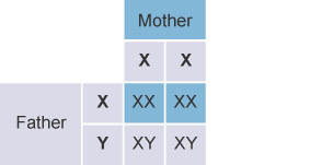
Sex Determination
- Process by which the genetic sex of an individual is established at fertilization.
Chromosomal Basis
- Humans have 46 chromosomes, including 2 sex chromosomes.
- Females: 46,XX
- Males: 46,XY
Role of the Gametes
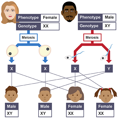
- The ovum always carries an X chromosome.
- Sperm may carry either:
- X chromosome → XX → genetic female
- Y chromosome → XY → genetic male
- Therefore, the sperm determines the genetic sex of the offspring.
Role of the Y Chromosome
- The Y chromosome contains the SRY gene (Sex-determining Region of Y).
- SRY produces testis-determining factor (TDF), which initiates differentiation of the primitive gonad into a testis.
- In the absence of functional SRY, the gonad normally develops along the ovarian pathway.
Determination vs. Differentiation Timeline
Sex determination occurs at fertilization (genetic programming), whereas sexual differentiation occurs later during embryonic development under the influence of genes and hormones (morphological execution).
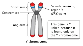
Sex-determining region Y (SRY) gene on Y chromosome
Sex Pre-selection or Chromosomal Basis of Sex Determination
- Human somatic cells normally contain 46 chromosomes (23 pairs):
- 44 autosomes (22 pairs)
- 2 sex chromosomes (1 pair)
- Of the 46 chromosomes:
- 23 are inherited from the mother
- 23 are inherited from the father
- Females normally have 44 autosomes + XX sex chromosomes (46,XX).
- Males normally have 44 autosomes + XY sex chromosomes (46,XY).
Formation of Gametes
- Male and female gametes develop from germ cells in the gonads:
- Spermatogonia in the testes give rise to spermatozoa.
- Oogonia in the ovaries give rise ultimately to mature ova (oocytes).
- Spermatogonia and oogonia are initially diploid (2n = 46 chromosomes).
- Through meiosis, the chromosome number is reduced by half, producing haploid gametes containing 23 chromosomes.
Sex Chromosomes in Gametes
- Each mature ovum contains 22 autosomes + X chromosome.
- Spermatozoa are of two types:
- 22 autosomes + X chromosome
- 22 autosomes + Y chromosome
- Therefore, the sperm determines the chromosomal sex of the offspring:
- X-bearing sperm + X ovum → XX → genetic female
- Y-bearing sperm + X ovum → XY → genetic male
Thus: Mother always contributes X → Father contributes X or Y → Sex is established at fertilization.
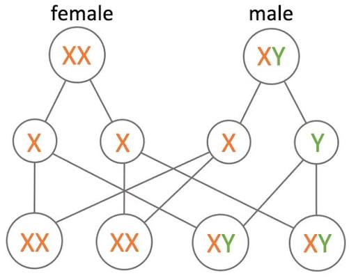
Role of Sperm in Sex Determination
- A single ejaculate contains sperm carrying either an X chromosome or a Y chromosome.
- Because the ovum always contributes an X chromosome, the chromosomal sex of the offspring is determined by whether an X-bearing or Y-bearing sperm fertilizes the ovum.
- X-bearing sperm + X ovum → XX → genetic female
- Y-bearing sperm + X ovum → XY → genetic male
Important Scientific Note
| Gamete Type | Physical Characteristics | Motility & Viability |
|---|---|---|
| Y-bearing sperm | Smaller, lighter | Faster swimming speed; shorter-lived |
| X-bearing sperm | Larger, heavier | Slower swimming speed; longer-lived |
- Father determines the chromosomal sex of the offspring because his sperm contributes either an X or a Y chromosome.
Shettles Hypothesis (Sperm Dynamics)
Y-Sperm: "Fast Sprinters" — lighter, rapidly reach the ampulla, but have low stamina and die quickly.
X-Sperm: "Endurance Marathoners" — larger DNA payload, slower pace, but resilient in acidic vaginal environments with prolonged survival.
Timing of Intercourse and Sex of the Offspring
- Therefore, intercourse very close to ovulation may favor conception of a male child because Y-bearing sperm were thought to move faster and reach the ovum first.
- Similarly, intercourse 2–3 days before ovulation was proposed to favor conception of a female child because X-bearing sperm were believed to survive longer in the female reproductive tract.
Fertile Period
- After ovulation, the oocyte remains capable of fertilization for approximately 12–24 hours.
- Sperm can remain viable in the female reproductive tract for up to about 5 days, particularly in favorable cervical mucus.
- Therefore, the fertile window extends from approximately 5 days before ovulation to about 1 day after ovulation.
Sexual differentiation in human
Determination of chromosomal sex and development of embryonic gonads
Sex Differentiation in Humans
- Series of developmental processes through which an embryo develops male or female reproductive structures and sexual characteristics.
- At fertilization, chromosomal sex is determined:
- XX → genetic female
- XY → genetic male
- During early embryonic development, the gonads are initially indifferent (bipotential) and can develop into either testes or ovaries.
- Gonadal differentiation is strongly influenced by the SRY gene (Sex-determining Region of the Y chromosome).
- SRY gene promotes formation of the testis-determining factor (TDF), which initiates differentiation of the indifferent gonad into a testis.
Therefore:
- Presence of functional SRY/TDF → testicular development → male sexual differentiation
- Absence of functional SRY/TDF → ovarian developmental pathway → female sexual differentiation
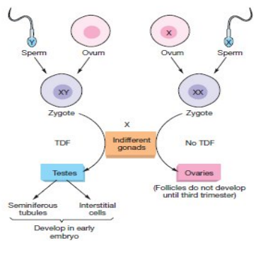
Determination of chromosomal sex and development of embryonic gonads
Main Stages of Sexual Differentiation
1. Gonadal Differentiation (Gonadogenesis)
- Process by which the gonads differentiate and mature into testes or ovaries.
- In early embryonic development, both male and female embryos possess indifferent (bipotential) gonads, which develop from the genital ridges.
- Up to about the 6th week of development, the gonads are morphologically similar in both sexes and have the potential to develop into either testes or ovaries.
- In a genetic male (46, XY), the SRY gene on the Y chromosome activates testis-forming pathways, leading to development of the testes and suppress pathways that favor ovarian development.
- In the absence of a functional SRY gene, ovarian-promoting pathways predominate and the indifferent gonad develops into an ovary.
- During ovarian development, oogonia proliferate, enter meiosis to form primary oocytes, and become enclosed by follicular cells to form primordial follicles.
Summary: Genital ridge → Indifferent gonad → SRY present → Testis | SRY absent → Ovary
2. Differentiation of the Internal Genital Ducts
- Two paired duct systems are initially present in the embryo:
- Mesonephric (Wolffian) ducts
- Paramesonephric (Müllerian) ducts
- In males, Wolffian ducts develop into major parts of the internal sex reproductive tract, while the Müllerian ducts regress.
- In females, Müllerian ducts develop into the uterine tubes, uterus, cervix, and upper part of the vagina, while the Wolffian ducts largely regress.
3. Differentiation of the External Genitalia and Urogenital Sinus
- External genitalia initially arise from common embryonic structures:
- Genital tubercle
- Urogenital folds
- Labioscrotal swellings
- These structures differentiate into male or female external genitalia under the influence of genetic and hormonal factors.
Embryological Hierarchy
Indifferent gonad → gonadal differentiation → internal genital duct differentiation → external genital differentiation
Genital tubercle, urogenital folds & labioscrotal swellings are external genital primordia, while the urogenital sinus is a separate embryological structure that contributes to parts of the lower urinary and reproductive tracts.
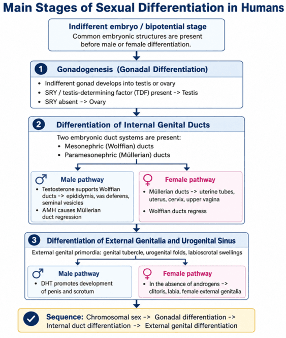
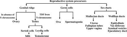
Gonadal Differentiation in Male and Female Embryos
Male
- In a genetically male embryo (46,XY), the indifferent gonad develops into a testis.
- Medulla of the genital ridge develops into testicular tissue, while the cortex largely regresses.
- Sertoli cells develop and secrete Anti-Müllerian Hormone (AMH/MIS).
- Leydig cells develop and secrete testosterone.
- These hormones promote further differentiation of the male internal reproductive system.
Female
- In a genetically female embryo (46,XX), testes do not develop;
- therefore, there are no fetal testicular Sertoli or Leydig cells producing AMH and testosterone.
- Cortex of the genital ridge develops into the ovarian tissue, while the medullary component largely regresses.
- The indifferent gonads therefore differentiate into ovaries.
Summary:
- Male (XY) → Testes → Sertoli cells → AMH + Leydig cells → Testosterone
- Female (XX) → No testes → No testicular AMH/testosterone → Ovarian development
Gonadal Origin & Secretion Mnemonics
Cortex vs. Medulla: Ovary develops from the Cortex (OC = "Ovarian Cortex"); Testes develop from the Medulla (MM = "Male Medulla").
Sertoli vs. Leydig: Sertoli cells Suppress Müllerian ducts (AMH/MIS) and support Sperm; Leydig cells produce Lipid-based Testosterone stimulated by LH.
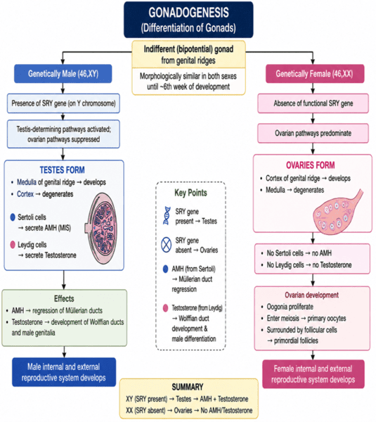
GONADOGENESIS (Differentiation of Gonads)
Differentiation of the Genital Ducts (Development of the Internal Accessory Sex Organs)
- Development of the internal reproductive organs depends on the selective development of one embryonic duct system and regression of the other.
- During early embryonic life, both male and female embryos possess two paired genital duct systems:
- Mesonephric (Wolffian) ducts
- Paramesonephric (Müllerian) ducts
- These ducts are present during the early weeks of development, giving the embryo the potential to develop either male or female internal reproductive structures.
- Around the 7th week of gestation, sexual differentiation of the genital ducts becomes evident under the influence of gonadal hormones.
In the Male
- Testosterone from Leydig cells promotes development of the Wolffian ducts into (epididymis or vas deferens, ejaculatory duct, seminal vesicles).
- Anti-Müllerian hormone (AMH/MIS) from Sertoli cells causes regression of the Müllerian ducts.
In the Female
- In the absence of significant fetal testicular testosterone, the Wolffian ducts regress.
- In the absence of AMH, the Müllerian ducts persist and develop into female internal reproductive organs (uterus, fallopian tubes & cervix).
| Sex | Wolffian (Mesonephric) Duct Fate | Müllerian (Paramesonephric) Duct Fate |
|---|---|---|
| Male (46,XY) | Develops: Epididymis, vas deferens, ejaculatory duct, seminal vesicles (stimulated by Testosterone) | Regresses: Inhibited by AMH / MIS from Sertoli cells |
| Female (46,XX) | Regresses: Passive involution due to absence of testosterone | Develops: Uterus, fallopian tubes, cervix, upper vagina (default; absence of AMH) |
Summary:
- Male: Testosterone → Wolffian duct develops; AMH → Müllerian duct regresses
- Female: No testicular testosterone → Wolffian duct regresses; No AMH → Müllerian duct develops
Derivatives of the Wolffian Duct ("SEED")
Wolffian structures develop exclusively under local testosterone stimulation: Seminal vesicles, Epididymis, Ejaculatory duct, and Ductus (vas) deferens. (The prostate arises from the urogenital sinus).
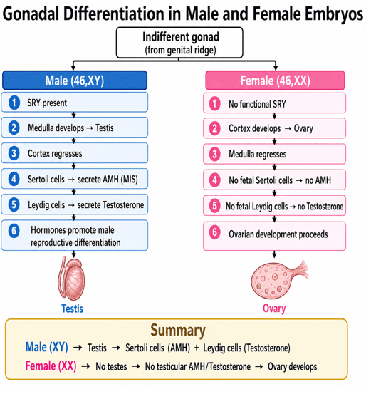
Gonadal Differentiation in Male and Female Embryos
Differentiation of the External Genitalia and Urogenital Sinus
- External genitalia initially develop from common embryonic structures present in both sexes:
- Genital tubercle
- Urogenital folds
- Labioscrotal swellings
- These structures are initially indifferent and can develop into either male or female external genitalia.
- Their differentiation is controlled mainly by genetic and hormonal influences.
Male
- Under the influence of androgens (testosterone) particularly dihydrotestosterone (DHT), 5α-reductase converts testosterone to dihydrotestosterone (DHT).
- Genital tubercle → penis
- Urogenital folds → ventral aspect of the penis/penile urethra
- Labioscrotal swellings → scrotum
Female
- In the absence of significant androgen action:
- Genital tubercle → clitoris
- Urogenital folds → labia minora
- Labioscrotal swellings → labia majora
| Embryonic Primordium | Male Adult Derivative (DHT Mediated) | Female Adult Derivative (Default / Estrogen) |
|---|---|---|
| Genital Tubercle | Penis (glans penis, corpora cavernosa, corpus spongiosum) | Clitoris (glans clitoridis, corpora cavernosa, bulbs of vestibule) |
| Urogenital Folds | Ventral aspect of penis / penile urethra | Labia minora |
| Labioscrotal Swellings | Scrotum | Labia majora |
| Urogenital Sinus | Prostate, bulbourethral glands, bladder, urethra | Skene glands, Bartholin glands, lower 2/3 of vagina, bladder |
Summary: Common embryonic structures → hormonal influence → male or female external genitalia
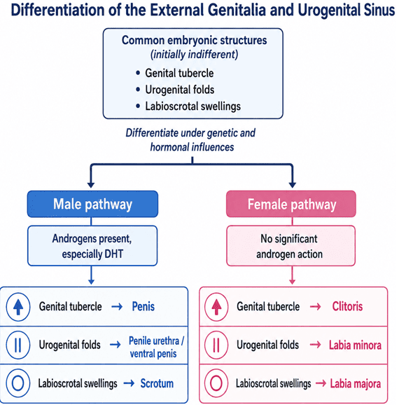
Differentiation of the External Genitalia and Urogenital Sinus
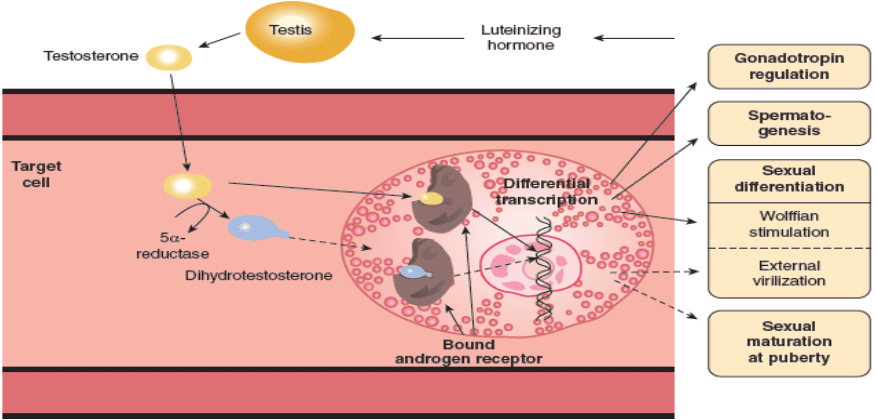
Diagram of the actions of testosterone (solid arrows) and dihydrotestosterone (dashed arrows). Note that they both bind to the same receptor, but DHT binds more effectively.
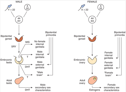
Summary of normal sex determination, differentiation, and development in humans: DHT: dihydrotestosterone, MIS: Müllerian inhibiting substance, T: testosterone.
Clinical Implications of Abnormal Sexual Differentiation
Abnormalities in genes, hormone synthesis, hormone receptors, or development of the genital ducts can result in:
- Disorders of sex development (DSDs) and
- Congenital abnormalities of the reproductive tract.
1. 5α-Reductase Deficiency
Pathophysiology: 5α-reductase converts testosterone to dihydrotestosterone (DHT). DHT is important for development of the male external genitalia and prostate.
In a 46,XY fetus (genetically male fetus), deficiency of 5α-reductase results in inadequate DHT production.
Clinical Presentation: Testes and testosterone-dependent internal male reproductive ducts may develop, but the external genitalia may be under virilized (under masculinized), ambiguous or female-appearing at birth.
Thus: 46,XY → Testes → Testosterone present → ↓ DHT → inadequate masculinization of external genitalia
2. Androgen Insensitivity Syndrome (AIS)
Etiology: Results from abnormalities of the androgen receptor, causing reduced or absent tissue responsiveness to testosterone and DHT.
In complete androgen insensitivity syndrome (CAIS):
- The individual is genetically male fetus (46,XY) and develops testes.
- Sertoli cells produce AMH, causing regression of the Müllerian ducts; therefore, the uterus and fallopian tubes are absent.
- Androgen action is absent, preventing normal development of the Wolffian ducts and male external genitalia.
- Hence, the external genitalia are typically female in appearance with a short blind-ending vagina.
Note: Epididymis, vas deferens, and seminal vesicles are mainly dependent on testosterone, whereas development of the prostate and male external genitalia is particularly dependent on DHT.
Comparison: 5α-Reductase Deficiency vs. Complete AIS
| Parameter | 5α-Reductase Deficiency (46,XY) | Complete AIS (46,XY) |
|---|---|---|
| Testosterone / DHT | Normal Testosterone / Decreased DHT | Normal/Elevated Testosterone / Normal DHT |
| Internal Duct Status | Male Wolffian structures present (SEED); No Müllerian (AMH normal) | No internal ducts (Wolffian fails via receptor defect; Müllerian regresses via AMH) |
| External Genitalia | Ambiguous/female at birth; virilizes at puberty | Female external phenotype; develops breasts at puberty, absent/sparse pubic hair |
3. Müllerian Duct Abnormalities in Genetic Females
Abnormal development, fusion or canalization of the Müllerian ducts in a genetically female (46,XX) individual may produce congenital abnormalities such as:
- Agenesis or atresia of the uterine tubes: congenital absence or blockage of the fallopian tubes.
- Septate uterus: uterus that is partly or completely divided into two cavities by a wall of tissue called a septum.
- Bicornuate uterus: uterine abnormality in which the upper part of the uterus is divided into two horn-like cavities, giving the uterus a heart-shaped appearance.
- Uterus didelphys: abnormality in which two separate uterine cavities develop, often with two cervices, because the two Müllerian ducts fail to fuse properly during fetal development.
- Uterine or vaginal agenesis: congenital absence or failure of development of the uterus and/or vagina.
- Cervical agenesis or atresia: means the cervix is absent or abnormally closed/blocked from birth.
Clinical Presentation: These abnormalities may present with infertility, recurrent pregnancy loss, pelvic pain, or menstrual abnormalities.
Mechanisms of Uterine Anomalies
Didelphys: Complete failure of Müllerian duct fusion → duplication of uterus and cervix.
Bicornuate: Partial failure of Müllerian duct fusion → heart-shaped indentation.
Septate: Failure of midline septum resorption after complete fusion → central avascular septum, high risk of early miscarriage.
4. Cervical and Vaginal Outflow Abnormalities
Failure of proper development or canalization may produce cervical stenosis, cervical atresia or vaginal obstruction.
Menstrual blood may accumulate above the obstruction after puberty, causing cyclical pelvic pain and primary amenorrhea.
5. Persistent Müllerian Duct Syndrome
Caused by deficiency of AMH or abnormalities of the AMH receptor in a genetically male individual (46,XY).
The testes develop and testosterone is produced, allowing male differentiation.
However, the Müllerian ducts fail to regress, so structures such as the uterus and fallopian tubes may persist in an otherwise male individual.
6. Congenital Adrenal Hyperplasia in a genetically female fetus (46,XX)
Pathophysiology: Excess fetal androgens, commonly from 21-hydroxylase deficiency, can virilize the external genitalia of a genetic female fetus (46,XX).
The ovaries, uterus, and fallopian tubes are usually present because there are no testes producing AMH.
In summary: Excess androgens in 46,XX fetus → virilized external genitalia, with normal internal female reproductive organs.
Summary
- Abnormal hormone synthesis → abnormal sexual differentiation
- Abnormal hormone receptors → impaired response to testosterone/DHT
- Abnormal AMH action → persistence of Müllerian structures in XY individuals
- Abnormal Müllerian development → congenital uterine, cervical, tubal, or vaginal abnormalities
- Excess androgens in XX fetus → virilization of external genitalia
