
Development of the face: embryology, innervation & congenital anomalies
On this page
Overview & facial primordia
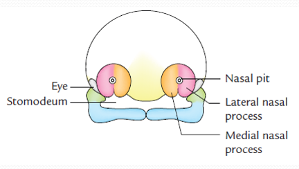
Craniofacial development is an intricate morphogenetic process taking place predominantly between the fourth and eighth weeks of human development. The human face forms around a central ectodermal depression termed the stomodeum (primitive mouth), governed by the coordinated proliferation, migration, merging, and fusion of five primary facial prominences (primordia).
The Five Primary Facial Swellings
- 1. Single Frontonasal Process: An unpaired median prominence lying immediately ventral to the developing forebrain vesicle (prosencephalon). It forms the upper anatomical boundary of the stomodeum.
- 2. Paired Maxillary Processes: Bilateral swellings derived from the dorsal portion of the first pharyngeal arch (mandibular arch). They flank the stomodeum laterally, forming the lateral borders of the primitive mouth.
- 3. Paired Mandibular Processes: Bilateral swellings derived from the ventral portion of the first pharyngeal arch. They form the caudal (inferior) margin of the stomodeum.
Neural crest & stomodeum
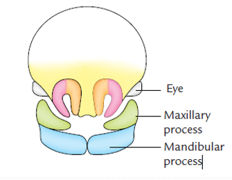
By the conclusion of the fourth embryonic week, the primitive face centers upon the stomodeum, bounded superiorly by the frontonasal prominence, laterally by the maxillary prominences, and inferiorly by the mandibular prominences. Deep within the stomodeum lies the oropharyngeal (buccopharyngeal) membrane, a bilaminar structure composed of surface ectoderm apposed directly to foregut endoderm without intervening mesoderm. At approximately day 26, this membrane breaks down, establishing anatomical continuity between the primitive amniotic cavity and the foregut.
Mesenchyme & Cranial Neural Crest Cell Migration
Histologically, each of the five facial primordia consists of a core of ectomesenchyme covered externally by a layer of surface ectoderm. Unlike the rest of the body where mesenchyme is derived from paraxial and lateral plate mesoderm, the mesenchyme of the facial prominences is derived primarily from cranial neural crest cells (CNCCs).
- Origin: During neurulation, neuroectodermal cells at the margins of the neural folds undergo an epithelial-to-mesenchymal transition (EMT) and migrate ventrolaterally beneath the surface ectoderm.
- Signaling Cascades: Migration and survival are governed by sonic hedgehog (SHH), fibroblast growth factors (FGF-8), bone morphogenetic proteins (BMP-4), and homeobox (HOX) gene expression gradients.
- Derivatives: Cranial neural crest cells give rise to the entire facial skeleton, including the maxilla, zygoma, mandible, palatine bones, vomer, ethmoid, auditory ossicles, cartilage, dental papillae, and dermis of the face.

Nasal placodes & processes
During the fifth week, bilateral oval thickenings of surface ectoderm appear on each side of the median plane in the lower, ventrolateral aspect of the frontonasal prominence. These are the olfactory (nasal) placodes.
Mesenchymal Reorganization & Horseshoe Elevations
- Placodal Invagination: The surface ectoderm of the olfactory placodes invaginates deeply into the underlying mesoderm, converting the flat placodes into depressed olfactory (nasal) pits. These pits initially communicate freely with the stomodeum below.
- Horseshoe-Shaped Elevations: Rapid proliferation of ectomesenchyme around the margins of each nasal pit creates elevated, horseshoe-shaped ridges:
- The medial rim of the horseshoe is the medial nasal process.
- The lateral rim of the horseshoe is the lateral nasal process.
- Globular Processes: As the medial nasal processes expand, they grow inferiorly and medially toward the stomodeum, developing expanded, bulbous caudal extremities known as the globular processes (of His). Initially, the two globular processes are separated in the midline by a shallow triangular notch.
Derivatives of facial swellings
Between weeks 6 and 7, differential growth causes the paired maxillary prominences to enlarge and expand medially, compressing the medial nasal processes toward the midline and reorganizing the anatomical landscape of the midface.

Step-by-Step Morphogenesis of Facial Regions
- Nasal Cavities & Primitive Septum: The olfactory pits burrow deeply in a dorsal and superior direction through the mesenchyme toward the forebrain, forming the primitive nasal cavities. The central remnant of the frontonasal prominence between the developing cavities thins out to form the primitive nasal septum.
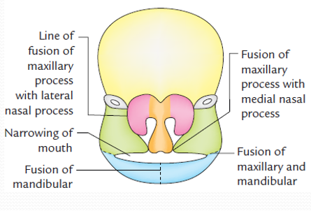
- Medial Merging of Globular Processes: The globular processes of the two medial nasal processes merge seamlessly across the midline. This fused intermaxillary block forms the philtrum of the upper lip, the premaxillary part of the upper jaw (carrying the four upper incisors), and the primary palate.
- Maxillary Process Expansion: The paired maxillary processes grow medially beneath the developing optic cups. They merge laterally with the lateral nasal processes (forming the cheek and lateral nasal boundary) and medially with the fused medial nasal processes, creating the lateral portions of the upper lip and the upper cheeks.
- Mandibular Merging: The two mandibular processes grow medially toward each other and merge in the midline during the end of the fourth week, obliterating the median groove to produce the mandible, chin, lower lip, and lower parts of the cheeks.
- The Oral Fissure: The primitive stomodeum narrows to form the definitive oral fissure (mouth aperture). The lateral angles of the mouth (labial commissures) are formed by the bilateral fusion of the maxillary and mandibular processes.
Development of the lips
The upper and lower lips develop through coordinated merging of ectomesenchyme accompanied by specialized ectodermal proliferation. Initially, the primitive jaws lack lips, consisting of continuous masses of mesenchyme bounded internally by the tongue and floor of the mouth.
1. Separation of the Lips from Jaws (Labiodental Sulcus)
Around the sixth week, a horse-shoe-shaped band of surface ectoderm called the labiodental lamina proliferates deeply into the underlying ectomesenchyme of both the maxilla and mandible. The central cells of this lamina undergo apoptosis and disintegrate, creating a deep cleavage trench: the labiogingival groove (sulcus). This groove physically separates the external lips and cheeks from the internal alveolar processes (gums). A persistent midline ectodermal strand remains intact to form the frenulum of the upper and lower lips.
2. Muscular Colonization of the Lips
While the connective tissue, dermis, and vascular channels of the lips arise from first arch and frontonasal neural crest mesenchyme, the facial muscles (including the orbicularis oris and buccinator) have a completely different embryological origin. Myoblasts migrate from the mesoderm of the second pharyngeal arch (hyoid arch) during the seventh week, invading the developing lips and cheeks. Because these muscle cells originate from Arch 2, they carry their own nerve supply with them: the motor branches of the facial nerve (CN VII).
The philtrum & sensory nerves
The embryological origin of the philtrum (the vertical midline groove of the upper lip) has historically been an area of debate, with significant clinical implications for neuroanatomy.
Classical Theory vs. Modern Evidence
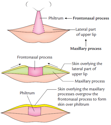
- The Classical Concept: Taught that the lateral portions of the upper lip are derived entirely from the mesenchyme and ectoderm of the maxillary processes, whereas the median portion (philtrum) is formed directly by the mesenchyme of the merged medial nasal processes (frontonasal prominence). In this model, the overlying ectoderm of the maxillary processes merely migrates across to overgrow the frontonasal mesenchyme.
- The Modern Embryological & Clinical Evidence: High-resolution scanning electron microscopy and clinical mapping of cleft lip repairs demonstrate that the upper lip is formed in its entirety from the maxillary processes. As the maxillary processes surge medially, their mesenchyme entirely compresses and covers the caudal ends of the medial nasal processes. The medial nasal processes become deeply embedded to form the underlying premaxillary bone and primary palate, while the superficial surface tissue (both mesenchyme and overlying skin of the philtrum) is formed by the medial extensions of the maxillary processes.
Embryological sensory innervation
The sensory innervation of the adult human face is divided cleanly among the three divisions of the trigeminal nerve (CN V). This arrangement directly reflects the embryonic derivation of each facial territory.

Anatomical Mapping & Pharyngeal Arch Relationships
- 1. Frontonasal Process → Ophthalmic Nerve (V1): All structures derived from the frontonasal prominence (forehead, upper eyelids, dorsum and bridge of the external nose) receive their cutaneous sensory innervation exclusively from the ophthalmic division of the trigeminal nerve (V1) (via the supraorbital, supratrochlear, infratrochlear, lacrimal, and external nasal nerves).
- 2. Maxillary Process → Maxillary Nerve (V2): The maxillary prominence is the dorsal subdivision of the first pharyngeal arch. All of its derivatives (upper cheeks, lower eyelids, lateral alae of the nose, and the entire upper lip including the philtrum) are innervated by the maxillary division of the trigeminal nerve (V2) (via the infraorbital, zygomaticofacial, and zygomaticotemporal nerves).
- 3. Mandibular Process → Mandibular Nerve (V3): The mandibular prominence represents the ventral subdivision of the first pharyngeal arch. All of its derivatives (lower lip, chin, lower cheeks, and lateral temporomandibular regions) are innervated by the mandibular division of the trigeminal nerve (V3) (via the mental, buccal, and auriculotemporal nerves).
Scroll the table sideways on a smaller screen.
| Component of Face | Embryonic Source of Development | Sensory Nerve Supply |
|---|---|---|
| Forehead | Frontonasal process | Ophthalmic division of Vth nerve (V1) |
| Nose (External) | Frontonasal process (bridge, dorsum, tip, alae) | Ophthalmic division of Vth nerve (V1) |
| Cheek: (a) Upper Part | Maxillary process (Arch 1) | Maxillary division of Vth nerve (V2) |
| Cheek: (b) Lower Part | Mandibular process (Arch 1) | Mandibular division of Vth nerve (V3) |
| Upper Lip | Fusion of maxillary processes of two sides with the frontonasal process* | Maxillary division of Vth nerve (V2) |
| Lower Lip | Fusion of mandibular processes of two sides | Mandibular division of Vth nerve (V3) |
*Clinical Developmental Note: Although classical anatomical descriptions cite frontonasal contribution, the sensory nerve supply of the philtrum via V2 demonstrates that maxillary ectomesenchyme completely covers the philtral area.
Nasolacrimal apparatus
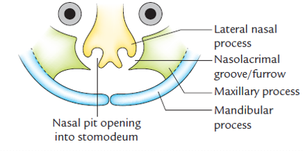
The development of the tear drainage conduit is tied to the fusion boundary between the lateral nasal prominence and the advancing maxillary prominence.
Three-Stage Morphogenesis
- 1. The Nasolacrimal Groove (Furrow): During the sixth week, the maxillary process merges with the lateral nasal process along an oblique line extending from the medial canthus of the eye to the primitive nasal pit. This junction forms a cleft-like furrow termed the nasolacrimal groove, lined by surface ectoderm.
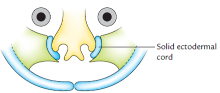
- 2. The Solid Ectodermal Cord: The ectoderm in the floor of the groove proliferates rapidly, forming a solid cylindrical epithelial cord (ectodermal cord) that sinks into the underlying mesenchyme. Shortly thereafter, the cord completely detaches from the surface ectoderm, and the adjacent maxillary and lateral nasal processes merge smoothly over it.
- 3. Canalization & Lacrimal Sac Formation: The solid epithelial cord undergoes central apoptosis (canalization) to establish a hollow tube: the nasolacrimal duct.
- The superior (cephalic) end widens to form the lacrimal sac, which connects with the lacrimal canaliculi emptying into the conjunctival sac at the medial canthus.
- The inferior (caudal) end burrows into the lateral wall of the nasal cavity, establishing secondary drainage into the inferior nasal meatus beneath the inferior nasal concha.
Development of external nose
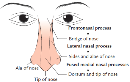
The external nose develops through the coordinated integration of five distinct embryonic facial prominences: the central frontonasal process, two medial nasal processes, and two lateral nasal processes.
Anatomical Components & Embryological Derivations
Morphogenetic Origin of Nasal Structures
- 1. Bridge of the Nose: Formed by the cranial, non-invaginated portion of the frontonasal process (giving rise to the paired nasal bones and frontal processes of the maxilla).
- 2. Dorsum & Tip (Apex) of the Nose: Formed by the midline fusion of the two medial nasal processes. Incomplete merging results in a bifid nose (median nasal cleft).
- 3. Sides & Alae (Wings) of the Nose: Formed by the two lateral nasal processes that migrate over the invaginating nasal pits, creating the lateral nasal cartilage and alar cartilages.
- 4. Nasal Columella & Septum: The columella is derived from the inferior fused portion of the medial nasal processes, while the internal nasal septum develops downward from the merged frontonasal mesenchyme to fuse with the palatine shelves.
Primary & secondary palatogenesis
Palatogenesis begins during the sixth embryonic week and is complete by week 12, separating the oral cavity from the developing nasal passages. The palate develops from two distinct embryological structures: the primary palate and the secondary palate.
1. Primary Palate (Median Palatine Process)
Formed by the deep merging of the two medial nasal processes in the midline, creating the wedge-shaped intermaxillary segment. The primary palate lies anterior to the incisive foramen, ossifying to form the premaxillary part of the maxilla, which houses the four upper incisor teeth.
2. Secondary Palate (Lateral Palatine Shelves)
The definitive hard and soft palate posterior to the incisive foramen develops from the secondary palate:
- Palatal Shelves: During the sixth week, two shelf-like mesenchymal projections—the lateral palatine processes (palatal shelves)—grow downward vertically from the internal aspects of each maxillary process, flanking the large developing tongue.
- Shelf Elevation (Week 7–8): With the elongation of the mandible, the embryonic tongue drops downward into the floor of the mouth. This frees the palatal shelves, which rapidly elevate into a horizontal orientation above the tongue.
- Midline Fusion (Week 8–12): The elevated shelves grow medially toward each other. The medial-edge epithelium undergoes cell death and desquamation, allowing mesenchymal continuity to fuse across the midline. The shelves also fuse anteriorly with the triangular primary palate and superiorly with the downgrowing nasal septum.
- Ossification: The anterior two-thirds of the secondary palate undergo endochondral and intramembranous ossification to form the bony hard palate, while the unossified posterior one-third forms the muscular soft palate and uvula.
Facial clefts & malformations
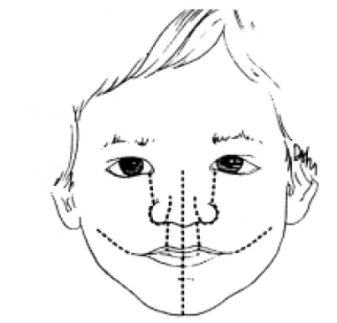
Facial clefts represent common human congenital malformations, occurring in approximately 1 in 700 live births. Their biological basis follows an established embryological rule:
Clinical Classification of Facial & Palatal Clefts
1. Cleft Lip (Cheiloschisis)
- Etiology: Failure of mesenchyme of the maxillary process to merge with the medial nasal process (globular process) on one or both sides.
- Unilateral Cleft Lip: The most common form (more frequent in males and on the left side). A gap extends through the lateral portion of the upper lip, often continuing into the nostril floor and alveolar ridge up to the incisive foramen.
- Bilateral Cleft Lip: Occurs when both maxillary processes fail to fuse with the intermaxillary segment. The central intermaxillary block (philtrum, premaxilla) projects forward without lateral osseous anchors (the “floating premaxilla”).
- Median Cleft Lip (“Harelip”): A rare midline defect caused by incomplete fusion of the two medial nasal processes with each other. Frequently associated with holoprosencephaly, where impaired sonic hedgehog (SHH) signaling prevents midline forebrain cleavage.
- Degrees: Complete clefts extend through the entire lip, nostril floor, and alveolus; incomplete clefts spare the nasal sill when partial mesenchymal bridging has taken place.
2. Cleft Palate (Palatoschisis)
- Etiology: Failure of the bilateral palatal shelves (lateral palatine processes) to elevate, meet, and fuse across the midline with each other or with the primary palate and nasal septum. More prevalent in females (secondary palate fusion occurs approximately one week later in female embryos).
- Anterior (Primary) Cleft Palate: Clefting located anterior to the incisive foramen, resulting from lack of fusion between the primary palate and the secondary palatine shelves.
- Posterior (Secondary) Cleft Palate: Clefting confined strictly posterior to the incisive foramen, involving the secondary hard palate, soft palate, or isolated uvula (bifid uvula).
- Complete Combined Cleft Lip & Palate: Complete failure of both primary and secondary palatogenesis, leaving the oral cavity, nasal cavity, and maxillary sinuses in direct communication. Impairs infant feeding due to an inability to generate negative intraoral suction, necessitating specialized cross-cut nipples (Haberman feeder) and multidisciplinary surgical repair (Millard rotation-advancement cheiloplasty around 3 months; palatoplasty at 9 to 12 months).
3. Oblique Facial Cleft (Tessier Cleft No. 3 / 4)
Results from failure of the maxillary process to merge with the lateral nasal process along the line of the nasolacrimal groove. Clinically presents as an open fissure extending from the upper lip at the corner of the mouth obliquely across the cheek directly into the medial canthus or floor of the orbit, exposing the nasolacrimal duct.
4. Lateral / Transverse Facial Cleft (Macrostomia / Tessier Cleft No. 7)
Results from incomplete merging between the maxillary process and the mandibular process of the first pharyngeal arch along the lateral angle of the oral fissure. Produces an abnormally wide oral aperture (macrostomia), which may extend bilaterally toward the tragus of the ear. Conversely, excessive fusion between these processes produces an abnormally small mouth (microstomia).
5. Median Mandibular Cleft
A rare anomaly resulting from failure of the paired mandibular processes to fuse across the ventral midline in the fourth week. Manifests as a deep midline cleft through the lower lip, mental symphysis, and body of the mandible.
Quick recap
References
The following authoritative medical embryology and anatomy reference texts support this curriculum study note:
- Langman’s Medical Embryology: Sadler, T.W. (2019) Langman’s Medical Embryology. 14th edn. Philadelphia: Wolters Kluwer. [Chapter 17: Head and Neck – Development of the Face and Palate, pp. 285–302].
- The Developing Human: Moore, K.L., Persaud, T.V.N. and Torchia, M.G. (2020) The Developing Human: Clinically Oriented Embryology. 11th edn. Philadelphia: Elsevier. [Chapter 9: Pharyngeal Apparatus, Face, and Neck, pp. 157–186].
- Larsen’s Human Embryology: Schoenwolf, G.C., Bleyl, S.B., Brauer, P.R. and Francis-West, P.H. (2021) Larsen’s Human Embryology. 6th edn. Philadelphia: Elsevier. [Chapter 12: Development of the Head, Neck, and Craniofacial Skeleton, pp. 347–382].
- Gray’s Anatomy: Standring, S. (2021) Gray’s Anatomy: The Anatomical Basis of Clinical Practice. 42nd edn. London: Elsevier. [Section 4: Head and Neck – Embryology of the Craniofacial Complex, pp. 412–428].