
Development of the ear: organogenesis, pharyngeal apparatus & congenital anomalies
On this page
Anatomical overview & timeline
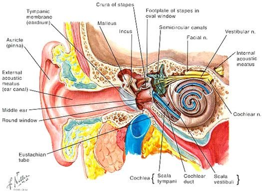
The human ear is a compound sensory organ functioning in audition (hearing) and vestibular equilibrium (balance). Macroscopically, it comprises three continuous anatomical regions arranged from lateral to medial:
- 1. External Ear: Consists of the acoustic collector funnel, the auricle (pinna), and the sound-conducting channel, the external auditory meatus (canal), terminating medially at the tympanic membrane.
- 2. Middle Ear (Tympanic Cavity): An air-filled, slit-like cavity carved within the petrous temporal bone containing three articulating mobile auditory ossicles (malleus, incus, and stapes), suspended by ligaments and attenuated by two middle ear muscles.
- 3. Internal Ear (Labyrinth): A neurosensory transducing complex embedded in the otic capsule of the temporal bone, comprising the fluid-filled membranous labyrinth housed within a protective bony labyrinth.
Developmental Milestones & Size at Birth
- Chronological Priority: Developmentally, the internal ear is the first to appear (beginning early in the 4th week of intrauterine life [IUL]), followed in sequence by the middle ear and the external ear.
- Adult Dimensions at Birth: Unlike most cranial structures, several critical ear structures attain full adult size at the time of birth:
- The internal ear (membranous and bony labyrinths).
- The tympanic cavity (middle ear space).
- The mastoid antrum.
- The auditory ossicles (malleus, incus, and stapes).
- Postnatal Maturation: Although the mastoid antrum is of adult size at birth, the mastoid air cells are entirely absent in the neonate. They develop by pneumatization starting at 2 years of age and continue expanding through puberty.
Embryological origins
Although the adult ear functions as a single biomechanical and neurosensory unit, its three principal components develop from three distinctly different primary germ layer sources interacting with the pharyngeal (branchial) apparatus:
Scroll the table sideways on a smaller screen.
| Ear Region | Embryonic Primordium | Primary Germ Layer | Adult Anatomical Derivatives |
|---|---|---|---|
| Internal Ear | Otic placode → Otic vesicle (otocyst) | Surface ectoderm & surrounding mesenchyme | Membranous labyrinth, sensory neuroepithelia, spiral and vestibular ganglia, bony otic capsule |
| Middle Ear | Tubotympanic recess (1st pharyngeal pouch); 1st and 2nd pharyngeal arch mesenchyme | Endoderm & cranial neural crest / paraxial mesoderm | Tympanic cavity, mastoid antrum, auditory tube, auditory ossicles (malleus, incus, stapes), and middle ear muscles |
| External Ear | 1st pharyngeal cleft (groove) & six auricular hillocks of His (1st & 2nd arches) | Surface ectoderm & cranial neural crest mesenchyme | External auditory meatus, outer layer of tympanic membrane, and auricle (pinna) |
Internal ear development
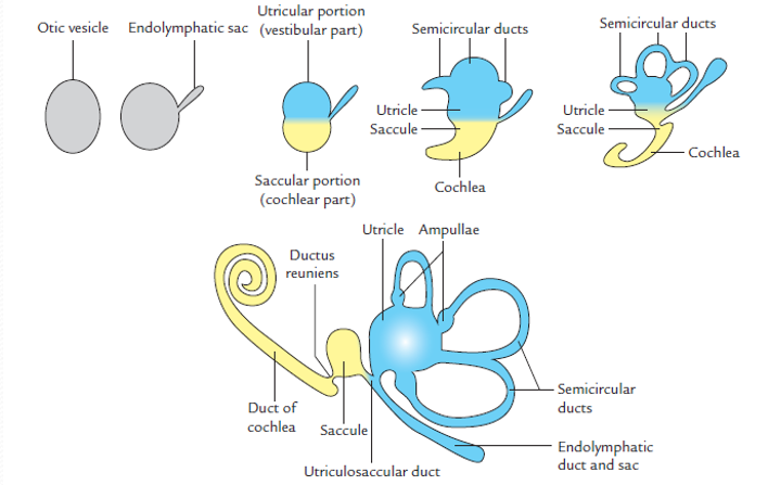
The internal ear is the first anatomical subdivision to initiate morphogenesis. Early in the fourth week of development (day 22), paired thickenings of the cephalic surface ectoderm appear on each side of the developing rhombencephalon (myelencephalon): the otic placodes.

From Otic Placode to Membranous Labyrinth
- Placode Invagination: Under inductive molecular signals from the notochord and paraxial mesoderm (principally FGF-3 and FGF-10), the otic placode invaginates into the underlying mesoderm to form a cup-shaped depression: the otic pit.
- Vesicle Closure & Detachment: The lateral margins of the otic pit approach each other, fuse, and detach from the overlying surface ectoderm, forming a hollow, fluid-filled epithelial sphere: the otic vesicle (otocyst).
- Divergence into Ventral and Dorsal Portions: As the otocyst elongates, differential cell growth separates it into two fundamental components:
- Ventral Saccular Portion (Cochlear Part): Gives rise to the saccule, the cochlear duct (housing the sensory organ of Corti), and neuroblasts that form the spiral ganglion of the vestibulocochlear nerve (CN VIII).
- Dorsal Utricular Portion (Vestibular Part): Gives rise to the utricle, the semicircular ducts, the endolymphatic duct and sac, and the vestibular ganglion of CN VIII.
Formation of the Otic Capsule & Perilymphatic Space
- Mesenchymal Condensation: The mesenchyme immediately surrounding the epithelial membranous labyrinth condenses and chondrifies into a hyaline cartilaginous shell: the cartilaginous otic capsule.
- Vacuolization & Perilymphatic Cavitation: Mesenchymal cells immediately adjacent to the membranous labyrinth undergo programmed apoptosis and fluid vacuolization. This forms the perilymphatic space.
- Ossification into Bony Labyrinth: Through endochondral ossification, the cartilaginous capsule converts into dense, compact petrous bone, forming the definitive bony labyrinth.
- Fluid Compartments:
- The perilymphatic space (between bony and membranous walls) is filled with perilymph, an extracellular-like fluid high in sodium (Na+) and low in potassium (K+).
- The interior of the membranous labyrinth is filled with endolymph, an intracellular-like fluid high in potassium (K+) secreted by the stria vascularis.
Cochlea, duct & organ of Corti
During the sixth week of gestation, the lower tubular pole of the saccule forms a tubular diverticulum: the cochlear duct. It penetrates the surrounding mesenchyme in a spiral trajectory until it completes 2.5 turns by the end of the eighth week. Its connection to the saccule narrows to form the slender ductus reuniens (duct of Hensen).
Compartmentalization of the Cochlear Canal
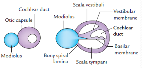
Vacuolization of the surrounding mesodermal otic capsule forms two large perilymphatic channels on either side of the cochlear duct (scala media):
- Scala Vestibuli: Positioned superiorly to the cochlear duct, separated from it by the thin, two-layered vestibular membrane (Reissner’s membrane). Communicates with the vestibule and terminates at the oval (vestibular) window.
- Scala Tympani: Positioned inferiorly to the cochlear duct, separated from it by the dense basilar membrane. Terminates at the secondary tympanic membrane sealing the round (cochlear) window.
- Helicotrema: The narrow apical aperture through which the scala vestibuli and scala tympani communicate at the apex of the cochlea.
- Modiolus & Spiral Ligament: The medial boundary of the cochlear duct is anchored to a central conical core of spongy bone: the modiolus (the future axis of the bony cochlea housing the spiral ganglion). The lateral wall is anchored to the surrounding bony capsule by the vascularized fibrous spiral ligament and the stria vascularis.
Morphogenesis of the Spiral Organ of Corti
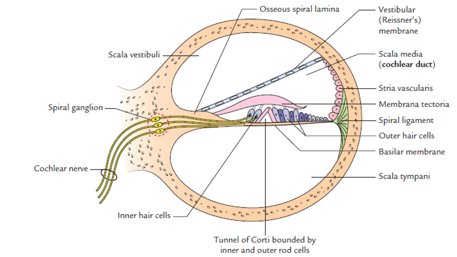
The neuroepithelial cells of the basilar membrane differentiate into two prominent longitudinal ridges:
- Inner Ridge: Differentiates into the spiral limbus, which secretes and provides an anchor for the acellular, gelatinous tectorial membrane (membrana tectoria).
- Outer Ridge: Differentiates into the sensory and supporting cells of the auditory system:
- One inner row of inner hair cells (IHCs): The primary sensory transducers of acoustic frequency.
- Three to four outer rows of outer hair cells (OHCs): Electromotile cochlear amplifiers that fine-tune frequency selectivity.
- Supporting cells: Deiters’ phalangeal cells, Hensen’s cells, Claudius’ cells, and inner/outer pillar cells bounding the fluid-filled tunnel of Corti.
Stereocilia protruding from the hair cells embed into the overlying tectorial membrane. Fluid pressure waves vibrating the basilar membrane shear these stereocilia, opening mechanosensitive K+ channels, generating receptor potentials, and transmitting impulses along the cochlear nerve to the brainstem.
Vestibular apparatus & canals
During the sixth week of gestation, the dorsal utricular portion of the otic vesicle gives rise to the organs of equilibrium: the semicircular ducts, the utricle, and the saccule.
Development of the Semicircular Ducts
- Disc-like Outpouchings: Three flat, disc-like diverticula evaginate from the wall of the utricle, positioned in three planes perpendicular to each other: the anterior (superior), posterior, and lateral (horizontal) semicircular ducts.
- Central Resorption: The central apposing epithelial walls of these flat pouches fuse and undergo apoptosis, leaving three open tubular peripheral loops: the semicircular ducts.
- Ampullae & Crus Commune:
- One end of each duct dilates to form the ampulla (crus ampullare), while the non-dilated opposite end remains narrow: the crus nonampullare.
- The nonampullary limbs of the anterior and posterior ducts fuse to form a shared conduit: the crus commune.
- Consequently, the three semicircular ducts communicate with the utricle through only five openings (three ampullary and two nonampullary).
Sensory Receptor Specializations: Balance & Equilibrium
Kinetic vs. Static Balance Receptors
- Crista Ampullaris (Kinetic / Dynamic Equilibrium): Sensory epithelial cells within each dilated ampullary crest differentiate into hair cells capped by a gelatinous protein-mucopolysaccharide dome called the cupula. The cristae detect angular acceleration and rotational head movement across the three spatial planes.
- Maculae (Static Equilibrium & Linear Acceleration): Sensory hair cells in the medial wall of the utricle (oriented horizontally) and the saccule (oriented vertically) develop into the maculae, capped by an otolithic membrane embedded with microscopic calcium carbonate crystals (otoliths/otoconia). The maculae detect gravity and linear acceleration (e.g., forward motion or riding in an elevator).
- Innervation: Impulses from both cristae and maculae are relayed via bipolar primary sensory neurons residing in the vestibular ganglion (of Scarpa), which project centrally through the vestibular division of the vestibulocochlear nerve (CN VIII) into the brainstem.
Middle ear cavity & tube
The Tubotympanic Recess
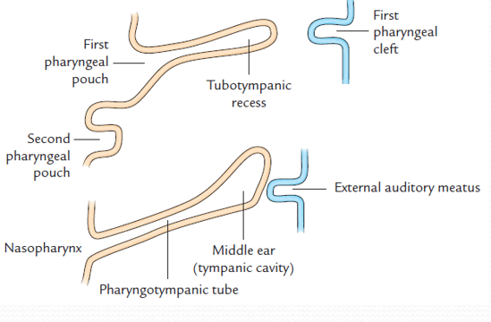
The middle ear cavity develops from the tubotympanic recess, an endodermal evagination that arises predominantly from the dorsal aspect of the first pharyngeal pouch, with a minor contribution from the second pouch:
- Distal Expansion: The blind distal portion of the recess expands laterally to form the primitive tympanic cavity, which envelops the auditory ossicles in late fetal life.
- Proximal Elongation: The proximal portion of the recess remains narrow and tubular, developing into the pharyngotympanic (auditory / Eustachian) tube, which connects the middle ear cavity to the nasopharynx to equalize middle ear pressure with atmospheric pressure.
- Mastoid Antrum & Air Cells: Posteriorsuperior expansion of the tympanic cavity invades the petrous bone to form the mastoid antrum. Following birth, epithelial extensions hollow out the mastoid process, forming the mastoid air cells beginning around 2 years of age.
Auditory ossicles & muscles
The three auditory ossicles (malleus, incus, and stapes) bridge the middle ear to transmit and amplify sound vibrations from the tympanic membrane to the oval window. They develop from cartilaginous bars of the first and second pharyngeal arches via endochondral ossification:
Scroll the table sideways on a smaller screen.
| Pharyngeal Arch | Cartilaginous Precursor | Ossicle Derivative | Associated Muscle | Cranial Nerve Supply |
|---|---|---|---|---|
| 1st Pharyngeal Arch (Mandibular) | Meckel’s cartilage (dorsal portion) | Malleus & Incus | Tensor tympani (inserts onto the handle of malleus) | Mandibular division of Trigeminal Nerve (CN V3) |
| 2nd Pharyngeal Arch (Hyoid) | Reichert’s cartilage (dorsal portion) | Stapes (head, crura, & lateral footplate) | Stapedius (inserts onto the neck of stapes) | Facial Nerve (CN VII) |
Mesenchymal Cavitation & Epithelial Envelopment
Throughout early and mid-fetal development, the auditory ossicles remain embedded within a dense mesenchymal gelatinous bed filling the middle ear:
- Late Mesenchymal Resorption: During the eighth and ninth gestational months, this surrounding mesenchyme undergoes programmed vacuolization, opening the air space of the tympanic cavity.
- Mesentery-like Folds: As the mesenchyme resorbs, the endodermal lining of the cavity extends over the ossicles, clothing them like visceral peritoneum clothes abdominal organs. The ossicles are suspended from the walls of the tympanic cavity by delicate mucosal folds and supporting ligaments.
Tympanic membrane (trinar origin)
The tympanic membrane (eardrum) forms by direct anatomical apposition of the first pharyngeal cleft ectoderm with the first pharyngeal pouch endoderm: the first pharyngeal membrane. It is one of the few structures in human embryology formed by all three primary germ layers:

The Three Definitive Tissue Layers
- 1. Outer Cuticular Layer (Stratum Cutaneum): Formed by stratified squamous keratinizing epithelium derived from the surface ectoderm lining the floor of the first pharyngeal cleft.
- 2. Intermediate Fibrous Layer (Lamina Propria): Formed by radial and circular collagenous connective tissue fibers derived from paraxial mesoderm and cranial neural crest mesenchyme intervening between the cleft and the pouch. The handle of the malleus is embedded into this fibrous layer.
- 3. Inner Mucosal Layer (Stratum Mucosum): Formed by simple cuboidal epithelium continuous with the middle ear lining, derived from the endoderm of the tubotympanic recess (first pharyngeal pouch).
External auditory meatus & pinna
Development of the External Auditory Meatus
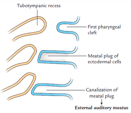
- First Pharyngeal Cleft: The canal develops from an inward invagination of the ectodermal first pharyngeal cleft.
- The Meatal Plug: Early in the third month, the ectodermal epithelial cells at the bottom of the funnel proliferate vigorously to form a solid epithelial plate: the meatal plug.
- Canalization: In the seventh month, central cells of the meatal plug undergo apoptosis, creating an open canal. Canalization is completed close to birth, establishing the definitive external auditory meatus leading to the external cuticular surface of the eardrum.
Development of the Auricle (Pinna)
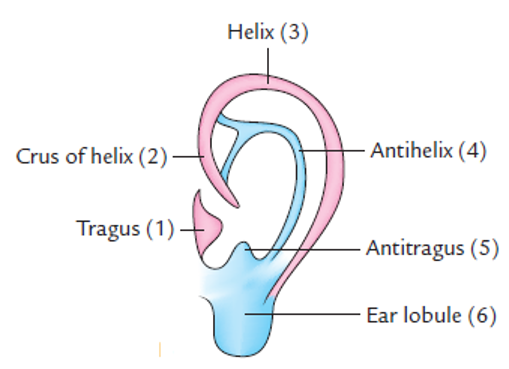
The auricle develops from six mesenchymal swellings (the auricular hillocks of His) that arise around the margins of the first pharyngeal cleft during the sixth week of gestation:
- Mandibular Arch (1st Arch): Three hillocks (1, 2, 3) develop along the caudal border of the first arch.
- Hyoid Arch (2nd Arch): Three hillocks (4, 5, 6) develop along the cephalic border of the second arch.

Scroll the table sideways on a smaller screen.
| Hillock Number | Embryonic Arch of Origin | Definitive Adult Auricular Structure |
|---|---|---|
| Hillock 1 | First Arch (Mandibular) | Tragus |
| Hillock 2 | First Arch (Mandibular) | Crus of Helix |
| Hillock 3 | First Arch (Mandibular) | Helix (ascending limb and apex) |
| Hillock 4 | Second Arch (Hyoid) | Antihelix |
| Hillock 5 | Second Arch (Hyoid) | Antitragus |
| Hillock 6 | Second Arch (Hyoid) | Ear Lobule (lobulus auriculae) |
Congenital anomalies of the ear
Anotia & Microtia
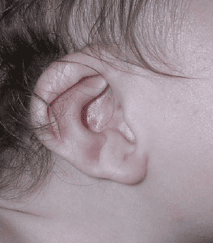
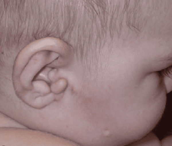
Anotia (complete absence of the auricle) and microtia (underdeveloped, rudimentary auricle) result from failure or suppression of mesenchymal proliferation within the auricular hillocks. They are frequently linked to First Arch Syndrome (Treacher Collins syndrome) and maternal ingestion of teratogens such as isotretinoin, thalidomide, or alcohol.
Preauricular Appendages (Skin Tags)
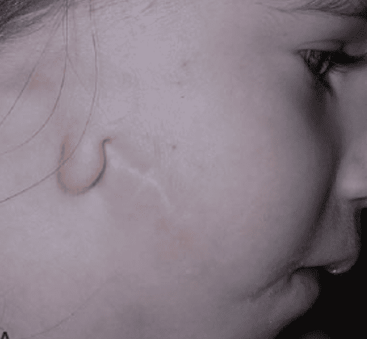
These are benign, fleshy, skin-covered fibrocartilaginous tags located anterior to the auricle, typically along an imaginary line between the tragus and the angle of the mouth. They develop from accessory auricular hillocks that differentiate independently of the main ear complex.
Preauricular Pits & Sinuses
These are small, shallow blind depressions or epithelial pits situated anterior to the ascending limb of the helix. They occur due to incomplete fusion or abnormal developmental folding of the auricular hillocks during closure of the first pharyngeal cleft. While often asymptomatic, they can become recurrently infected, requiring surgical excision.
Atresia of the External Auditory Meatus
Results from failure of the solid ectodermal meatal plug to canalize in late fetal life. Can occur as an isolated fibrous membrane or as complete bony atresia. It causes severe conductive hearing loss and is commonly accompanied by ossicular and first arch craniofacial dysplasias.

Congenital hearing impairment
Congenital hearing loss affects roughly 1 to 3 per 1,000 live births, classified anatomically into conductive hearing loss, sensorineural hearing loss (SNHL), or mixed deficits.
Etiological Classification
- Structural & Conductive Malformations: Results from abnormal development of the sound-conduction apparatus:
- Failure of the meatal plug to canalize (atresia of external auditory meatus).
- Hypoplasia or congenital bony fixation of the auditory ossicles (e.g., stapes footplate ankylosis).
- Absence or hypoplasia of the tympanic cavity.
- Genetic Causes: More than 50% of congenital deafness cases are hereditary. Mutations in the GJB2 gene encoding the gap junction protein Connexin 26 account for half of all autosomal recessive non-syndromic sensorineural deafness. Connexin 26 regulates potassium recycling across cochlear supporting cells, a pathway essential for sustaining the endocochlear potential.
- Intrauterine Infectious Teratogens (TORCH Complex):
- Congenital Rubella Syndrome: Maternal rubella infection during the critical organogenesis window (first trimester) infects the otic vesicle epithelium, damaging the cochlear duct and destroying the organ of Corti.
- Cytomegalovirus (CMV): The most common non-genetic cause of sensorineural hearing loss in infants, causing progressive, asymmetrical neuroepithelial destruction.
Summary of ear derivatives
Scroll the table sideways on a smaller screen.
| Ear Region | Embryonic Structure / Primordium | Definitive Adult Anatomical Derivative |
|---|---|---|
| Internal Ear | Saccular portion of Otic Vesicle | Saccule and Cochlear Duct (Organ of Corti) |
| Utricular portion of Otic Vesicle | Utricle, Semicircular Ducts, Endolymphatic Duct and Sac | |
| Middle Ear | First Pharyngeal Arch (Meckel’s cartilage) | Malleus, Incus, and Tensor Tympani muscle |
| Second Pharyngeal Arch (Reichert’s cartilage) | Stapes (head, neck, crura) and Stapedius muscle | |
| First Pharyngeal Pouch (Tubotympanic recess) | Pharyngotympanic (auditory) tube and Middle Ear cavity (tympanum) | |
| First Pharyngeal Membrane | Tympanic Membrane (eardrum) | |
| External Ear | First Pharyngeal Cleft | External Auditory Meatus (canal) |
| Auricular Hillocks (1st & 2nd pharyngeal arches) | Auricle (Pinna) |
Quick recap
References
Verified embryological and anatomical literature supporting this study note:
- Langman’s Medical Embryology: Sadler, T.W. (2019) Langman’s Medical Embryology. 14th edn. Philadelphia: Wolters Kluwer. [Chapter 19: Ear].
- The Developing Human: Moore, K.L., Persaud, T.V.N. and Torchia, M.G. (2020) The Developing Human: Clinically Oriented Embryology. 11th edn. Philadelphia: Elsevier. [Chapter 18: Development of the Eyes and Ears].
- Larsen’s Human Embryology: Schoenwolf, G.C., Bleyl, S.B., Brauer, P.R. and Francis-West, P.H. (2021) Larsen’s Human Embryology. 6th edn. Philadelphia: Elsevier. [Chapter 17: Development of the Ears and Eyes].
- Gray’s Anatomy: Standring, S. (2021) Gray’s Anatomy: The Anatomical Basis of Clinical Practice. 42nd edn. London: Elsevier. [Chapter 37: External and Middle Ear; Chapter 38: Inner Ear].