Cellular Basis of Disease: Cellular Injury, Adaptive Responses, and Cell Death
Core Educational Competencies
- Structural and Functional Alterations: Delineating how pathological states induce macroscopic and microscopic shifts in tissue architecture.
- Etiological Categorization: Classifying injurious stimuli (intrinsic vs. extrinsic) alongside their representative systemic diseases.
- Cellular Reactive Patterns: Characterizing the continuum of responses exhibited by parenchymal and stromal cells exposed to hostile microenvironments.
- Intercellular and Matrix Dynamics: Evaluating the role of cell-cell adhesions and cell-extracellular matrix (ECM) scaffolds in defining clinical disease topographies.
- Pathological Outcomes: Analyzing downstream sequelae following acute and chronic cellular insults.
- Molecular Pathophysiology: Unpacking the biochemical mechanisms driving organellar failure, energy depletion, oxidative stress, and cell death.
Systematic Module Framework
| Module Phase | Topic Area | Primary Mechanistic & Morphologic Focus |
|---|---|---|
| Phase 1 | The Fundamental Living Cell | Structural baselines, functional compartmentalization, and specialized cell types. |
| Phase 2 | Etiology of Cell Damage | Intrinsic genetic mutations vs. extrinsic environmental, toxic, and hemodynamic stressors. |
| Phase 3 | Cascade of Pathological Events | Progression from initial physiological disturbance to structural degeneration. |
| Phase 4 | Mechanisms of Injury & Death | Bioenergetic failure, calcium dysregulation, ROS propagation, and membrane breakdown. |
| Phase 5 | Adaptive Adjustments to Stress | Cellular size, numerical, and phenotypic transformations (hypertrophy, hyperplasia, atrophy, metaplasia). |
| Phase 6 | Intracellular Accumulations | Pathological pooling of lipids, carbohydrates, proteins, and pigments. |
| Phase 7 | Abnormal Mineralization | Dystrophic precipitation in injured tissues vs. metastatic calcification under hypercalcemia. |
| Phase 8 | Biological Senescence | Mitochondrial decay, oxidative wear-and-tear, DNA repair errors, and progeroid phenotypes. |
1. Fundamental Biology of the Living Cell
The cell serves as the indispensable structural and physiological unit of all biological entities. The human body is built from a diverse spectrum of differentiated cells. Through progressive cellular lineage commitment and terminal differentiation, each cell variant develops distinct architectural configurations and metabolic machinery designed to execute specialized tasks.
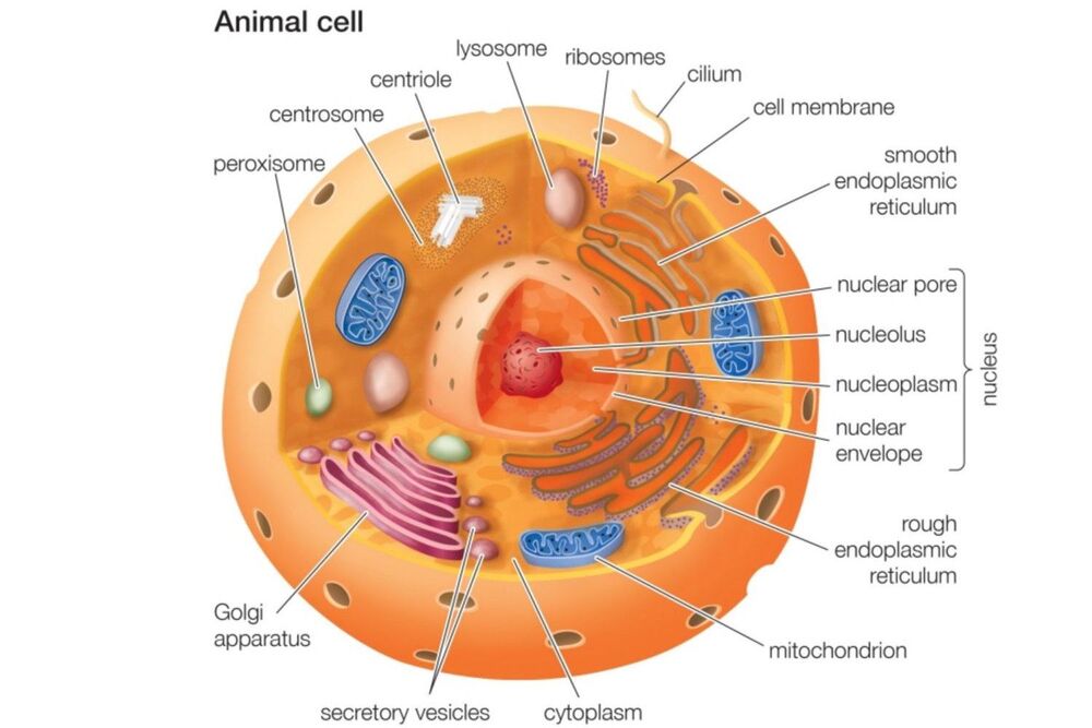
Ultrastructural Compartmentalization of the Eukaryotic Cell
2. Intercellular Junctions and Matrix Anchors
Multicellular tissue architecture relies heavily on specialized junctional complexes that link adjacent cells together or tether cells to the underlying non-cellular basement membrane:
| Junction Classification | Specific Complex | Structural & Functional Contribution |
|---|---|---|
| Cell-to-Cell Interactions | Tight Junctions (Zonula Occludens) | Establish impermeable paracellular seals; maintain apical-basolateral polarity. |
| Adherens Junctions (Zonula Adherens) | Anchor adjacent microfilaments (actin cytoskeleton) via cadherin bridges. | |
| Desmosomes (Macula Adherens) | Fasten intermediate filaments between neighboring cells to distribute mechanical shear stress. | |
| Gap Junctions | Form connexin-lined aqueous channels permitting rapid metabolic and electrical coupling. | |
| Cell-to-Matrix Interactions | Hemidesmosomes | Anchor cytoplasmic intermediate filaments to basal lamina collagen through integrin heterodimers. |
| Focal Adhesions | Connect internal actin stress fibers to extracellular matrix components; act in mechanotransduction. |
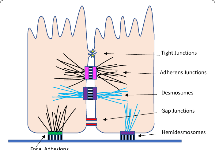
Topography and Spatial Organization of Cell Junctional Complexes
Clinical Significance of Junctional Disruption
Junctional complexes preserve tissue boundaries, transduce vital extracellular biochemical signals, and provide structural integrity. Pathologic breakdown of these linkages occurs during hypoperfusion/ischemia, inflammatory cytokine release, autoimmune attacks, abrasive mechanical tension, and neoplastic progression.
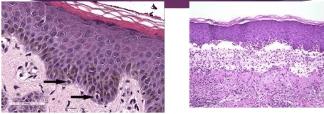
Dermatopathology: Normal Stratified Squamous Epidermis vs. Bullous Pemphigoid
3. Comparative Pathology: Tissue Architecture in Disease
Histopathologic examination highlights structural changes between normal parenchyma and diseased or transformed tissue:
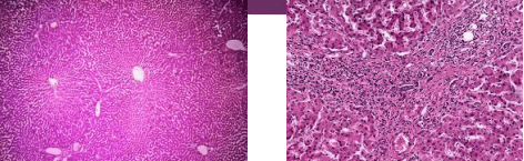
Hepatic Remodeling: Normal Hepatic Architecture vs. End-Stage Cirrhosis
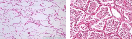
Pulmonary Parenchyma: Aerated Alveoli vs. Inflammatory Consolidation in Pneumonia
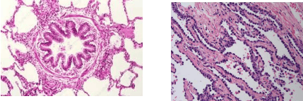
Bronchial Histology: Normal Bronchiolar Lining vs. Adenocarcinoma in Situ
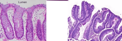
Colonic Mucosa: Normal Crypt Architecture vs. Dysplastic Tubular Adenoma
4. The Nature and Evolution of Disease
Key Definitions: What Constitutes Disease?
Disease represents the dynamic morphological and functional reactions mounted by individual cells, specialized tissues, or integrated organ systems when confronted with an injurious insult. Cellular injury operates by:
- Disrupting essential biochemical cycles (e.g., ATP generation, ionic homeostasis, protein synthesis).
- Directly damaging subcellular structures (plasma membrane, nuclear genome, or organellar integrity).
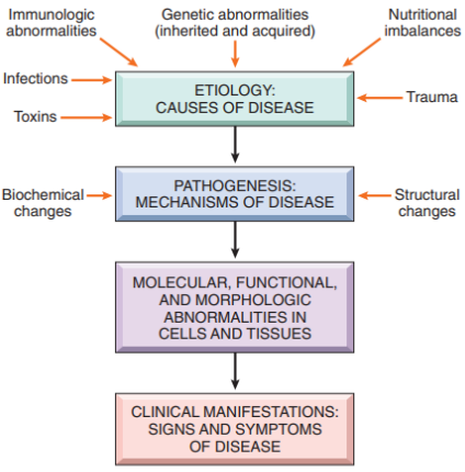
Sequential Stages in the Evolution and Clinical Expression of Disease
5. Spectrum of Injurious Agents
Cell damage stems from a broad range of intrinsic and extrinsic environmental stressors:
| Category | Specific Etiological Driver | Clinical Conditions & Representative Pathologies |
|---|---|---|
| Intrinsic Factors | Infectious Pathogens | Infective endocarditis (perivalvular abscess), myocarditis, Lyme disease, diphtheria, toxoplasmosis, Chagas disease. |
| Infiltrative & Storage Conditions | Sarcoidosis, systemic amyloidosis, hemochromatosis, inborn errors of metabolism, glycogen storage diseases. | |
| Autoimmune & Collagen Vascular Diseases | Rheumatoid arthritis, systemic sclerosis (scleroderma), systemic lupus erythematosus (SLE), dermatomyositis. | |
| Iatrogenic & Surgical Interventions | Coronary artery bypass grafting (CABG), valve replacement/TAVI, cardiac transplantation, therapeutic radiation, iatrogenic AV nodal blocks. | |
| Extrinsic Factors | Metabolic & Physical Extremes | Hypothyroidism, severe anorexia, profound hypoxia, systemic acidosis, hypothermia, hyperthermia, ionizing radiation. |
| Electrolyte Imbalances | Hypokalemia, hyperkalemia, hypercalcemia, hypermagnesemia, osmotic shifts. | |
| Neurological Conditions | Elevated intracranial pressure (ICP), CNS neoplasms, temporal lobe epilepsy, obstructive sleep apnea. | |
| Chemicals & Toxic Substances | Corrosive acids, concentrated alkalis, free radicals, xenobiotics, adverse pharmaceutical reactions. | |
| Genetic Defects | Chromosomal & Inherited Mutations | Down syndrome (Trisomy 21), inborn errors of metabolism, cystic fibrosis. |
| Acquired Somatic Mutations | Proto-oncogene activation, tumor suppressor inactivation (p53), chemical/radiation carcinogenesis. |
6. Determinants of Tissue Vulnerability
The ultimate morphological and clinical consequence of an injurious agent depends on the type of tissue, injury severity, and duration of exposure:
| Tissue Proliferative Class | Replication Capacity | Anatomical Examples & Response to Insult |
|---|---|---|
| Labile Tissues | Continuous, rapid stem-cell driven division | Epidermis, oral mucosa, gastrointestinal lining, hematopoietic bone marrow. Readily reconstitute after mild to moderate damage. |
| Stable Tissues | Quiescent (G0 phase); retain capacity to divide when stimulated | Parenchymal organs (hepatocytes, renal tubular epithelium, pancreatic acini) and mesenchymal fibroblasts/osteoblasts. |
| Permanent Tissues | Terminally differentiated; lack post-natal mitotic capacity | Skeletal muscle fibers, cardiac myocytes, central nervous system neurons. Injury heals through collagenous scar deposition, not regeneration. |
Pathological Definitions: Degeneration vs. Accumulation vs. Deposition
- Degenerations: Reversible metabolic derangements and structural changes occurring inside metabolically active parenchymal cells (e.g., cloudy swelling, hydropic degeneration, fatty change). Does not include stromal phagocytes.
- Accumulations: Pathologic pooling of exogenous or endogenous substrates (carbohydrates, lipids, proteins) within the cytoplasm or nucleus of epithelial or mesenchymal cells due to metabolic defects (e.g., alpha-1-antitrypsin aggregates, storage diseases).
- Depositions: Infiltration and precipitation of substances outside cells into interstitial spaces, basement membranes, or extracellular scaffolds (e.g., amyloid fibrils, dystrophic calcification in necrotic debris, organized thrombi).
7. Homeostasis and Cellular Adaptations to Stress
Normal cells operate within a narrow range of physiologic parameters termed homeostasis. When subjected to physiological stresses or pathological stimuli, cells mount reversible adaptations in size, number, metabolic activity, or phenotype to preserve viability.
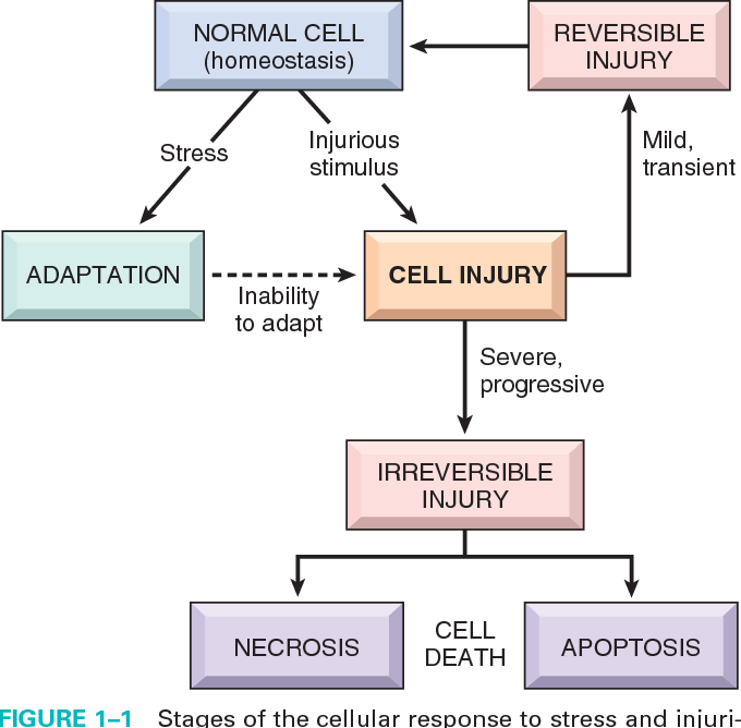
General Categories of Cellular Reactions to Stress and Toxic Stimuli
| Adaptive Mode | Primary Mechanism | Physiological Examples | Pathological Examples |
|---|---|---|---|
| Hyperplasia | Increase in cell number driven by growth factors in cells capable of mitotic division. |
1. Hormonal: Proliferation of breast glandular epithelium at puberty and during pregnancy. 2. Compensatory: Bone marrow erythroid hyperplasia following acute blood loss; partial hepatectomy regeneration. |
1. Endometrial hyperplasia: Unopposed estrogen in repeated anovulatory cycles (precancerous). 2. Benign Prostatic Hyperplasia (BPH): Androgen/estrogen sensitivity. 3. Lymphoid hyperplasia: Reactive follicles during persistent antigenic stimulation. |
| Hypertrophy | Increase in cell size and mass due to synthesis of more structural proteins and metabolic organelles. |
1. Skeletal muscle enlargement in athletes undergoing resistance training. 2. Estrogen-stimulated myometrial smooth muscle hypertrophy in the pregnant uterus. |
1. Adaptive: Concentric left ventricular hypertrophy (LVH) secondary to systemic hypertension or aortic valve stenosis; urinary bladder detrusor hypertrophy secondary to bladder neck/prostatic stricture. 2. Compensatory: Hypertrophy of the remaining kidney after unilateral nephrectomy. |
| Atrophy | Reduction in cell size and functional volume through increased ubiquitin-proteasome degradation and autophagic vacuolization. |
1. Localized: Involution of the thymus gland post-puberty; postmenopausal atrophy of the endometrium and breasts. 2. Generalized: Senile involution with diminished organ functional reserve. |
1. Generalized: Cachexia in advanced malignancy, chronic starvation/protein-energy malnutrition. 2. Endocrine: Adrenal/thyroid/gonadal atrophy following pituitary failure or exogenous steroid therapy. 3. Vascular: Renal atrophy from renal artery atherosclerotic narrowing. 4. Pressure: Vertebral body erosion by expanding aortic aneurysms. 5. Neuropathic: Skeletal muscle denervation in poliomyelitis. 6. Disuse: Limb immobilization in a plaster cast. |
| Metaplasia | Reprogramming of stem cells to replace one adult differentiated cell type with another better equipped to withstand a hostile microenvironment. | Generally absent as a normal homeostatic process; reflects an adaptive response to persistent irritation. |
1. Respiratory Squamous Metaplasia: Replacement of ciliated columnar epithelium with stratified squamous epithelium in chronic smokers. 2. Urothelial Metaplasia & Leukoplakia: Bladder transitional to keratinized squamous epithelium in chronic Schistosoma hematobium infection. 3. Intestinal Metaplasia: Gastric mucosa converting to goblet-cell rich intestinal epithelium in chronic H. pylori gastritis. 4. Mesenchymal: Myositis ossificans (bone formation in skeletal muscle following hematoma/injury). |
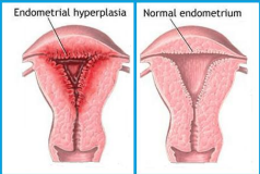
Gross Morphology: Normal Endometrium vs. Pathologic Endometrial Hyperplasia
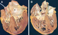
Pathologic Left Ventricular Hypertrophy Induced by Chronic Pressure Overload
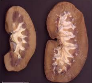
Compensatory Hypertrophy in a Solitary Functioning Kidney
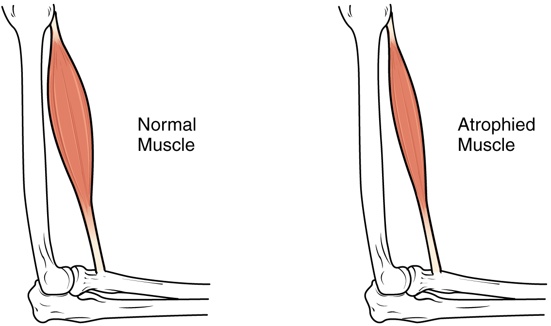
Skeletal Muscle: Normal Muscular Contour vs. Pathologic Muscle Atrophy
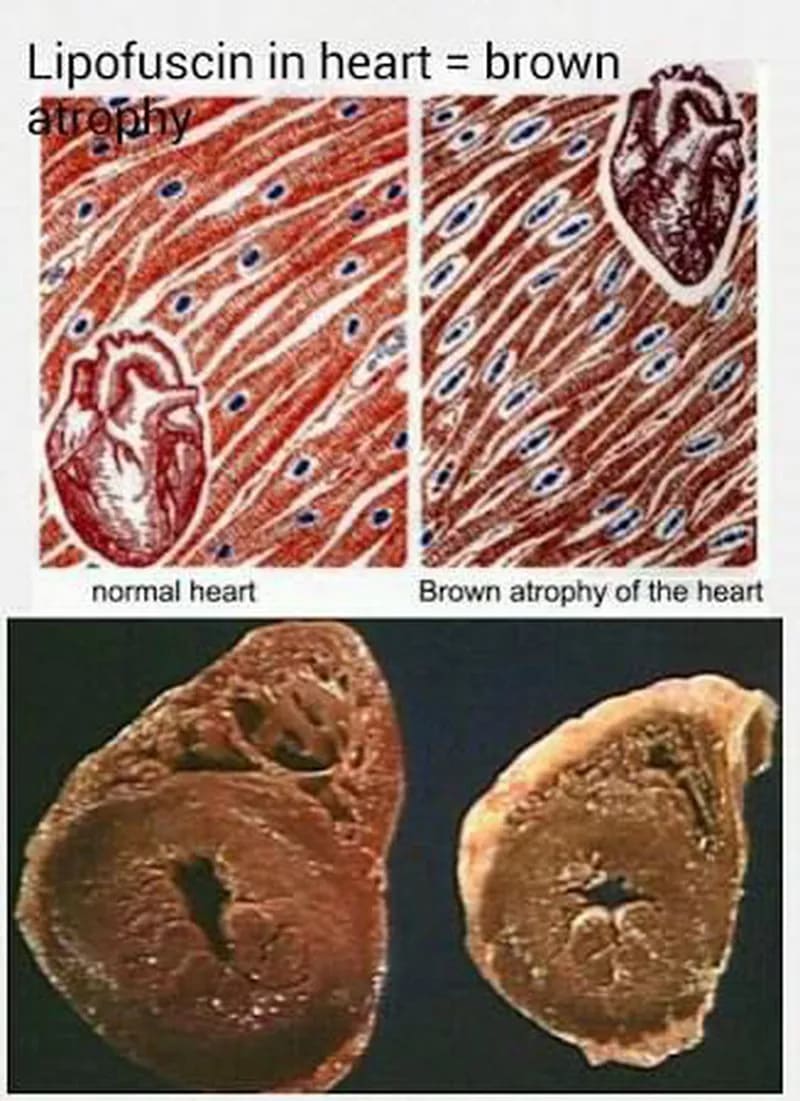
Morphological Hallmarks of Cardiac Brown Atrophy in Senility
Clinical Features: Brown Atrophy of the Heart
Arises from prolonged senescent wear-and-tear leading to accumulation of lipochrome (lipofuscin) pigment within atrophied cardiac myocytes.
- Macroscopic (Gross): Marked reduction in heart mass and dimensions; loss of subepicardial adipose tissue replaced by translucent gelatinous/edematous material; coronary arteries follow a tortuous, snake-like course over the shrunken ventricles; overall dark-brown hue.
- Microscopic: Myocytes are narrowed and separated; intracellular deposits of golden-yellow to brown, granular, insoluble lipid-peroxidation pigment cluster at the nuclear poles (perinuclear halos).
8. Biology of Cellular Aging
True Aging vs. Accelerated Progeria
A. True Biological Aging: Senescence operating under minimal baseline environmental stress. Controlled by intrinsic genetic drivers, notably progressive mitochondrial DNA (mtDNA) mutations. This leads to diminished oxidative phosphorylation, decreased ATP synthesis, reduced metabolic enzyme production, and depleted cellular receptors needed for growth signals.
Extrinsic Accelerators:
- Cumulative oxidative stress from free radicals (aggravated by ionizing radiation exposure).
- Exhaustion of antioxidant protective systems (e.g., systemic depletion of vitamin E or glutathione peroxidase).
- Declining cell-mediated and humoral immunity (immunosenescence).
B. Premature Aging (Progeria): Pathological syndromes caused by specific genetic mutations that disrupt DNA replication and nucleotide repair machinery, resulting in accelerated nuclear instability and premature cellular loss.
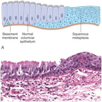
Respiratory Epithelium: Ciliated Columnar Architecture Transitioning to Squamous Metaplasia
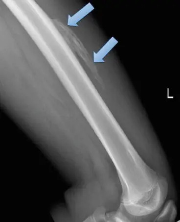
Skeletal Radiography: Mesenchymal Heterotopic Bone Formation in Myositis Ossificans
9. Biochemical Mechanisms of Cell Injury
Diverse etiologic insults converge on common biochemical pathways inside the cell:
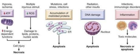
Principal Molecular Mechanisms and Organellar Targets in Cell Injury
10. The Hypoxic-Ischemic Cascade
Interruption of arterial perfusion (ischemia) or reduced oxygen tension (hypoxia) disrupts mitochondrial oxidative phosphorylation. This initiates a sequence of biochemical events:
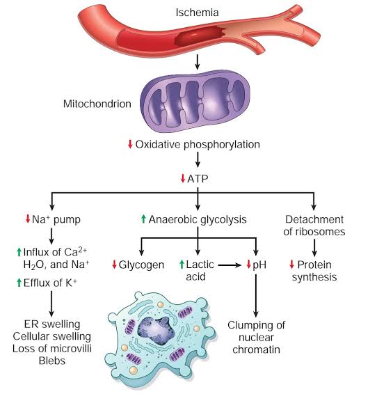
Pathophysiologic Cascade of Ischemia and Cellular Bioenergetic Collapse
Clinical Correlation: Lactic Acidosis and Shock Resuscitation
Under tissue hypoperfusion (hypovolemic, cardiogenic, or distributive septic shock), oxygen-deprived cells shift from aerobic metabolism to anaerobic glycolysis. This generates excess lactic acid, which enters the systemic circulation and produces anion-gap metabolic acidosis. Serial point-of-care measurements of serum lactate and calculations of lactate clearance rates serve as key clinical metrics to gauge the adequacy of volume resuscitation, inotropic support, and end-organ perfusion.
11. Subcellular Targets and Organellar Responses
| Target Organelle | Structural Response to Injury | Functional Sequelae & Clinical Examples |
|---|---|---|
| Mitochondria (First Line of Injury) |
Proliferation / Increased Number | Seen in cellular hypertrophy to meet higher bioenergetic demands. |
| Involution / Decreased Number | Seen in cellular atrophy due to lower metabolic activity. | |
| Megamitochondria / Swelling | Marked high-amplitude swelling seen in hydropic degeneration and oncocytomas (benign neoplasms packed with mitochondria). | |
| Rough Endoplasmic Reticulum (RER) | Ribosome detachment & cisternal dilatation | Impairs apoprotein and structural protein synthesis; dilating cisternae produce microscopic vacuolar change. |
| Smooth Endoplasmic Reticulum (SER) (Second Line of Injury) |
Hypertrophy and enzymatic induction | Mixed-function cytochrome P450 oxidase expansion produces pharmacological tolerance (e.g., barbiturate adaptation); triggers stress (heat-shock) protein release. |
| Disrupted lipid metabolism | Intracellular lipid accumulation; underlies steatosis (fatty change) and inborn storage diseases. | |
| Cytoskeleton | Microtubule & intermediate filament defects | Distorts cell architecture; impairs inflammatory chemotaxis and decreases macrophage phagocytic efficiency. |
| Impaired axonemal / spindle transport | Ciliary immobility in respiratory epithelium causes recurrent sinopulmonary infections; abnormal meiotic/mitotic spindles in gametogenesis cause infertility; neurofibrillary tau tangles in Alzheimer's disease replace functional neuropil. | |
| Lysosomes | Phagolysosomal fusion & incomplete catabolism | Endocytosed or autophagocytosed debris forms secondary lysosomes, generating residual bodies (tertiary lysosomes) with lipofuscin or indigestible exogenous pigment (tattoos). |
| Acid hydrolase enzyme deficiencies | Inability to degrade macromolecular substrates causes genetic Lysosomal Storage Diseases (e.g., Gaucher, Tay-Sachs, Niemann-Pick). | |
| Plasma & Nuclear Membranes (Final Irreversible Step) |
Loss of selective permeability & membrane rupture | Loss of environmental signal reception, ionic gradients, and structural boundary integrity leads directly to irreversible cell death (necrosis). |
12. Free Radical Mediated Injury and Reperfusion Stress
Definition: Free Radicals and Reactive Oxygen Species (ROS)
A free radical is an independent chemical species with a single unpaired valence electron in its outer atomic orbital. This configuration makes it highly reactive, unstable, and capable of triggering auto-catalytic chain reactions:
Free Radical + Biological Molecule → Radical Intermediate + New Free Radical Generation
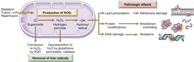
Pathways of Reactive Oxygen Species (ROS) Generation, Degradation, and Pathological Damage
Clinical Entity: Ischemia-Reperfusion Injury
While restoring arterial blood flow to ischemic tissue is essential to prevent infarction, the sudden re-entry of molecular oxygen can paradoxically accelerate cell injury:
- Oxygen influx reacts with damaged mitochondrial electron transport chains and activated xanthine oxidase to produce massive bursts of superoxide and hydroxyl free radicals.
- Ischemic endothelial cells and recruited neutrophils release inflammatory mediators: platelet-activating factor (PAF), inducible nitric oxide synthase (iNOS / NO•), and complement fragments.
- Severe intracellular calcium overload causes opening of the mitochondrial permeability transition pore (MPTP), triggering necrosis and apoptosis.
13. Endoplasmic Reticulum (ER) Stress and the Unfolded Protein Response
Accumulation of misfolded or unfolded proteins inside the ER lumen induces cellular stress. Contributing factors include gene mutations, metabolic deprivation, oxidative stress, senescent decline, and viral infections:
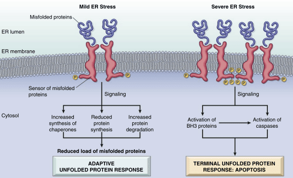
Mechanisms of the Endoplasmic Reticulum (ER) Stress Response: Adaptation vs. Apoptosis
14. Morphological Features of Reversible Cell Injury
Before an injury becomes lethal, stressed cells display characteristic microscopic changes indicating reversible damage:
- Cellular Swelling (Hydropic Change): The initial manifestation of cellular injury, caused by failure of ATP-dependent membrane ion pumps (Na+/K+ ATPase), leading to sodium influx, potassium efflux, and water accumulation within the cytoplasm.
- Steatosis (Fatty Change): Accumulation of lipid vacuoles within parenchymal cells involved in fat metabolism (especially hepatocytes and cardiac myocytes).
- Myelin Figures: Large, whorled concentric phospholipid masses derived from damaged organellar membranes.
- Endoplasmic Reticulum Dilatation: Detachment of surface ribosomes accompanied by vesicular expansion of cisternae.
- Plasma Membrane Alterations: Blebbing of the outer cell contour, distortion or blunting of specialized microvilli, and loosening of intercellular attachment complexes.
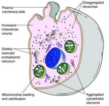
Subcellular and Organellar Architecture in Reversible Cellular Swelling
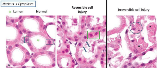
Renal Tubular Histopathology: Normal Architecture vs. Reversible Swelling vs. Irreversible Tubular Necrosis
15. Irreversible Injury and Cell Death: Necrosis
Definition and General Pathogenesis of Necrosis
Necrosis is the morphological expression of cell death within living host tissue, resulting from progressive enzymatic breakdown of cellular components and protein denaturation following severe, unresolvable injury. Necrosis is invariably pathologic and triggers an acute inflammatory reaction characterized by leukocyte infiltration (especially polymorphonuclear neutrophils).
16. Nuclear Changes in Irreversible Cell Injury
Nuclear breakdown follows a characteristic sequence during necrosis:
- Pyknosis: Nuclear shrinkage and progressive chromatin condensation; the nucleus appears as a dense, shrunken, dark basophilic sphere.
- Karyorrhexis: Destructive fragmentation of the pyknotic nucleus into dispersed basophilic granular debris throughout the cytoplasm.
- Karyolysis: Enzymatic dissolution of chromatin by cellular endonucleases; the nucleus fades and completely dissolves, leaving a pale eosinophilic "ghost" cell.
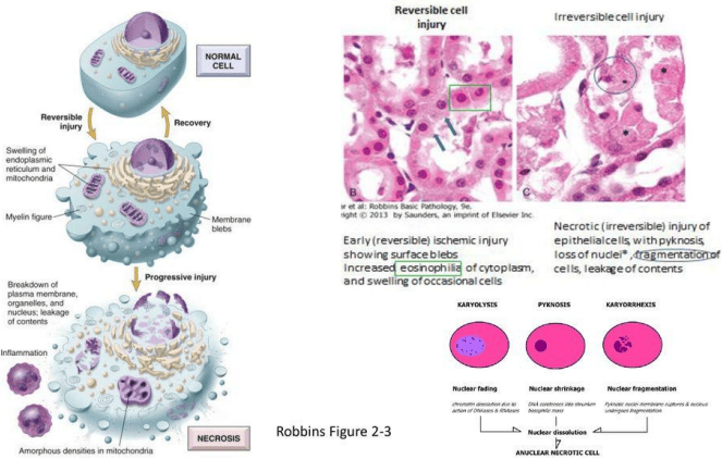
Sequential Morphologic Evolution of Cell Death and Nuclear Dissolution Patterns
17. Histologic Patterns and Types of Necrosis
| Type of Necrosis | Etiology & Mechanism | Histopathological Characteristics | Clinical Context & Organ Sites |
|---|---|---|---|
| 1. Coagulative Necrosis | Severe acute ischemia and hypoxia. Acidosis denatures structural proteins and autolytic enzymes, blocking immediate proteolysis. | Ghost-like cellular outlines are preserved for days. Loss of nuclei with diffuse cytoplasmic eosinophilia. | Infarction of all solid visceral organs (myocardium, renal cortex, spleen), except the central nervous system. |
| 2. Liquefactive Necrosis | Rapid enzymatic digestion of dead cells by hydrolytic enzymes from bacteria or recruited neutrophils. | Complete loss of tissue architecture; rapid transformation into a viscous, liquid, amorphous mass of debris and dead leukocytes. |
1. Bacterial and fungal infections resulting in abscess cavities (pus formation). 2. Hypoxic/ischemic infarcts of the brain (cerebral encephalomalacia). |
| 3. Caseous Necrosis | Host cell-mediated (Type IV hypersensitivity) immune response driven by activated T-lymphocytes and macrophages against persistent pathogens. | Grossly soft, friable, cheese-like appearance. Microscopically shows structureless, amorphous, eosinophilic granular debris surrounded by a granulomatous rim of epithelioid macrophages, Langhans giant cells, and lymphocytes. | Tuberculous infections (Mycobacterium tuberculosis) and specific systemic fungal diseases (e.g., histoplasmosis, coccidioidomycosis). |
| 4. Gangrenous Necrosis | Clinical term for ischemic coagulative necrosis involving multiple tissue planes across an extremity or hollow viscus. |
1. Dry Gangrene: Sterile ischemic mummification; coagulative pattern predominates. 2. Wet Gangrene: Superimposed bacterial infection triggers liquefactive digestion, foul odor, and rapid tissue destruction. |
Lower extremity ischemia in diabetes mellitus and severe atherosclerosis; strangulated bowel; acute ischemic cholecystitis. |
| 5. Enzymatic Fat Necrosis | Extravasation of activated pancreatic lipases and esterases into peripancreatic and mesenteric adipose depots. | Enzymes hydrolyze triglycerides into free fatty acids, which complex with calcium ions (saponification). Produces chalky white deposits grossly; microscopic shadowy necrotic adipocytes with basophilic calcium precipitates. | Acute necrotizing pancreatitis and trauma to adipose-rich tissue (e.g., traumatic breast fat necrosis). |
| 6. Fibrinoid Necrosis | Antigen-antibody (immune complex) deposition in vascular walls accompanied by leakage and polymerization of plasma proteins (including fibrin). | Microscopic finding showing bright pink, amorphous, refractile, smudgy proteinaceous deposits within arterial and arteriolar walls. | Immune-mediated vasculitis (e.g., polyarteritis nodosa), severe malignant hypertension, and preeclampsia/placental vascular lesions. |
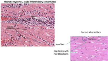
Cardiac Histopathology: Acute Myocardial Infarction Demonstrating Coagulative Necrosis
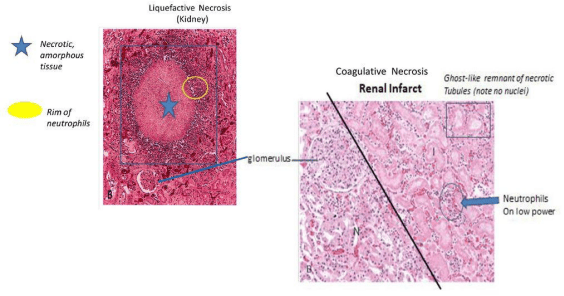
Renal Pathology: Suppurative Liquefactive Abscess vs. Ischemic Coagulative Infarction
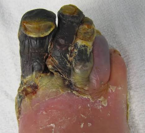
Clinical Manifestation of Gangrenous Necrosis of the Lower Extremity
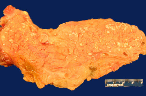
Enzymatic Fat Saponification within Mesenteric Adipose Tissue in Acute Pancreatitis
Diagnostic Serum Biomarkers of Tissue Necrosis
Irreversible plasma membrane disruption allows intracellular enzymes and proteins to leak into the extracellular fluid and enter the peripheral circulation. Quantifying these released biomarkers serves as a primary tool for diagnosing organ-specific necrosis:
- Cardiac Myocyte Necrosis: Cardiac troponins (cTnI and cTnT) and creatine kinase-MB (CK-MB) for diagnosing acute myocardial infarction.
- Hepatocellular Necrosis: Serum transaminases: aspartate aminotransferase (AST) and alanine aminotransferase (ALT) for viral, toxic, or ischemic hepatitis.
- Exocrine Pancreatic Necrosis: Serum amylase and lipase for diagnosing acute pancreatitis.
18. Programmed Cell Death: Apoptosis
Definition of Apoptosis
Apoptosis is a highly regulated, energy-dependent (ATP-requiring) pathway of programmed cell death in which an intrinsic or extrinsic genetic suicide program is activated to eliminate individual cells. Key characteristics:
- The plasma membrane remains intact without releasing intracellular contents into the extracellular space.
- No secondary inflammatory response is elicited.
- The cell condenses, fragments into membrane-bound apoptotic bodies, and is rapidly recognized and engulfed by neighboring cells and resident macrophages.
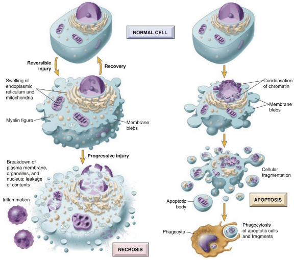
Morphologic Progression of Apoptotic Cleavage vs. Necrotic Lysis
19. Molecular Pathways and Regulation of Apoptosis
Apoptotic signaling operates via two converging mechanisms that activate an initiator and executioner caspase cascade:
| Pathway Element | Intrinsic (Mitochondrial) Pathway | Extrinsic (Death Receptor) Pathway | Cytotoxic T-Lymphocyte Alternative |
|---|---|---|---|
| Initiating Trigger | DNA damage (radiation, free radicals), growth factor deprivation, cellular stress, misfolded protein accumulation. | Ligation of cell-surface death receptors belonging to the Tumor Necrosis Factor receptor family (Fas [CD95], TNFR1). | Recognition of foreign viral or neoplastic antigens by cytotoxic CD8+ T cells. |
| Key Sensor / Regulatory Molecules |
Anti-apoptotic: Bcl-2, Bcl-xL. Pro-apoptotic: Bax, Bak. Sensors: BH3-only proteins (Bad, Bim, Bid, Puma, Noxa). |
FasL (Fas Ligand), TNF-alpha, and adapter proteins containing a cytoplasmic death domain: FADD (Fas-Associated Death Domain). | Direct release of perforin (creates transmembrane pores) and granzyme B. |
| Initiator Caspase | Loss of Bcl-2/xL allows Bax/Bak channels to form → Cytochrome c leaks into cytosol → binds Apaf-1 → forms apoptosome → activates Caspase-9. | Receptor trimerization recruits FADD → forms Death-Inducing Signaling Complex (DISC) → autocatalytic cleavage activates Caspase-8 and Caspase-10. | Granzyme B directly cleaves and activates executioner caspases, bypassing upstream receptors. |
| Execution Phase |
Initiator caspases activate executioner Caspase-3 and Caspase-6. These cleave structural cytoskeletal proteins, degrade transcription factors, and activate CAD endonucleases to cause internucleosomal DNA fragmentation. |
||
| Phagocytic Clearance | Inversion of membrane phosphatidylserine to the outer leaflet serves as an "eat-me" signal for macrophage clearance without triggering inflammation. | ||
Molecular Feature: p53 as the Guardian of the Genome
Severe DNA damage (from UV radiation, ionizing radiation, or chemical carcinogens) activates the TP53 tumor suppressor gene, upregulating the p53 protein. p53 halts the cell cycle at the G1/S transition via p21 induction to facilitate nucleotide excision repair. If the genomic damage cannot be repaired, p53 triggers apoptosis by directly upregulating the pro-apoptotic proteins Bax and Apaf-1.
20. Physiological and Pathological Contexts of Apoptosis
- Physiological Settings:
- Programmed tissue remodeling during normal embryogenesis (e.g., interdigital webbing involution).
- Hormone-dependent physiologic involution in adults (e.g., endometrial sloughing during the menstrual cycle, post-lactation mammary regression).
- Homeostatic cell turnover in proliferating populations (e.g., continuous shedding of enterocytes from gastrointestinal crypts).
- Elimination of self-reactive lymphocytes during thymic maturation (negative selection) to prevent autoimmunity.
- Pathological Settings:
- Genotoxic stress and DNA damage induced by therapeutic radiation or cytotoxic chemotherapy.
- Elimination of cells infected by intracellular viruses (e.g., apoptotic Councilman/acidophil bodies in viral hepatitis).
- Pathologic atrophy in duct-obstructed parenchymal organs (e.g., pancreas, parotid gland).
- Progressive loss of neuronal populations in neurodegenerative diseases (Alzheimer, Parkinson, Huntington disease).
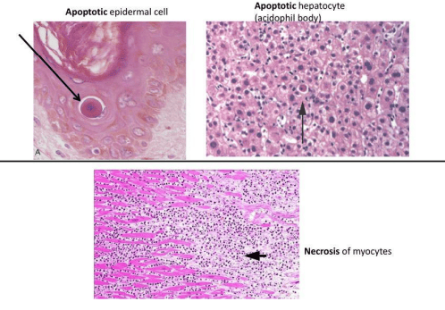
Microscopic Appearances: Apoptotic Keratinocyte, Councilman Body in Hepatitis, and Myocardial Necrosis
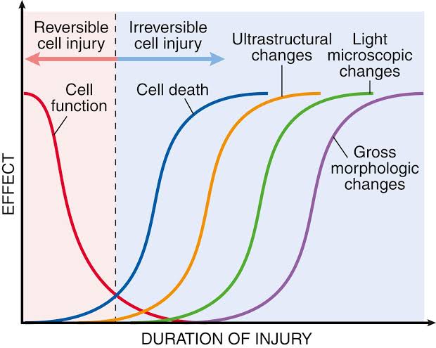
Temporal Relationship Between Functional Loss, Biochemical Death, and Structural Findings in Cell Injury
21. Systematic Comparison: Necrosis vs. Apoptosis
| Histological / Biological Feature | Apoptosis | Necrosis |
|---|---|---|
| Definition & Nature | Programmed, strictly regulated cell destruction. | Accidental, unregulated cell death from severe injury. |
| Mechanism & Energy Needs | Active process; requires ATP; driven by caspase cascades. | Passive process; no ATP required; driven by bioenergetic failure and membrane lysis. |
| Cellular Volume & Morphology | Cell shrinkage; cytoplasmic blebbing; chromatin condensation; apoptotic body formation. | Cell swelling (oncosis); organellar breakdown; plasma membrane lysis. |
| Membrane Integrity | Preserved; altered lipid symmetry (phosphatidylserine flip). | Disrupted; cellular contents spill into interstitial space. |
| Host Inflammatory Response | Absent; no secondary inflammation. | Present; marked neutrophilic and mononuclear infiltration. |
| Physiological vs. Pathological Role | Both; functions in normal development, tissue remodeling, and pathology. | Invariably pathological. |
| Clinical Examples | Embryonic digit separation, endometrial shedding, Councilman bodies in hepatitis. | Myocardial infarction, acute tubular necrosis, dry/wet gangrene, bacterial abscesses. |
22. Intracellular Accumulations
Mechanisms Driving Abnormal Accumulations
Substances pool within cells transiently or permanently due to four main metabolic derangements:
- Overproduction or Normal Load with Slower Clearance: Normal endogenous substances produced at normal or elevated rates with inadequate catabolism (e.g., triglycerides in hepatic steatosis).
- Defective Protein Folding and Chaperone Transport: Mutations that cause abnormal folding and impaired packaging (e.g., alpha-1-antitrypsin deficiency).
- Inherited Enzyme Deficiencies: Inborn errors of metabolism where lacking lysosomal enzymes causes substrate accumulation (e.g., Gaucher, Tay-Sachs, Niemann-Pick).
- Deposition of Insoluble Exogenous Materials: Inability of cells to degrade phagocytosed foreign particles (e.g., inhaled silica, carbon anthracosis).
23. Spectrum of Intracellular and Extracellular Deposits
| Material Class | Type & Site of Accumulation | Pathogenesis & Histochemical Staining | Associated Disease Entities |
|---|---|---|---|
| Proteins | Intracellular |
1. Reabsorption droplets: Proximal renal tubular epithelial cells in heavy proteinuria. 2. Excess synthesis: Immunoglobulin-packed ER in plasma cells producing eosinophilic Russell bodies (rhinoscleroma, multiple myeloma); cytokeratin aggregate condensation producing Mallory-Denk bodies in alcoholic hepatitis. |
Nephrotic syndrome, rhinoscleroma, multiple myeloma, alcoholic steatohepatitis. |
| Extracellular | Glassy, amorphous, eosinophilic protein deposited in connective tissue or vascular basements. |
1. Dense collagen hyalinization in mature scars. 2. Hyaline arteriolosclerosis: Extravasated plasma proteins and basement membrane thickening in systemic hypertension and diabetes mellitus. 3. Amyloidosis: Pathologic beta-pleated fibrillar protein. Stains pale pink with H&E; shows salmon-pink appearance under Congo Red with diagnostic apple-green birefringence under polarized light. |
|
| Mucin (Carbohydrates) | Intracellular (Mucoid Change) | Mucin distends the cell, pushing the nucleus to the periphery, creating a characteristic signet-ring appearance. | Catarrhal respiratory inflammation, mucinous cystadenoma of the ovary, signet-ring cell adenocarcinoma of the stomach. |
| Extracellular (Myxomatous Change) | Mucinous material pooling within connective tissue ground substance. Grossly gelatinous; microscopically shows stellate cells in a loose basophilic network. | Mesenchymal soft tissue neoplasms (myxomas), myxoid neurofibromas, mitral valve prolapse (myxomatous degeneration). | |
| Glycogen | Intracellular | Excessive accumulation within liver, skeletal muscle, or myocardium. Stains intensely magenta with Periodic Acid-Schiff (PAS) and digests with diastase. | Glycogen Storage Diseases: Type I (von Gierke disease; glucose-6-phosphatase defect), Type II (Pompe disease; lysosomal alpha-glucosidase defect), Type V (McArdle syndrome; muscle glycogen phosphorylase defect). |
| Lipids (Neutral Fats & Complex Lipids) | Intracellular |
1. Triglycerides (Steatosis): Clear vacuoles displacing the hepatocyte nucleus; stains positive with Oil Red O or Sudan IV on frozen sections. 2. Sphingolipids/Gangliosides: Lysosomal storage defect in macrophages/histiocytes. |
Alcoholic and non-alcoholic fatty liver disease (NAFLD), obesity, diabetes; Gaucher disease (glucocerebrosidase defect; "crumpled tissue paper" histiocytes), Niemann-Pick disease (sphingomyelinase defect; foamy histiocytes). |
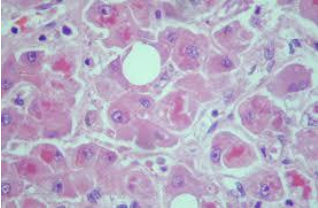
Hepatic Steatosis (Fatty Liver): Gross Morphology and Microscopic Macrovesicular Lipid Vacuolation
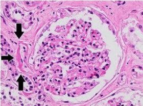
Renal Amyloid Deposition: Amorphous Glomerular Obliteration and Congo Red Staining
24. Pigment Accumulations: Endogenous vs. Exogenous
| Pigment Type | Color & Biochemical Origin | Pathophysiologic Mechanism & Distribution | Diagnostic Significance |
|---|---|---|---|
| Lipofuscin | Golden-yellow to brown; lipid-protein complexes derived from free-radical peroxidation of polyunsaturated lipids. | "Wear-and-tear" pigment of aging. Accumulates in tertiary lysosomes of permanent and long-lived cells (cardiac myocytes, hepatocytes, neurons). | Marker of cellular aging, prolonged starvation, and brown atrophy of the heart. |
| Melanin | Dark brown to black; synthesized from tyrosine oxidation by tyrosinase inside melanocytes. | Transferred to keratinocytes and taken up by dermal macrophages (melanophores). Stimulated by UV light and pituitary MSH; inhibited by adrenocortical hormones. |
1. Localized hyperpigmentation: Melanocytic nevi, malignant melanoma. 2. Generalized hyperpigmentation: Addison disease (loss of adrenocortical feedback → high ACTH/MSH), suntan, chloasma of pregnancy. 3. Hypopigmentation: Albinism (tyrosinase deficiency), vitiligo/leukoderma (autoimmune melanocyte destruction). |
| Hemosiderin | Golden-yellow to brown, granular, iron-containing ferritin aggregate. |
Forms when local or systemic iron levels exceed ferritin storage capacity. 1. Local: Erythrocyte extravasation in bruises; alveolar macrophage phagocytosis in chronic pulmonary venous congestion ("heart failure cells"). 2. Visceral (Hemosiderosis): Iron accumulation in Kupffer cells, splenic cords, and bone marrow from hemolytic anemias or repeated blood transfusions. |
Demonstrated histochemically by the Prussian blue (Perls) reaction, turning iron bright blue. |
| Bilirubin | Yellow-green; non-iron-containing breakdown product of heme catabolism. | Heme is converted to biliverdin, then unconjugated bilirubin. Transported via albumin to the liver, conjugated to glucuronic acid, and excreted into bile. Excess accumulation causes systemic jaundice (icterus). | Identifies hemolytic processes, biliary tract obstruction (gallstones, pancreatic cancer), and parenchymal hepatic injury. |
| Exogenous Pigments | Carbon (Coal Dust) | Inhaled carbon particulates phagocytosed by alveolar macrophages and drained to hilar and mediastinal lymph nodes (Anthracosis). | Blackens lung parenchyma; can cause coal workers' pneumoconiosis. |
| Silica | Inhaled crystalline stone/quartz dust triggers macrophage activation and chronic fibroblastic proliferation (Silicosis). | Grey fibrotic pulmonary nodules; increases tuberculosis susceptibility. | |
| Heavy Metals | Chronic ingestion or exposure to lead, mercury, or silver causes heavy metal precipitation in oral mucosal margins. | Blue gingival lines (e.g., Burtonian line in chronic lead poisoning; Argyria with silver). | |
| Carotenoids & Tattoos | Excessive beta-carotene ingestion causes yellow-orange skin discoloration (carotenemia, sparing the sclerae). Injected tattoo ink is phagocytosed by dermal macrophages and persists for life. | Benign cosmetic and dermatologic indicators. |
Clinical Entity: Hemochromatosis ("Bronzed Diabetes")
An inherited autosomal recessive metabolic disorder (commonly HFE gene mutations, C282Y) that causes uncontrolled duodenal iron absorption. Plasma transferrin saturation increases, overwhelming ferritin storage capacity and depositing iron in parenchymal organs:
- Endocrine Pancreas: Hemosiderin accumulation in beta-islet cells with interstitial fibrosis causes secondary insulin-dependent diabetes mellitus.
- Liver: Progressive iron loading in hepatocytes and Kupffer cells causes micronodular cirrhosis and increases hepatocellular carcinoma risk.
- Integument: Dermal hemosiderin deposition around adnexal sweat glands combined with reactive melanogenesis produces characteristic bronze hyperpigmentation.
- Myocardium: Intracytoplasmic hemosiderin accumulation in myocytes leads to dilated or restrictive cardiomyopathy and cardiac arrhythmias.
- Mesenteric Lymph Nodes & Joints: Pigment deposition in reticuloendothelial cells, chondrocalcinosis, and polyarthropathy.
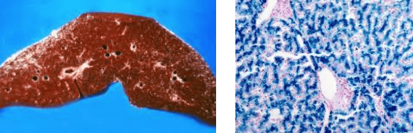
Hepatic Hemochromatosis: Gross Rust-Brown Remodeling and Prussian Blue Staining
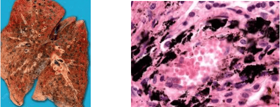
Pulmonary Anthracosis: Pleural Carbon Deposition and Particulate-Laden Macrophages
25. Pathologic Calcification
Definition of Pathologic Calcification
The abnormal tissue deposition of calcium salts, along with smaller amounts of iron, magnesium, and other mineral salts, occurring in tissues other than bone and teeth. Classifies into two distinct forms based on serum calcium levels and tissue viability:
| Parameter | A. Dystrophic Calcification | B. Metastatic Calcification |
|---|---|---|
| Tissue Status | Occurs in dead, dying, or degenerate tissues. | Occurs in normal, living tissues. |
| Serum Calcium Levels | Normal serum calcium and normal phosphate metabolism. | Elevated (hypercalcemia secondary to altered calcium/phosphate metabolism). |
| Underlying Etiologies |
1. Caseous necrosis in chronic tuberculosis. 2. Saponification plaques in enzymatic fat necrosis. 3. Advanced calcific aortic stenosis in senescent/bicuspid valves. 4. Atherosclerotic plaques in damaged arterial walls. 5. Longstanding organized thrombi (phleboliths). 6. Retained dead fetus (lithopedion). 7. Psammoma bodies in papillary thyroid or serous ovarian carcinomas. |
1. Hyperparathyroidism: Primary (parathyroid adenoma) or secondary (chronic kidney disease). 2. Bone Destruction: Multiple myeloma, extensive osteolytic metastases, immobilization. 3. Vitamin D Disorders: Hypervitaminosis D, sarcoidosis (macrophage-mediated 1-alpha-hydroxylase activation). 4. Renal Failure: Phosphate retention causing secondary hyperparathyroidism. |
| Predominant Anatomical Sites | Localized to foci of prior necrosis, scarring, or chronic inflammation. | Interstitial tissues of organs that lose acid and maintain an internal alkaline environment: gastric mucosa, renal tubular basements, pulmonary alveolar septa, systemic arteries, pulmonary veins. |
| Morphologic Appearance |
Gross: Hard, gritty, chalky-white to grey-white deposits. Microscopic: Deeply basophilic (blue-purple), irregular, granular intracellular (mitochondrial) or extracellular deposits with H&E staining. |
|
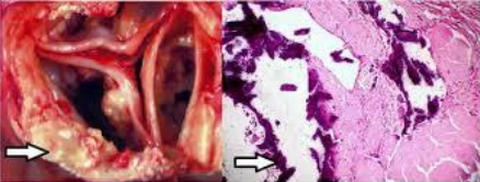
Dystrophic Calcification of a Stenotic Aortic Valve
26. Case Inquiries & Self-Assessment
Case-Based Practice Prompts
Clinical Case 1: A 72-year-old male develops acute ischemic injury to the cerebral hemisphere secondary to acute right internal carotid artery occlusion. What specific pattern of necrosis occurs in the affected parenchyma?
Answer: Liquefactive Necrosis. While ischemic necrosis in solid viscera produces coagulative necrosis, cerebral ischemia triggers liquefactive necrosis due to high hydrolytic enzyme and lipid content and the lack of a stabilizing extracellular collagen matrix.
Clinical Case 2: A diabetic patient is diagnosed with "wet gangrene" of the right lower extremity. What pathophysiologic events distinguish this from dry gangrene?
Answer: While dry gangrene is primarily sterile ischemic coagulative necrosis, wet gangrene develops when a bacterial infection superimposes on the dead tissue. Bacterial and neutrophil hydrolytic enzymes trigger rapid liquefaction, creating foul-smelling, purulent, rapidly progressing tissue destruction.
Clinical Case 3: A 57-year-old man presents with nausea, vomiting, and scleral icterus following acute Hepatitis A infection. A liver biopsy shows scattered acidophilic (Councilman) bodies. What cellular process does this represent?
Answer: Apoptosis. Acidophil bodies are rounded, shrunken, intensely eosinophilic hepatocytes with condensed or fragmented nuclear remnants that have been cleaved by caspases during cytotoxic T-cell elimination of virus-infected cells.
Clinical Case 4: An endocervical biopsy in a 23-year-old woman shows stratified squamous epithelium instead of the expected single layer of mucin-secreting columnar cells. What adaptation has occurred, and what drives it?
Answer: Squamous Metaplasia. Driven by chronic irritation and local inflammation (e.g., chronic cervicitis, acidic vaginal pH changes), leading to stem-cell reprogramming into a stratified squamous lining better suited to resist chemical and mechanical irritation.
27. Multiple Choice Review
Question 1: Reperfusion Pathology
A 63-year-old male with a history of unstable angina suffers an acute myocardial infarction. He receives intravenous tissue plasminogen activator (tPA) to restore coronary patency. Despite successful recanalization, myocardial cell injury worsens due to which cellular abnormality?
- A. Loss of intermediate cytoskeletal filaments
- B. Reduced intracellular pH from anaerobic glycolysis
- C. Increased free radical formation
- D. High-amplitude mitochondrial swelling
- E. Nuclear chromatin clumping
- F. Reduced translation and protein synthesis
Correct Answer: C — Re-introducing molecular oxygen into previously ischemic tissue paradoxically triggers bursts of reactive oxygen species (superoxide, hydrogen peroxide, hydroxyl radicals) from damaged mitochondrial respiratory chains and infiltrating neutrophils, exacerbating membrane and lipid damage (ischemia-reperfusion injury).
Question 2: Physiological Adaptation in Pregnancy
A 28-year-old woman delivers her first child. She successfully breastfeeds the infant for almost a year without complication. Which cellular adaptation initiated in her breasts during gestation made prolonged lactation possible?
- A. Stromal hypertrophy
- B. Steatocyte atrophy
- C. Ductal epithelial metaplasia
- D. Lobular hyperplasia
Correct Answer: D — Gestational mammary enlargement is driven by estrogen, progesterone, and prolactin stimulation, which increases the total number of secretory glandular and ductular epithelial cells (hormonal hyperplasia) to prepare the breast for lactation.
Question 3: Acute Abdominal Pathology
A 40-year-old woman presents with the sudden onset of excruciating epigastric pain radiating to her back. Physical examination shows diffuse abdominal tenderness with guarding and muscular rigidity. Laboratory workup reveals marked elevation of serum lipase at 415 U/L. Contrast CT confirms pancreatic inflammation and peripancreatic fluid collections. Which form of necrosis characteristically accompanies these findings?
- A. Coagulative necrosis
- B. Dry gangrene
- C. Fat necrosis
- D. Apoptosis
- E. Liquefactive necrosis
Correct Answer: C — Acute pancreatitis causes pancreatic lipases to leak into peripancreatic and mesenteric fat. These enzymes split triglycerides into free fatty acids that complex with calcium salts, generating grossly visible chalky-white saponification foci (enzymatic fat necrosis).