Acute Inflammation
1. Fundamental Definitions and Core Concepts
Key Definitions: Inflammation
- Inflammation: A complex, physiological, protective response executed by vascularized living tissue to combat noxious stimuli, including pathogenic invasion, physical trauma, or chemical injury.
- Biological Purpose: Serves to mobilize humoral immune factors and circulating defensive leukocytes from systemic microvessels directly to the afflicted extravascular focus to destroy the offending agent, clear necrotic debris, and initiate structural repair.
- Vascular Exclusivity: The inflammatory cascade is restricted exclusively to viable, perfused tissues. Non-vascularized structures (e.g., central articular cartilage or the optical cornea) and non-viable necrotic debris cannot autonomously mount an acute inflammatory reaction.
From an operational standpoint, host inflammation represents an orderly, multi-step defensive strategy designed to:
- Detect and identify foreign molecular antigens or non-viable native cellular fragments.
- Recruit circulating leukocytes and plasma-derived humoral proteins across the microvascular endothelium.
- Neutralize, destroy, or sequester the offending physical, chemical, or biological agent.
- Enzymatically digest and phagocytose non-viable parenchymal cells and cellular debris.
- Activate regenerative cascades, leading either to parenchymal regeneration or fibroblastic scar deposition.
2. Clinical Distinction: Infection versus Inflammation
A common diagnostic mistake is to consider infection and inflammation interchangeable. They represent distinct, though often overlapping, pathophysiological entities:
| Entity | Pathological Definition | Clinical Relationship |
|---|---|---|
| Infection | The invasion, colonization, and parasitic multiplication of exogenous micro-organisms (bacteria, viruses, fungi, or protozoa) within host tissues. | Serves as a frequent inciting etiology of acute inflammation. |
| Inflammation | The cellular and microvascular host defense reaction mounted against noxious stimuli, cell injury, or antigenic foreign matter. | Occurs in response to infection, but frequently develops under totally sterile, non-microbial conditions. |
Clinical Subsets of Non-Infectious (Sterile) Inflammation
- Post-Ischemic Tissue Necrosis: Sterile neutrophilic response following acute myocardial infarction.
- Thermal, Mechanical, and Radiation Injuries: Epidermal burn blister formation, soft-tissue crush contusions, and solar ultraviolet erythema.
- Crystalline Deposition Arthropathies: Acute gouty arthritis driven by intra-articular monosodium urate monohydrate crystals.
- Autoimmune Pathologies: Endogenous tissue destruction seen in rheumatoid arthritis or systemic lupus erythematosus.
- Type I Hypersensitivity: Acute allergic reactions triggered by otherwise innocuous environmental allergens.
- Exogenous Foreign Body Reactions: Inflammatory cuffing around surgical sutures, talc particulates, or wood splinters.
3. Physiological Purpose and Evolutionary Imperative
Acute inflammation is essential for vertebrate survival. In its absence, minor superficial abrasions could allow uncontrolled microbial proliferation, leading to systemic bacteremia and fatal septic shock. Furthermore, damaged tissues would fail to undergo enzymatic debridement, preventing wound closure, matrix deposition, and clinical healing.
However, acute inflammation is not a benign process. While physiological inflammation is self-limited, excessive microvascular permeability, extensive cellular recruitment, and the extracellular release of reactive oxygen species and proteases can cause significant collateral injury to normal host tissues.
4. Primary Cellular and Molecular Participants
| Component | Constituents & Cell Subsets | Primary Inflammatory Responsibility |
|---|---|---|
| Microvasculature | Post-capillary venules, terminal arterioles, and true capillary beds. | Undergoes smooth muscle relaxation (vasodilation) and endothelial contraction to permit plasma exudation and leukocyte diapedesis. |
| Circulating Leukocytes |
• Neutrophils: Dominant initial responders (0–24 hours). • Monocytes: Recruited subsequently, differentiating into tissue macrophages (24–48 hours). • Eosinophils: Characteristic of helminthic infections and allergic states. • Lymphocytes: Recruited in persistent, viral, or autoimmune reactions. |
Phagocytosis of foreign pathogens, oxidative and non-oxidative killing, secretion of amplification cytokines, and scavenger debridement. |
| Tissue Sentinel Cells | Resident histiocytic macrophages, dermal and mucosal dendritic cells, and perivascular mast cells. | Continuous environmental surveillance via pattern recognition receptors; immediate release of preformed histamine, leukotrienes, TNF-α, and IL-1. |
| Plasma Protein Systems | The complement cascade (C3a, C5a, C3b), coagulation factors, fibrinolytic system, and the plasma kinin system (bradykinin). | Circulate as inactive pro-enzymes; undergo sequential cleavage upon endothelial leakage to drive chemotaxis, opsonization, vasodilation, and algogenic signaling. |
| Chemical Mediators | Vasoactive amines, eicosanoids (prostaglandins, leukotrienes, lipoxins), cytokines, chemokines, platelet-activating factor (PAF), and nitric oxide. | Molecular orchestrators that initiate, regulate, amplify, and coordinate the active termination of the vascular and cellular response. |
5. The 5 R's of the Inflammatory Response
- Recognition: Detection of pathogens or damaged host cells by sentinel macrophages, dendritic cells, and mast cells.
- Recruitment: Trans-endothelial mobilization of circulating leukocytes and plasma exudates into the extravascular space.
- Removal: Phagocytic internalization, intracellular destruction of microbes, and enzymatic degradation of necrotic debris.
- Regulation & Termination: Downregulation of pro-inflammatory signals and switching of eicosanoid classes to anti-inflammatory mediators (e.g., lipoxins, resolvins) as neutrophils undergo apoptosis.
- Repair: Fibroblast proliferation, angiogenesis, and re-epithelialization leading to parenchymal restoration or scar formation.
6. The Cardinal Signs of Inflammation
Documented historically by Aulus Cornelius Celsus (first 4 signs) and Rudolf Virchow (fifth sign), the cardinal manifestations reflect local microvascular dynamics and sensory nerve activation:
| Clinical Sign | Latin Nomenclature | Underlying Pathophysiological Mechanism |
|---|---|---|
| Redness | Rubor | Mediator-induced arteriolar vasodilation (via histamine, prostaglandins, and nitric oxide), resulting in increased regional perfusion and capillary engorgement (active hyperemia). |
| Heat | Calor | Increased local delivery of warm, core systemic blood through dilated microcirculatory beds. (Note: Systemic fever is a centrally mediated response coordinated by hypothalamic PGE2 under the influence of pyrogenic cytokines like IL-1, TNF-α, and IL-6). |
| Swelling | Tumor | Increased microvascular permeability in post-capillary venules causing trans-endothelial extravasation of protein-rich fluid into the interstitium (inflammatory exudate / edema). |
| Pain | Dolor | Direct chemical sensitization of nociceptors by bradykinin and prostaglandins (PGE2, PGI2), combined with physical mechanical tension on sensory nerve terminals caused by interstitial edema. |
| Loss of Function | Functio Laesa | Reflex muscle spasm and protective mechanical splinting secondary to pain, combined with physical tissue swelling, structural parenchymal destruction, or cellular necrosis. |
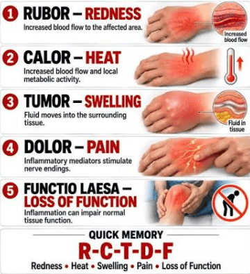
The Cardinal Signs of Inflammation
Clinical Vignette: Odynophagia in Acute Tonsillitis
Clinical Scenario: An adolescent presents with sudden-onset throat pain, marked dysphagia, high fever, and tonsillar swelling covered by exudate.
Pathophysiological Correlation:
- Nociceptive Sensitization: Locally released inflammatory mediators (particularly prostaglandins and bradykinin) directly sensitize unmyelinated C-fibers and lightly myelinated A-delta fibers supplying the pharyngeal mucosa.
- Mechanical Compression: During swallowing, the bolus exerts physical pressure across inflamed, edematous pharyngeal lymphoid tissue, causing pain and functional limitation (dysphagia/functio laesa).
7. Distinction: Acute versus Chronic Inflammation
| Pathological Feature | Acute Inflammation | Chronic Inflammation |
|---|---|---|
| Temporal Onset | Rapid (within seconds, minutes, or a few hours). | Insidious and slow (days to weeks). |
| Duration of Response | Transient (hours to several days). | Protracted (weeks, months, years, or lifelong). |
| Predominant Infiltrate | Polymorphonuclear neutrophils (PMNs). | Mononuclear cells: Macrophages, lymphocytes, and plasma cells. |
| Vascular Dynamics | Prominent: Marked arteriolar vasodilation, vascular stasis, and prominent venular leakage. | Less prominent; characterized instead by ongoing neovascularization (angiogenesis). |
| Interstitial Edema | Extensive, protein-rich exudative edema. | Minimal to absent. |
| Tissue Destruction | Generally mild, localized, and self-limited. | Severe, progressive, and sustained. |
| Fibrotic Remodeling | Absent initially (unless progressing to abscess or organization). | Extensive (fibroblastic activation and scar deposition). |
Case Analysis: Post-Injection Thigh Abscess
Clinical Scenario: A 19-year-old male presents with severe pain, swelling, and localized fluctuance on the anterior thigh four days after receiving an intramuscular injection from an unlicensed provider. On physical exam: erythema, warmth, severe tenderness, fluctuance, and an oral temperature of 38.7°C.
Pathological Explanations:
- Cardinal Signs Demonstrated: Rubor (erythema), Calor (warmth), Tumor (swelling), Dolor (tenderness), and Functio Laesa (impaired lower-limb ambulation).
- Significance of Fluctuance: Confirms a localized liquid center within a walled-off cavity, indicating liquefactive tissue necrosis and pus collection (abscess).
- Predominant Leukocyte: Neutrophils, which release hydrolytic granule enzymes that liquefy host tissue and bacteria.
- Nature of Fever (38.7°C): A systemic manifestation mediated by circulating cytokines (IL-1, TNF-α) acting on the anterior hypothalamic preoptic area.
- Necessity of Surgical Drainage: The core of an established abscess is avascular and packed with necrotic debris, low pH, and high tissue pressure. Systemic antibiotics cannot effectively penetrate this dead space; physical incision and drainage are required to decompress the cavity and remove the bacterial load.
8. Comprehensive Etiologies of Acute Inflammation
| Etiological Trigger | Cellular & Morphological Pattern | Clinical Manifestations & Examples |
|---|---|---|
| Pyogenic Bacteria | Neutrophil-dense suppurative/purulent exudate; tissue liquefaction. | Lobar pneumonia, acute appendicitis, bacterial meningitis, pyelonephritis, osteomyelitis, cellulitis. |
| Fungal Pathogens | Variable: from neutrophilic suppuration to mixed granulomatous inflammation. | Invasive candidiasis, cryptococcosis, pulmonary histoplasmosis, aspergillosis. (Severely neutropenic patients often exhibit extensive fungal invasion with minimal inflammatory response). |
| Parasitic Helminths | Eosinophil-rich inflammatory infiltrate driven by IgE and IL-5. | Tissue-invasive schistosomiasis, lymphatic filariasis, hydatid cysts (Echinococcus), visceral larva migrans. |
| Microbial Toxins | Endothelial injury, microvascular leakage, toxic shock, and necrosis. | Lipopolysaccharide (LPS endotoxin from Gram-negative bacteria), Staphylococcal toxic shock syndrome toxin-1 (TSST-1), Clostridial enterotoxins. |
| Tissue Necrosis (Sterile) | Sterile acute inflammation; neutrophilic influx followed by macrophage clearance. | Myocardial infarction, ischemic renal cortical necrosis, severe frostbite, deep thermal burns, ionizing radiation. |
| Foreign Bodies | Foreign-body giant cell reaction, persistent macrophage activation, fibrosis. | Retained surgical sutures, talc granulomas, silicosis, glass fragments, wood splinters. |
| Endogenous Crystals | Acute neutrophilic synovitis driven by inflammasome activation. | Monosodium urate crystals in acute gout, cholesterol crystals in atheromatous plaques, calcium pyrophosphate in pseudogout. |
| Immune / Allergic | Mast cell degranulation, eosinophilia, or complement/immune-complex vasculitis. | Bronchial asthma, allergic rhinitis, acute urticaria, autoimmune hemolytic anemia, systemic lupus erythematosus. |
Case Study: Podagra in Acute Gouty Arthritis
Clinical Scenario: A 45-year-old male awakes with excruciating pain, erythema, warmth, and marked swelling over his right first metatarsophalangeal (MTP) joint. He has no puncture wound, no fever, and no history of mechanical injury. Synovial fluid aspiration shows needle-shaped, negatively birefringent crystals, but Gram stain and cultures show no organisms.
Diagnostic Insights:
- Etiology: This is a sterile inflammatory arthropathy driven by monosodium urate (MSU) crystals. Infection is not required to provoke acute inflammation.
- Mechanism of Crystal-Mediated Inflammation: Monosodium urate crystals are recognized by synovial macrophages as DAMPs, activating the intracellular NLRP3 inflammasome, causing active cleavage and release of mature IL-1β, which drives rapid neutrophilic recruitment.
9. Cellular Sentinel Networks and Recognition Receptors
The host inflammatory cascade begins only after noxious agents are detected. Sentinel cells (macrophages, dendritic cells, and mast cells) are positioned at environmental interfaces (skin, respiratory tract, gastrointestinal mucosa) to monitor tissues using specialized Pattern Recognition Receptors (PRRs).
PAMPs versus DAMPs: Structural Comparison
- Pathogen-Associated Molecular Patterns (PAMPs): Conserved microbial molecular structures essential for pathogen survival that are absent in host tissues (e.g., LPS, flagellin, viral dsRNA, unmethylated CpG motifs).
- Damage-Associated Molecular Patterns (DAMPs): Endogenous intracellular molecules released into the extracellular space following cell rupture, necrosis, or cellular stress (e.g., extracellular ATP, uric acid, high-mobility group box 1 [HMGB1], heat-shock proteins, genomic DNA, and loss of intracellular potassium).
| Feature | PAMPs | DAMPs |
|---|---|---|
| Full Name | Pathogen-Associated Molecular Patterns | Damage-Associated Molecular Patterns |
| Primary Source | Exogenous microbes (bacteria, viruses, fungi, parasites). | Injured, ischemic, or necrotic host cells. |
| Examples | LPS, peptidoglycan, lipoteichoic acid, flagellin, ssRNA/dsRNA. | Extracellular ATP, uric acid crystals, HMGB1, mitochondrial DNA, reduced K+. |
| Clinical Implication | Indicates potential or active microbial infection. | Indicates sterile trauma, ischemic necrosis, or chemical toxicity. |
| Receptors | Cell-surface and endosomal PRRs (TLRs, C-type lectins). | Cytosolic PRRs (NLRs) and cell-surface purinergic receptors. |
| Inflammasome Activation | Yes (provides priming signals via NF-κB). | Yes (triggers assembly of the NLRP3 complex). |
10. Toll-Like Receptors (TLRs) and Target Specificity
Named after the Drosophila Toll gene, TLRs are membrane-spanning signaling receptors found on the cell surface (for extracellular pathogens) or inside endosomal membranes (for ingested viral and bacterial nucleic acids):
| Toll-Like Receptor | Subcellular Location | Target Microbial Ligand |
|---|---|---|
| TLR2 | Plasma Membrane | Bacterial lipoproteins, peptidoglycans, lipoteichoic acid (Gram-positive bacteria). |
| TLR3 | Endosomal Membrane | Double-stranded viral RNA (dsRNA). |
| TLR4 | Plasma Membrane (with CD14 & MD-2) | Lipopolysaccharide (LPS endotoxin) of Gram-negative bacteria. |
| TLR5 | Plasma Membrane | Bacterial flagellin. |
| TLR7 & TLR8 | Endosomal Membrane | Single-stranded viral RNA (ssRNA). |
| TLR9 | Endosomal Membrane | Unmethylated cytidine-phosphate-guanosine (CpG) bacterial and viral DNA motifs. |
Clinical Reasoning: Endosomal Localization of Nucleic Acid Receptors
Receptors that detect viral and bacterial nucleic acids (TLR3, TLR7, TLR8, TLR9) are located within endosomes rather than on the cell surface. This spatial segregation prevents accidental recognition of normal extracellular host DNA and RNA, helping to prevent autoimmune activation.
11. The Inflammasome Pathway
The inflammasome is a cytosolic multiprotein oligomer responsible for generating active interleukin-1β (IL-1β). The standard activation cascade involves:
- Priming: Microbial ligands bind cell-surface PRRs (like TLR4), activating NF-κB, which upregulates transcription of pro-IL-1β and NLRP3 sensor proteins.
- Sensing: Intracellular danger signals (e.g., phagocytosed urate crystals, silica, cholesterol crystals, extracellular ATP, or potassium efflux) activate cytoplasmic sensor proteins (such as NLRP3).
- Assembly: The sensor protein aggregates with apoptosis-associated speck-like protein containing a CARD (ASC) and pro-caspase-1 to form the mature inflammasome complex.
- Enzymatic Cleavage: Active caspase-1 cleaves inactive pro-IL-1β into mature, bioactive IL-1β.
- Secretion & Amplification: Mature IL-1β is released extracellularly, where it acts on endothelial cells to promote leukocyte adhesion, recruitment, and fever induction.
12. Hemodynamic Alterations, Exudates, and Transudates
Microvascular reactions in acute inflammation involve transient arteriolar vasoconstriction (seconds), followed by prolonged arteriolar vasodilation, elevated hydrostatic pressure, and endothelial cell contraction in post-capillary venules. The resulting separation of endothelial cells allows fluid and high-molecular-weight plasma proteins to escape into the extravascular space.
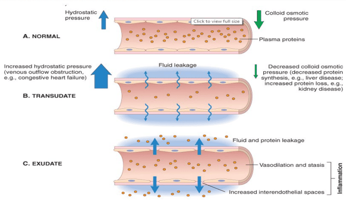
Pathophysiological Mechanics of Fluid Leakage Across Capillaries
| Diagnostic Feature | Exudate (Inflammatory) | Transudate (Non-Inflammatory) |
|---|---|---|
| Pathogenetic Basis | Increased microvascular permeability due to interendothelial gap formation. | Hemodynamic imbalance: elevated hydrostatic pressure or reduced plasma oncotic pressure. |
| Endothelial Integrity | Disrupted, retracted, or injured. | Intact and anatomically normal. |
| Protein Content | High (> 3.0 g/dL; pleural fluid/serum protein ratio > 0.5). | Low (< 3.0 g/dL; pleural fluid/serum protein ratio < 0.5). |
| Specific Gravity | Elevated (> 1.020). | Low (< 1.012). |
| Cellular Content | Abundant leukocytes (neutrophils, macrophages) and necrotic cellular debris. | Acellular or minimal shed mesothelial cells; no neutrophils. |
| Gross Appearance | Turbid, cloudy, purulent, or fibrinous. | Clear, straw-colored, or serous. |
| Clinical Etiologies | Bacterial pneumonia (parapneumonic effusion), appendicitis, abscesses, burns, pleuritis. | Congestive heart failure, hepatic cirrhosis (ascites), nephrotic syndrome, severe malnutrition. |
Terminology Distinctions in Vascular Pathology
- Edema: Excessive collection of interstitial or serous cavity fluid. Can be either an inflammatory exudate or an acellular hemodynamic transudate.
- Pus (Purulent Exudate): A thick inflammatory exudate composed of dead and dying neutrophils, liquefied parenchymal cells, protein-rich edema, and microorganisms.
- Hyperemia versus Congestion: Hyperemia is an active, regulated process mediated by arteriolar dilation that increases forward blood flow. Congestion is a passive, pathological state caused by obstructed venous outflow, leading to blue-red deoxygenated stasis.
13. The Leukocyte Extravasation Cascade
To eliminate offending agents, circulating leukocytes must leave the central axial blood flow and cross the vessel wall in post-capillary venules. This occurs through an orderly sequence of events:
- Margination & Rolling: As post-capillary venules dilate, blood flow slows (vascular stasis). Heavier white blood cells are pushed out of the central stream toward the endothelial surface. Leukocytes roll along the endothelium via transient low-affinity interactions between Selectins (P-selectin, E-selectin on activated endothelium; L-selectin on leukocytes) and their carbohydrate sialyl-Lewis X ligands.
- Firm Adhesion: Chemokines presented on the luminal endothelial surface bind leukocyte receptors, triggering a conformational shift in leukocyte Integrins (LFA-1, Mac-1, VLA-4) from a low- to a high-affinity state. These bind firmly to endothelial immunoglobulin superfamily receptors (ICAM-1, VCAM-1).
- Transmigration (Diapedesis): Leukocytes crawl between endothelial junctions, primarily using homophilic interactions with PECAM-1 (CD31), and secrete matrix metalloproteinases to breach the vascular basement membrane.
- Chemotaxis: Once in the extravascular space, leukocytes migrate along an increasing chemical concentration gradient toward the site of injury. Chemoattractants include:
- Exogenous bacterial products (N-formylmethionine peptides).
- Complement cleavage products (C5a, C3a).
- Eicosanoids (Leukotriene B4 [LTB4]).
- Chemokines (e.g., Interleukin-8 [IL-8] targeting neutrophils).
- Leukocyte Activation: Upon arrival, binding of chemotactic and microbial ligands to leukocyte cell-surface receptors triggers an intracellular calcium influx, activating the cell for phagocytosis, degranulation, and oxidative burst.
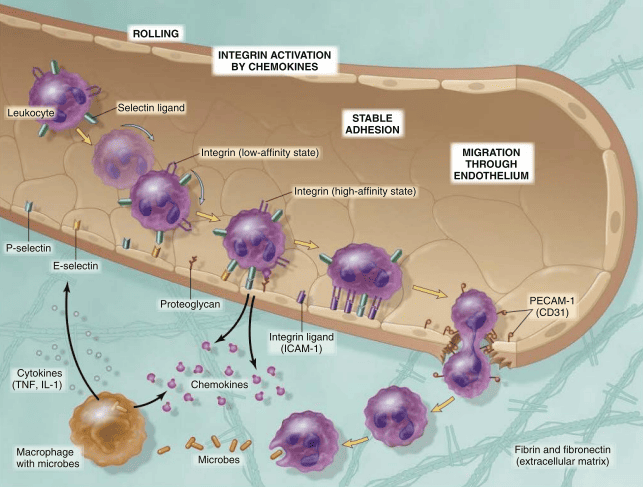
Leukocyte migration from blood to tissues
14. Phagocytosis and Opsonization
Phagocytosis ("cell eating") is the process by which specialized professional phagocytes (neutrophils and macrophages) recognize, bind, engulf, and degrade particulate matter, microbial pathogens, and necrotic debris.
The Biological Role of Opsonins
While un-opsonized particles can bind directly to macrophage scavenger or mannose receptors with low affinity, coating pathogens in host serum proteins (opsonization) markedly enhances recognition, binding speed, and phagocytic efficiency.
| Opsonin | Physiological Origin | Phagocytic Receptor | Functional Impact |
|---|---|---|---|
| Immunoglobulin G (IgG) | B-lymphocyte derived plasma cells. | FcγRI (CD64) on neutrophils and macrophages. | High-affinity binding of the Fc portion triggers cross-linking, membrane evagination, and engulfment. |
| Complement Fragment C3b | Proteolytic cleavage of plasma complement C3. | Complement Receptors CR1, CR3, CR4. | Covalent fixation to bacterial surfaces promotes rapid attachment and ingestion. |
| Mannose-Binding Lectin (MBL) | Hepatic-derived circulating plasma collectin. | C1q receptor / collectin receptors. | Recognizes terminal microbial mannose residues, coats the target, and activates the lectin complement pathway. |
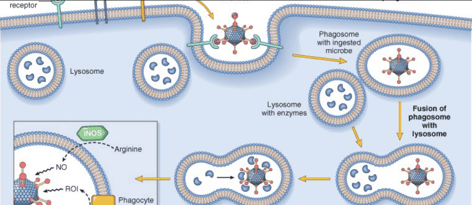
Receptor Recognition, Engulfment, and Phagolysosomal Fusion
15. Step-by-Step Mechanisms of Phagocytic Killing
- Receptor Binding: Opsonized pathogen binds to FcγR, CR1, or scavenger receptors on the phagocyte.
- Cytoskeletal Rearrangement: Receptor clustering triggers focal actin polymerization beneath the plasma membrane.
- Pseudopod Extension: Pseudopodial projections flow around the attached target.
- Phagosome Internalization: The pseudopod arms meet and fuse, pinching off a vesicle called the phagosome.
- Phagolysosomal Fusion: The phagosome travels along microtubules toward the cell interior, where it fuses with azurophilic and specific lysosomes, forming a phagolysosome.
- Intraluminal Digestion: Pathogens are killed within this sealed compartment, which concentrates hydrolytic enzymes and toxic oxidants while protecting the host cell's own cytoplasm.
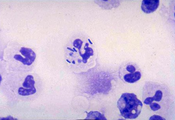
Histological Confirmation of Bacterial Engulfment by Neutrophils
16. Oxygen-Dependent versus Oxygen-Independent Killing
| Enzymatic / Chemical System | Biochemical Reaction Pathway | Primary Microbicidal Product | Mechanism of Lethality |
|---|---|---|---|
| NADPH Oxidase (Phagocyte Oxidase / NOX2) | 2O₂ + NADPH → 2O₂•⁻ + NADP⁺ + H⁺ | Superoxide Anion (O₂•⁻) | Initiates the oxidative respiratory burst; damages microbial lipids, proteins, and DNA. |
| Superoxide Dismutase (SOD) | 2O₂•⁻ + 2H⁺ → H₂O₂ + O₂ | Hydrogen Peroxide (H₂O₂) | Moderately microbicidal; serves as the critical substrate for downstream conversion into stronger oxidants. |
| Myeloperoxidase (MPO) System | H₂O₂ + Cl⁻ → HOCl + OH⁻ | Hypochlorous Acid (HOCl) | The most potent microbicidal oxidant produced by human neutrophils; acts like hypochlorite bleach via protein oxidation and halogenation. |
| Fenton Reaction | H₂O₂ + Fe²⁺ → Fe³⁺ + •OH + OH⁻ | Hydroxyl Radical (•OH) | Extremely reactive free radical; causes immediate lipid peroxidation and membrane rupture. |
| Inducible Nitric Oxide Synthase (iNOS) | L-Arginine + O₂ → NO + Citrulline; NO + O₂•⁻ → ONOO⁻ |
Peroxynitrite (ONOO⁻) | Reactive nitrogen species; nitration of protein tyrosines, iron-sulfur cluster disruption, and microbial metabolic arrest. |
| Oxygen-Independent Granule Enzymes | Enzymatic and biophysical disruption. | Lysozyme, Defensins, Lactoferrin, BPI, Acid Hydrolases. | Cleave bacterial peptidoglycan walls (lysozyme), form pore channels in membranes (defensins), chelate essential free iron (lactoferrin), and lower intraluminal pH (acid hydrolases). |
Clinical Vignette: Chronic Granulomatous Disease (CGD)
Clinical Scenario: A 4-year-old boy presents with recurrent submandibular lymphadenitis, hepatic abscesses, and osteomyelitis. His leukocyte counts are normal, and neutrophils show appropriate chemotaxis and normal bacterial phagocytosis. However, nitroblue tetrazolium (NBT) or dihydrorhodamine (DHR) flow cytometry shows defective superoxide production.
Pathological Explanations:
- Enzymatic Defect: Congenital deficiency in any of the subunits of NADPH oxidase (phagocyte oxidase), most commonly X-linked gp91phox.
- Mechanism of Vulnerability: Phagocytes cannot generate superoxide (O₂•⁻) or downstream H₂O₂ and HOCl.
- Catalase-Positive Microbial Susceptibility: Catalase-negative bacteria produce metabolic H₂O₂ that the host phagocyte can use via myeloperoxidase to kill the organism. However, catalase-positive pathogens (e.g., Staphylococcus aureus, Aspergillus fumigatus, Nocardia, Serratia marcescens, Burkholderia cepacia) degrade their own H₂O₂, leaving the phagocyte with no substrate for oxidative killing.
Clinical Vignette: Myeloperoxidase (MPO) Deficiency
Pathology: An autosomal recessive deficiency of myeloperoxidase within the azurophilic granules of neutrophils.
Diagnostic Insights: In contrast to CGD, the respiratory burst and NADPH oxidase are normal, and H₂O₂ production is intact. However, conversion of H₂O₂ to HOCl is impaired. Most individuals are clinically asymptomatic due to compensatory alternative oxidative and non-oxidative pathways, though diabetics with MPO deficiency face an increased risk of severe mucosal and systemic Candida albicans infections.
17. Host Antioxidant Defense Systems
Because reactive oxygen species (ROS) can leak into the extracellular environment and injure normal host tissues, human cells maintain enzymatic and non-enzymatic antioxidant defenses:
| Antioxidant Defense | Primary Cellular Location | Biochemical Mechanism of Action |
|---|---|---|
| Superoxide Dismutase (SOD) | Cytoplasm (Cu/Zn-SOD) and Mitochondria (Mn-SOD) | Converts toxic superoxide into less reactive hydrogen peroxide (2O₂•⁻ + 2H⁺ → H₂O₂ + O₂). |
| Catalase | Peroxisomes | Decomposes hydrogen peroxide into water and molecular oxygen (2H₂O₂ → 2H₂O + O₂). |
| Glutathione Peroxidase | Cytosol and Mitochondria | Converts H₂O₂ to water (H₂O₂ + 2GSH → GSSG + 2H₂O) and reduces toxic lipid hydroperoxides. |
| Metal Carrier Proteins | Serum (Transferrin, Ferritin, Ceruloplasmin) | Chelate free iron (Fe²⁺) and copper (Cu⁺), preventing the Fenton reaction that generates toxic hydroxyl radicals. |
18. Principal Chemical Mediators of Acute Inflammation
| Chemical Mediator | Primary Cellular or Plasma Source | Primary Physiological & Pathological Actions |
|---|---|---|
| Histamine | Mast cell granules, basophils, circulating platelets. | Immediate arteriolar vasodilation; transient endothelial cell contraction in post-capillary venules forming interendothelial gaps. |
| Prostaglandins (PGE2, PGD2, PGI2) | Mast cells, endothelial cells, leukocytes (via cyclooxygenase pathway). | Arteriolar vasodilation; augments edema formation; sensitizes nociceptors to pain; induces hypothalamic fever. |
| Leukotrienes (LTC4, LTD4, LTE4) | Leukocytes, mast cells (via 5-lipoxygenase pathway). | Marked venular hyperpermeability; severe bronchospasm; arteriolar vasoconstriction. |
| Leukotriene B4 (LTB4) | Neutrophils and macrophages. | Potent leukocyte chemoattractant; stimulates neutrophil aggregation, adhesion, and generation of ROS. |
| Lipoxins (LXA4, LXB4) | Platelet-leukocyte transcellular biosynthetic interactions. | Anti-inflammatory resolution mediators; inhibit neutrophil chemotaxis and promote macrophage uptake of apoptotic cells. |
| Tumor Necrosis Factor (TNF) & IL-1 | Activated tissue macrophages, dendritic cells, endothelial cells. | Upregulate endothelial adhesion molecules (E-selectin, ICAM-1); induce systemic acute-phase reactions (fever, somnolence, anorexia, hypotension/shock). |
| Chemokines (e.g., IL-8) | Macrophages, endothelial cells, fibroblasts. | Direct directional leukocyte migration (chemotaxis) and activate integrin binding affinity. |
| Complement Anaphylatoxins (C3a, C5a) | Proteolytic cleavage of plasma complement proteins. | Direct mast cell degranulation (histamine release); C5a is a potent neutrophil chemoattractant and activator. |
| Bradykinin | High-molecular-weight kininogen via plasma kallikrein. | Arteriolar vasodilation; increased venular permeability; extravascular smooth muscle contraction; direct activation of nociceptive pain fibers. |
| Platelet-Activating Factor (PAF) | Leukocytes, mast cells, endothelial cells. | Platelet aggregation; potent bronchoconstriction; increases vascular permeability up to 10,000 times more than histamine. |
19. Morphological Variants of Acute Inflammation
While all acute inflammatory responses share core microvascular features (vasodilation, fluid exudation, and neutrophilic infiltration), specific anatomical sites and inciting etiologies produce characteristic morphological patterns:
| Pattern | Exudate Composition | Pathological Mechanism | Typical Clinical Examples |
|---|---|---|---|
| Serous Inflammation | Thin, watery, cell-poor fluid derived from plasma or mesothelial secretions. | Mild vascular permeability without large endothelial gaps; fibrinogen does not cross the barrier. | Cutaneous burn blisters, viral vesicles (Herpes simplex), early serous pleural effusions. |
| Fibrinous Inflammation | Fibrin-rich exudate that polymerizes into a dense, pink meshwork. | Marked vascular leakage allows large molecules (fibrinogen) to escape and convert to fibrin via local procoagulant factors. | Uremic or post-infarct pericarditis ("bread-and-butter"), acute fibrinous pleuritis. |
| Purulent / Suppurative | Pus: necrotic host cells, abundant neutrophils, and liquefied debris. | Pyogenic bacterial infection causes extensive neutrophilic recruitment and liquefactive tissue necrosis. | Acute phlegmonous appendicitis, purulent bacterial meningitis, acute pyelonephritis. |
| Abscess Formation | Localized, walled-off pool of thick purulent exudate. | Deep pyogenic bacterial inoculation causes localized liquefactive tissue loss, encircled by a fibrous and vascular rim. | Staphylococcal furuncles, deep intramuscular abscesses, cerebral abscesses, amebic abscesses. |
| Ulceration | Fibrinopurulent exudate covering an excavated base of granulation tissue. | Sloughing and shedding of inflamed, necrotic epithelial mucosa or cutaneous surface. | Peptic ulcer disease (gastric/duodenal), diabetic foot ulcers, chronic decubitus pressure ulcers. |
20. Visual and Histopathological Correlates
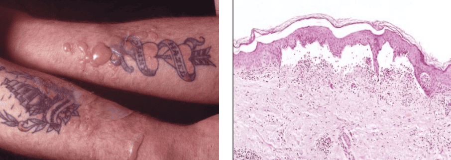
Morphological Features of Cutaneous Serous Inflammation
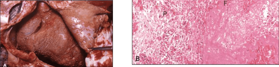
Gross and Histopathological Features of Fibrinous Pericarditis
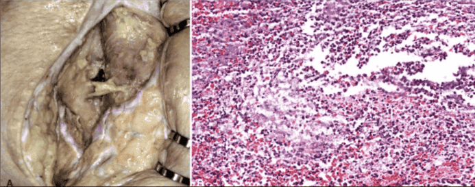
Gross and Microscopic Suppurative (Purulent) Inflammation
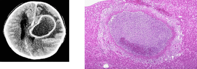
Contrast CT and Histopathology of a Localized Abscess
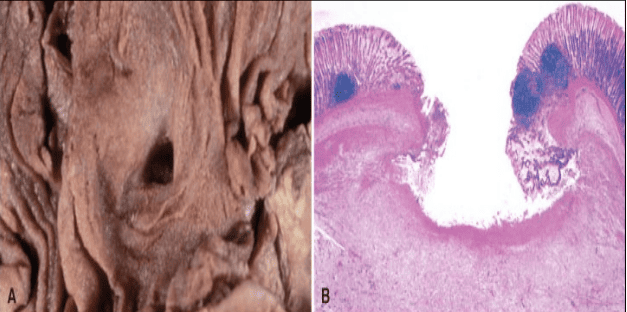
Gross Anatomy and Histopathological Layers of a Peptic Ulcer
21. Natural Outcomes of the Acute Inflammatory Cascade
The clinical course of acute inflammation depends on the nature of the inciting stimulus, the extent of tissue destruction, the organ involved, and host immune competence. There are four primary outcomes:
- Complete Resolution: The ideal outcome, seen when the injury is limited or short-lived, with minimal parenchymal loss in tissues capable of regeneration. Involves neutralization and decay of chemical mediators, vascular normalization, neutrophil apoptosis, and clearance of dead cells by macrophages.
- Healing by Connective Tissue Replacement (Fibrosis / Organization): Occurs after substantial tissue destruction, in organs composed of non-dividing permanent cells (e.g., myocardium), or when extensive fibrinous exudate cannot be cleared by fibrinolysis. Granulation tissue grows into the exudate, depositing collagen and creating an organized fibrous scar.
- Abscess Formation: Develops predominantly with pyogenic bacterial infections (e.g., Staphylococcus aureus). Marked localized liquefactive necrosis forms a pus cavity that typically requires surgical drainage.
- Progression to Chronic Inflammation: Occurs when the offending stimulus cannot be cleared (e.g., persistent mycobacterial infection, retained foreign body, or ongoing autoimmune attack), leading to progressive parenchymal destruction, mononuclear infiltration, and simultaneous fibrosis.
Clinical Case Analysis: Fibrinous Pericarditis
Clinical Scenario: A 58-year-old male with end-stage renal disease presents with positional retrosternal chest pain and a pericardial friction rub. Postmortem examination shows a rough, shaggy pericardial surface covered by pink fibrillary material.
Pathological Explanations:
- Morphological Pattern: Fibrinous pericarditis.
- Composition of Fibrillary Material: Polymeric fibrin meshes formed from plasma-derived fibrinogen that crossed severely damaged endothelial gaps and clotted in the pericardial space.
- Potential Outcomes:
- Resolution: Endogenous tissue plasminogen activator drives fibrinolysis, and macrophages clear the debris, restoring normal serosal anatomy.
- Organization: Ingrowth of capillary sprouts and fibroblasts converts the fibrin mesh into dense, fibrous adhesions, potentially causing constrictive pericarditis that impairs diastolic ventricular filling.
Integrated Case Analysis: Acute Bronchial Asthma
Clinical Scenario: A 16-year-old with atopic asthma develops severe dyspnea and wheezing after inhaling household dust. Sputum analysis reveals eosinophils, mucus plugs, and airway edema.
Pathological Explanations:
- Primary Bronchospastic Mediators: Histamine (from mast cells) and cysteinyl leukotrienes (LTC4, LTD4, LTE4), which cause prolonged airway smooth muscle contraction and hypersecretion of mucus.
- Eosinophil Recruitment: Driven by the Th2 cytokine IL-5 and the specialized CC chemokine eotaxin (CCL11).
- Mechanism of Leukotriene Receptor Antagonists (e.g., Montelukast): Competitively block CysLT1 receptors on bronchial smooth muscle cells, preventing leukotriene-induced bronchoconstriction, vascular leakage, and mucus hypersecretion.
Integrated Case Analysis: Gram-Negative Septic Shock
Clinical Scenario: A 35-year-old female develops high fever, marked tachypnea, warm extremities, confusion, and refractory hypotension (blood pressure 70/40 mmHg) following an untreated ascending pyelonephritis. Blood cultures grow Escherichia coli.
Pathological Explanations:
- Trigger Molecule: Bacterial Lipopolysaccharide (LPS endotoxin) released from the outer membrane of Gram-negative bacilli.
- Receptor System: Binds circulating LPS-binding protein (LBP) and attaches to TLR4 / CD14 / MD-2 complexes on macrophages and endothelial cells.
- Key Cytokines: Activates NF-κB, inducing high-level secretion of TNF-α, IL-1, and IL-6.
- Hypotension Mechanism: High systemic levels of TNF-α and IL-1 trigger widespread expression of inducible nitric oxide synthase (iNOS) across vascular smooth muscle cells. Massive nitric oxide production leads to systemic arteriolar vasodilation, peripheral pooling, increased vascular permeability, and fluid extravasation, culminating in septic distributive shock.