Doctors Revision

Pathology

Chronic Inflammation
Pathology

Chronic Inflammation

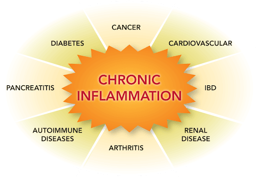
Chronic Inflammation Chronic Inflammation Learning Objectives By the end of this detailed study guide, you should be able to clearly understand and explain: The precise definition of chronic inflammation and identify the key cellular mediators involved. The specific mechanisms and different types of granulomatous inflammation (foreign body vs. immune). The various examples of giant cells, distinguishing between those that occur naturally (physiological) and those that indicate disease (pathological). Introduction to Chronic Inflammation Defining Chronic Inflammation Chronic inflammation is defined as a physiological response of prolonged duration—lasting for weeks or months (and sometimes even years). Unlike acute inflammation, which is a rapid, short-lived response characterized by fluid leakage and neutrophil infiltration, chronic inflammation is entirely different in its nature. The defining characteristic of chronic inflammation is that three distinct processes occur simultaneously at the site of the lesion: Active Inflammation: The immune system continues to fight. Tissue Injury: The ongoing battle causes collateral damage to the host’s own tissues. Attempts at Repair: The body simultaneously tries to heal the damage, usually through scarring (fibrosis) and the creation of new blood vessels. Onset and Progression Chronic inflammation does not always start the same way. It can initiate in two main ways: Following Acute Inflammation: If an initial acute inflammatory response fails to clear the offending agent (like a persistent bacterial infection), the body transitions the response into a chronic state. Insidious (Sneaky) Onset: It may begin as a low-grade, smoldering response from the very beginning. In these cases, there are absolutely no manifestations of a preceding acute reaction. The patient might not even realize it is happening until significant tissue damage has already occurred (e.g., in rheumatoid arthritis or atherosclerosis). Causes of Chronic Inflammation The immune system generally wants to clear a threat quickly and return to normal. Chronic inflammation only occurs when an offending agent is highly stubborn or when the immune system becomes misdirected. A. Persistent Infections Certain microorganisms are extremely difficult for the immune system to eradicate. They possess unique defense mechanisms that allow them to survive inside the body for long periods. Examples include: Mycobacteria: The bacteria responsible for Tuberculosis and Leprosy. They have waxy cell walls that resist being digested by immune cells. Treponema pallidum: The spiral-shaped bacterium that causes Syphilis. Certain Viruses, Fungi, and Parasites: These pathogens can hide within host cells or evade immune detection, leading to a constant, unresolved immune battle. B. Immune-Mediated Inflammatory Diseases (Hypersensitivity) Sometimes, the immune system inappropriately attacks the body’s own tissues or harmless environmental substances. Because the eliciting antigens (the targets of the attack) cannot be eliminated—since they are part of the body itself or constantly present in the environment—these disorders tend to be chronic and highly intractable (hard to treat). Autoimmune diseases: Conditions like Rheumatoid Arthritis or Systemic Lupus Erythematosus. Allergic diseases: Chronic asthma is a prime example of continuous inflammation driven by environmental antigens. C. Prolonged Exposure to Potentially Toxic Agents Continuous exposure to toxic substances over a long period forces the body into a state of chronic inflammation. These agents can be from outside the body (exogenous) or inside the body (endogenous). Exogenous Materials (Outside the body): An example is inhaled particulate silica. When inhaled, silica dust cannot be degraded by the lungs’ immune cells. The constant presence of these sharp, indestructible particles induces a chronic inflammatory response known as silicosis. Endogenous Agents (Inside the body): An example is chronically elevated plasma lipid components (high blood cholesterol). The persistent presence of toxic oxidized lipids in the blood vessel walls triggers a relentless inflammatory response that contributes heavily to the development of atherosclerosis (hardening of the arteries). Morphologic Features (What it looks like under a microscope) If a pathologist examines a tissue sample experiencing chronic inflammation under a microscope, they will observe three hallmark features that clearly distinguish it from acute inflammation. Infiltration with Mononuclear Cells: Acute inflammation is dominated by polymorphonuclear cells (neutrophils). In contrast, chronic inflammation is dominated by mononuclear cells—cells with a single, round, or un-lobed nucleus. These specifically include macrophages, lymphocytes, and plasma cells. Tissue Destruction: There is visible damage and destruction of the normal tissue architecture. This destruction is induced directly by the persistent offending agent (like a toxin or virus) or indirectly as collateral damage caused by the highly potent chemicals released by the accumulating inflammatory cells. Attempts at Healing: Because the tissue is being destroyed, the body desperately tries to patch the holes. This connective tissue replacement of damaged tissue is accomplished by two simultaneous processes: Angiogenesis: The sprouting and creation of new, fragile blood vessels to bring nutrients to the healing area. Fibrosis: The massive deposition of fibrous connective tissue (collagen) by fibroblasts, resulting in heavy scarring. Cells and Mediators of Chronic Inflammation The chronic inflammatory response is orchestrated by a specific cast of cellular characters. The major players include Macrophages, Lymphocytes, Plasma cells, Eosinophils, Neutrophils (in specific ongoing scenarios), and Mast cells. A. Macrophages: The Dominant Cells Macrophages are the undisputed heavyweights and dominant cells of chronic inflammation. They are large, highly capable tissue cells derived from circulating blood white blood cells called monocytes. After monocytes emigrate from the bloodstream into the tissues, they transform into macrophages. The Mononuclear Phagocyte System Macrophages are normally diffusely scattered in most connective tissues throughout the body to act as local guards. Together, these cells comprise the mononuclear phagocyte system (older name: reticuloendothelial system). Depending on the organ they reside in, they are given special names: Kupffer cells: Found in the Liver. Sinus histiocytes: Found in the Spleen and Lymph nodes. Microglial cells: Found in the Central Nervous System (Brain and Spinal Cord). Alveolar macrophages: Found in the Lungs. In all tissues, these cells act as essential filters (eating particulate matter, microbes, and dead/senescent cells) and as sentinels to alert the specific components of the adaptive immune system (T and B lymphocytes) to injurious stimuli. Lifecycle and Activation of Macrophages Migration: The half-life of circulating blood monocytes is very short, about 1 day. Under the influence of chemical signals (adhesion molecules
cell injury death
Pathology

Cell Injury & Death

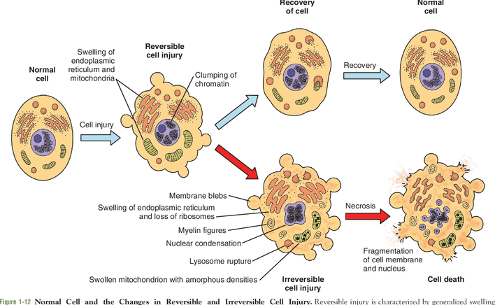
Cell Injury: Adaptation & Death Cell Injury, Death, and Adaptation Learning Objectives By the end of this comprehensive guide, you will be deeply conversant with: The different cellular responses to injury and the specific agents that cause them. The various types and stages of cell injury (Reversible vs. Irreversible). The underlying biochemical mechanisms and distinct morphological changes observed in cell injury. The defining characteristics of Cell Death (Necrosis vs. Apoptosis) and other emerging pathways. The different forms of cellular adaptations (Hypertrophy, Hyperplasia, Atrophy, Metaplasia) and cellular depositions, along with their clinicopathological relevance. Cellular Responses to Stress Cells are active, dynamic participants in their environment. They do not merely exist; they constantly adjust their internal structure and function to accommodate changing physiological demands and extracellular stresses. Under normal conditions, cells maintain a steady, balanced state called homeostasis, where the intracellular environment is kept within a highly regulated, narrow range of physiologic parameters. However, when a cell encounters stress or a pathologic stimulus, it follows a specific progression of responses: Adaptation: The cell’s first response is to adapt. This allows the cell to achieve a “new steady state,” preserving its viability and function in a hostile environment (e.g., a muscle cell growing larger to handle heavy lifting). Reversible Cell Injury: If the adaptive capability is exceeded, or if the external stress is inherently harmful but mild/transient, the cell sustains injury. However, up to a certain point, this injury is reversible, and the cell can return to a stable baseline if the stress is removed. Irreversible Cell Injury and Cell Death: If the injurious stimulus is severe, persistent, or rapid in onset, the cell passes a “point of no return.” It suffers irreversible injury and ultimately dies via Necrosis or Apoptosis. Clinical Example The Myocardium (Heart Muscle) Adaptation: In a patient with chronic high blood pressure, the heart must work harder to pump blood. In response to this increased hemodynamic load, the heart muscle cells enlarge (Hypertrophy). This adaptation helps short-term but increases the oxygen/metabolic demand of the heart. Reversible Injury: If a coronary artery narrows and the blood supply becomes inadequate (ischemia), the muscle first suffers reversible injury. The cells may swell and stop contracting efficiently, but they are not dead yet. Irreversible Injury: Unless the blood supply is rapidly restored (e.g., via a stent or clot-busting drug), the cells cross the threshold into irreversible injury, leading to cell death (Myocardial Infarction / Heart Attack). Causes of Cell Injury The agents that can injure cells range from the very large (physical trauma) to the submicroscopic (genetic mutations). They are generally grouped into seven categories: 1. Oxygen Deprivation (Hypoxia & Ischemia) Hypoxia is a deficiency of oxygen, which fundamentally injures cells by reducing aerobic oxidative respiration (halting ATP production). It is an extremely important and common cause of cell injury and death. Ischemia (Arterial obstruction): The most common cause of hypoxia. Ischemia is a loss of blood supply. Elaboration: Ischemia is actually worse than pure hypoxia because it not only deprives the tissue of oxygen but also deprives it of metabolic substrates (glucose) and fails to remove toxic metabolic waste products (like lactic acid). Inadequate oxygenation of the blood: Due to cardiorespiratory failure (e.g., severe pneumonia or heart failure). Decreased oxygen-carrying capacity: Seen in severe anemia, severe blood loss, or Carbon Monoxide (CO) poisoning (where CO irreversibly binds hemoglobin, blocking oxygen). 2. Physical Agents Physical forces can physically tear, burn, or irradiate cells. Examples include: Mechanical trauma (crush injuries, lacerations). Extremes of temperature (burns causing protein coagulation, deep cold causing ice crystal formation). Sudden changes in atmospheric pressure (decompression sickness). Radiation (ionizing radiation directly breaks DNA and creates free radicals). Electric shock (causes cardiac arrhythmias and severe tissue burning). 3. Chemical Agents and Drugs The list of chemicals that may produce cell injury defies compilation; almost any substance can be toxic in the wrong amount. Harmless substances in excess: Simple chemicals like glucose or salt in hypertonic concentrations can cause cell injury directly by deranging electrolyte and fluid balance, drawing water out of cells and killing them. Even pure oxygen at high concentrations is toxic (causing oxidative stress). Poisons: Strong acids and alkalis directly destroy cell membranes. Toxins and Drugs: Trace amounts of poisons (cyanide, arsenic), environmental pollutants (asbestos, carbon monoxide), social drugs (ethanol/alcohol, cigarette smoke), and even therapeutic drugs (chemotherapy) can injure cells. 4. Infectious Agents Biological pathogens attack cells either by directly invading them or by releasing toxins. These range from submicroscopic viruses (which hijack the cell’s DNA/RNA machinery) to bacteria, fungi, protozoa, and metazoa (tapeworms several feet in length). 5. Immunologic Reactions While the immune system defends against pathogens, friendly fire can cause massive tissue damage. Autoimmune diseases: Injurious reactions to endogenous self-antigens (the body attacks its own cells). Allergic reactions: Exaggerated immune responses to external agents (viruses, environmental substances, allergens) are important causes of tissue injury. 6. Genetic Abnormalities Errors in the DNA code can lead to cell death by preventing the creation of essential proteins. Deficient protein function: Such as enzyme defects leading to inborn errors of metabolism. Accumulation of damage: Misfolded proteins or damaged DNA trigger cell death (apoptosis) if they are beyond repair. Chromosomal abnormalities: Examples include congenital malformations associated with Down Syndrome (Trisomy 21). 7. Nutritional Imbalances A major global cause of cell injury. Deficiencies: Protein-calorie deficiencies cause an appalling number of deaths, chiefly among low-income populations. Specific vitamin deficiencies (e.g., Scurvy from Vitamin C deficiency) are not uncommon even in developed countries. Excesses: Nutritional excesses, such as obesity and high-fat diets, are significant factors in cellular injury leading to atherosclerosis, diabetes, and cancer. Factors Affecting Cell Response to Injury Why does one cell survive a stress while another dies? The outcome depends on two sets of variables: Variables of the Injurious Agent Nature of the injury: (Is it a mild toxin or a severe physical crush?) Duration of injury: (Did the ischemia last for 5 minutes or 50 minutes?) Severity: (A low dose vs. a massive overdose of a drug). Variables of the

Routes of Administration
Anatomy, Pathology

Routes of Administration

Routes of Drug Administration Routes of Administration Fundamental Definitions and Concepts What is a Drug? In the strictest scientific sense, a drug is defined as any chemical agent or substance which affects, alters, or modifies any biological process within a living organism. It is important to realize that the body does not distinguish between a “therapeutic medication,” an “environmental toxin,” or a “recreational substance”—to the body’s cells, they are all simply foreign chemicals (xenobiotics) that bind to biological targets and induce a change. What is Pharmacology? Pharmacology is the comprehensive scientific study of exactly how these drugs affect biological systems. It investigates the entire lifecycle of a drug interaction: from how the drug is manufactured and sourced, to how it travels through the bloodstream, how it binds to microscopic cellular receptors, and ultimately, how the body destroys and removes it. The Five Major Branches of Pharmacology To fully understand drug action, pharmacology is systematically divided into distinct domains: 1 Pharmacokinetics “What the body does to the drug.” This encompasses the four pillars of drug biodisposition: Absorption (getting in), Distribution (moving around), Metabolism/Biotransformation (being broken down by enzymes), and Excretion (leaving the body). 2 Pharmacodynamics “What the drug does to the body.” This looks at the microscopic level: drug receptors (the protein locks that drugs fit into), the physiological effects of the drug, cellular responses, and potential toxicity or adverse effects. 3 Pharmacotherapeutics The clinical study of the strictly medical use of drugs to prevent, diagnose, or treat diseases. 4 Pharmacognosy The highly specialized study of identifying, extracting, and isolating crude materials from natural sources to be used as drugs. 5 Toxicology The study of the poisonous, adverse, and toxic effects of chemicals on living systems. Sources of Drugs and Forms of Medication Sources of Drugs (Pharmacognosy) Historically, all drugs came from nature. Today, we source drugs from five primary categories: Plants: The oldest source of medicine. Examples include Morphine (from the opium poppy), Digoxin (from the foxglove plant for heart failure), and Quinine (from the cinchona tree bark for malaria). Animals: Historically, many hormones were extracted from slaughtered livestock. Examples include Insulin (previously extracted from pig and cow pancreases) and Heparin (a blood thinner extracted from pig intestines). Minerals: Earthly elements used directly for health. Examples include Iron (for anemia), Lithium (for bipolar disorder), and Magnesium (as an antacid or laxative). Synthetic: Today, the vast majority of drugs are entirely synthesized in chemistry laboratories. This allows for massive scaling, precise purity, and the structural modification of natural drugs to reduce side effects. Microbes: Many life-saving drugs are produced by harnessing bacteria and fungi. The most famous example is Penicillin (produced by the Penicillium fungus). Forms of Medication Medications are practically never pure, raw chemicals. They are carefully formulated into specific “preparations” or “dosage forms.” The form of the medication strictly dictates its route of administration. The composition of the medicine is intricately designed by pharmaceutical scientists to enhance its absorption, dictate its metabolism rate, and ensure patient compliance. Common forms include: Tablet: A solid dosage form made by highly compressing powdered drug and inactive binders into a hard pill. Capsule: A drug enclosed within a gelatin shell. They dissolve quickly in the stomach, releasing the powder or liquid inside. Elixir: A clear, sweetened, hydro-alcoholic liquid intended for oral use, perfect for drugs that do not dissolve easily in pure water. Enteric-coated: A specially designed tablet with an acid-resistant shell. It passes through the highly acidic stomach unharmed and only dissolves when it reaches the alkaline environment of the small intestine. This protects sensitive drugs from destruction and protects the stomach from irritating drugs. Suppository: A solid, bullet-shaped mass that is inserted into a body cavity (rectum or vagina) which is explicitly designed to melt at exact human body temperature (37°C) to release the drug. Suspension: A liquid preparation containing undissolved solid drug particles. Because the drug settles at the bottom, these must be shaken vigorously before administration. Transdermal patch: An adhesive patch placed on the skin that delivers a specific, slow, and continuous dose of medication through the skin and directly into the bloodstream. Routes of Drug Administration A route of administration is the specific anatomical path by which a drug, fluid, poison, or other substance is brought into contact with the body. Routes of administration are broadly classified into three main channels based on whether they act locally or systematically, and whether they involve the digestive tract: Enteral: Through the gastrointestinal tract (Oral, Sublingual, Buccal, Rectal). Parenteral: Bypassing the gastrointestinal tract, usually via injection (IV, IM, SC, etc.). Topical: Applied to a specific surface for a localized effect (Skin, eyes, ears, lungs). Enteral Routes of Administration The term Enteral comes from the Greek word enteron, meaning intestine. It refers to anything involving the alimentary tract, from the mouth down to the rectum. A. Oral Route or Per Os (P.O.) The oral route involves swallowing a drug. It is the most common, oldest, and generally most universally accepted route of administration. It utilizes the body’s natural machinery used for digesting food, absorbing nutrients, and eliminating wastes. Advantages of the Oral Route: It is safe: Because absorption is relatively slow, there is a window of opportunity to induce vomiting or pump the stomach in case of an accidental overdose. It is convenient: Patients can take it themselves anywhere. It is cheap: Tablets and capsules do not require sterile manufacturing conditions like injectable fluids do. No skilled personnel required: The patient does not need a nurse or doctor to administer the dose. Disadvantages of the Oral Route: Unpalatable drugs: Bitter or foul-tasting drugs can cause severe irritation to the intestinal tract, resulting in nausea, vomiting, and diarrhea. Destruction by enzymes and acid: Some drugs are completely annihilated by stomach acid (HCl) or digestive enzymes before they can be absorbed. For example, Insulin is a protein; if swallowed, the stomach digests it just like a piece of meat, destroying its therapeutic value. Not suitable for emergencies: It takes time for a pill

Introduction to Basic Pharmacology
Anatomy, Pathology

Introduction to Basic Pharmacology

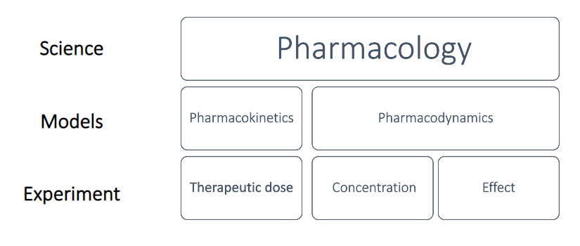
Pharmacology Intro: Basic & Practicals Introduction to Basic Pharmacology and Pharmacology Practicals (Instrumentation) Learning Outcomes of the Lecture By the end of this comprehensive guide, students should be fully equipped to: Define pharmacology and clearly outline its major branches and sub-disciplines. Distinguish definitively between the concepts of pharmacodynamics and pharmacokinetics. Explain the critical importance of pharmacology practicals and laboratory experiments in medical and scientific training. Identify and understand the major instruments and equipment used in modern and historical pharmacology laboratories. Describe the specific functions of organ bath systems, transducers, and recording devices in depth. Recognize and apply ethical considerations (such as the 3Rs) in experimental pharmacology. Introduction to Pharmacology Pharmacology is broadly defined as the rigorous scientific study of drugs and their interactions with living systems. Derived from the Greek words pharmakon (drug or poison) and logos (study), it is a vast field that examines every aspect of how drugs produce their physiological effects, how the human (or animal) body processes these foreign substances, and how these chemicals can be utilized therapeutically to treat disease, or experimentally to understand biological processes. Definition: A Drug A drug, in the context of pharmacology, can be defined as any chemical substance (natural, synthetic, or endogenous) that modifies physiological or biochemical functions when administered to a living organism. This includes everything from life-saving antibiotics to everyday pain relievers, as well as substances of abuse and environmental toxins. The Multidisciplinary Nature of Pharmacology Pharmacology does not exist in isolation. It acts as a bridge between the physical sciences and the biological sciences. It integrates core knowledge from several crucial disciplines, including: Physiology: Understanding normal body functions is essential before one can understand how a drug alters those functions. Biochemistry: Provides the foundation for understanding the chemical basis of drug action at the enzymatic and metabolic levels. Molecular Biology: Helps in understanding how drugs interact with genetic material, intracellular signaling, and protein synthesis. Medicinal Chemistry: Focuses on the structural design, synthesis, and optimization of pharmaceutical drugs. Toxicology: The study of the adverse or poisonous effects of chemicals, closely tied to drug safety. Clinical Medicine: The ultimate application of pharmacological knowledge to diagnose, prevent, and treat illnesses in human patients. Major Branches of Pharmacology To fully grasp pharmacology, the field is traditionally divided into several distinct, yet deeply interconnected, branches. 1. Pharmacodynamics Pharmacodynamics essentially studies what the drug does to the body. It delves into the specific biochemical and physiological effects of drugs and their mechanisms of action. Key aspects of pharmacodynamics include: Mechanism of Drug Action: Exactly how a drug produces its effect at the cellular level. Drug–Receptor Interactions: How drugs bind to specific protein targets (receptors) to initiate or block a biological response. Cellular Targets: Drugs typically exert their effects by interacting with four main regulatory proteins: Ion Channels: Drugs can act as openers (increasing ion influx) or blockers (preventing ion passage). Enzymes: Drugs often act as inhibitors, preventing the enzyme from converting a substrate into a product (e.g., aspirin inhibiting COX enzymes). Transporters: Drugs can act as transport inhibitors, preventing the movement of molecules across cell membranes. Receptors: Drugs can be Agonists (activating the receptor for signal transduction) or Antagonists (blocking the receptor and preventing activation). Dose–Response Relationships: The mathematical and graphical relationship between the amount of drug given (dose) and the magnitude of the effect produced. As the log of the drug concentration increases, the effect typically increases until a maximum plateau is reached. Therapeutic and Toxic Effects: Determining the primary intended effects versus unintended side effects. Example of Pharmacodynamics How β-blockers reduce heart rate: A beta-blocker (like Atenolol) acts as an antagonist. It specifically targets and blocks β1-adrenergic receptors located in the heart muscle. By blocking these receptors, it prevents adrenaline from binding, which structurally and functionally reduces the heart rate and blood pressure (this is what the drug does to the body). 2. Pharmacokinetics Pharmacokinetics studies what the body does to the drug. It traces the journey of a drug molecule from the moment it enters the body until it is completely removed. It involves four major, continuous processes, universally remembered by the acronym ADME: A – Absorption The movement of a drug from its site of administration (e.g., gut, muscle, skin) into the systemic blood circulation. Factors like route of administration, lipid solubility, and pH heavily influence this. D – Distribution The reversible transfer of a drug from one location to another within the body, typically from the bloodstream into tissues, organs, and intracellular spaces. It is affected by blood flow, tissue binding, and membrane permeability (e.g., the blood-brain barrier). M – Metabolism (Biotransformation) The chemical modification or breakdown of drugs, primarily occurring in the liver. The body attempts to make the drug more water-soluble so it can be easily excreted. E – Excretion The irreversible elimination of the drug and its metabolites from the body. The kidneys (via urine) are the primary route, but drugs can also be excreted through bile, feces, sweat, saliva, tears, and lungs (exhaled air). Example of Pharmacokinetics First-pass metabolism of drugs like propranolol: When propranolol is taken orally, it is absorbed by the digestive tract and carried directly to the liver via the hepatic portal vein. The liver highly metabolizes (destroys) a large portion of the drug before it ever reaches the systemic circulation. This “first-pass effect” drastically reduces the bioavailability of the drug, which is an example of what the body does to the drug. 3. Therapeutics (Clinical Pharmacology/Pharmacotherapeutics) This branch focuses strictly on the clinical use of drugs to prevent, diagnose, or treat diseases. It is the practical application of pharmacology in a healthcare setting, emphasizing evidence-based medicine, rational prescribing, and patient care. Antihypertensive therapy: Using drugs to lower high blood pressure and prevent cardiovascular events. Antidiabetic therapy: Managing blood sugar levels using insulin or oral hypoglycemic agents. Antimicrobial therapy: Utilizing antibiotics, antivirals, or antifungals to eradicate infections while minimizing harm to the host. 4. Toxicology Toxicology is the study of the harmful, adverse, or toxic effects of drugs, chemicals, and

intracellular accumulation
Anatomy, Pathology

Intracellular Accumulation & Acute Inflammation

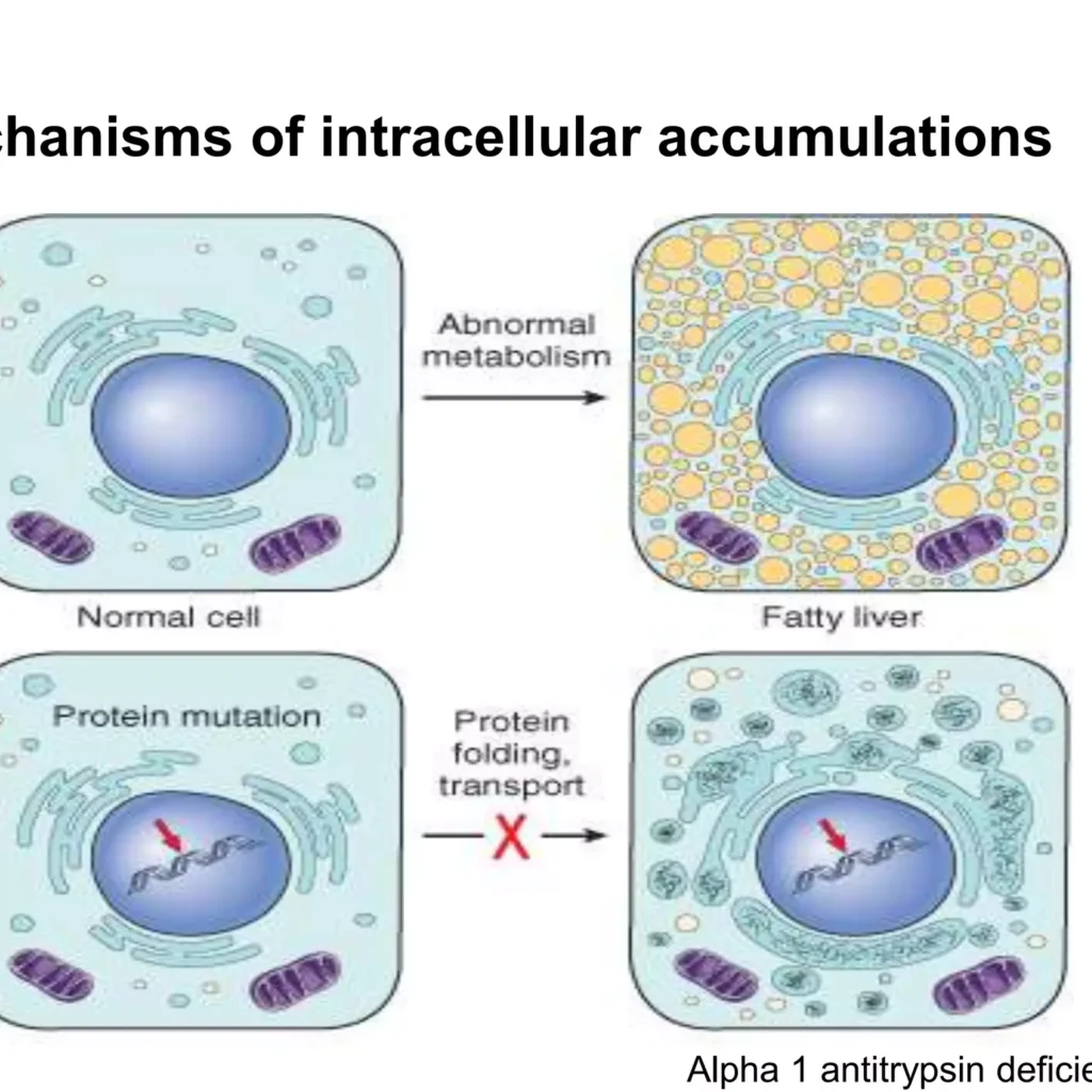
Intracellular Accumulation & Acute Inflammation Intracellular Accumulation & Acute Inflammation Intracellular Accumulations Intracellular accumulations are the buildup of substances—such as lipids, proteins, glycogen, or pigments—within cells due to metabolic derangements, genetic defects, or environmental factors. These accumulations occur in the cytoplasm or nucleus, ranging from harmless to severely toxic, causing reversible or irreversible cell injury. Key mechanisms include increased production, defective metabolism/transport, or lack of enzymes to break down substances. General Principles Cells often act as reservoirs for metabolic products or exogenous substances. These accumulations represent a sign of metabolic derangement. Subcellular Localization Cytoplasm: Most common (e.g., Fatty change, Glycogen). Organelles: Specifically within Lysosomes (e.g., Pompe disease) or the Endoplasmic Reticulum (e.g., Protein folding defects). Nucleus: Rare, but seen in certain viral infections or lead poisoning. The Four Pathological Mechanisms Abnormal Metabolism: A normal endogenous substance (like water, lipids, or proteins) is produced at a normal or increased rate, but the metabolic rate is inadequate for its removal (e.g., Steatosis). Defect in Protein Folding/Transport: Genetic mutations or acquired defects cause proteins to misfold. These “garbage” proteins build up because they cannot be exported or degraded (e.g., α1-antitrypsin deficiency). Enzymatic Deficiency: An inherited lack of a vital enzyme (usually lysosomal) means a specific substrate cannot be broken down, leading to massive buildup—known as Storage Diseases. Inability to Degrade Exogenous Material: The cell encounters a substance (like carbon or silica) for which it has no natural enzymes to digest. Examples of Abnormal Accumulations 1. Fatty Change (Steatosis) The abnormal accumulation of triglycerides within parenchymal (functional) cells. Organ Involvement: Primarily the Liver (yellow, greasy, enlarged). It is also significant in the Heart (where it can cause “Tiger effect” banding) and the Kidneys. Etiology (The “Why”): Toxins: Most notably Alcohol, which is a mitochondrial toxin that impairs fat oxidation. Protein Malnutrition: Lack of “apoproteins” needed to carry fat out of the liver. Anoxia: Lack of oxygen prevents the oxidation (burning) of fatty acids. Diabetes Mellitus & Obesity: Causes an oversupply of free fatty acids to the liver. 2. Cholesterol and Cholesteryl Esters Pathology: Unlike triglycerides, cholesterol is usually stored in macrophages or smooth muscle cells. Atherosclerosis: The most critical clinical result. Phagocytic cells in the large arteries become overloaded with lipid, forming “Foam Cells.” These accumulate in the intimal layer of arteries, leading to yellow fatty streaks and eventually plaques. 3. Proteins Morphology: Appear as rounded, eosinophilic (bright pink) droplets, vacuoles, or aggregates. Clinical Examples: Nephrotic Syndrome: Excess protein leaks into the kidney tubules; the cells reabsorb it, creating pink protein droplets. Russell Bodies: Found in plasma cells (overproduction of immunoglobulins). Misfolded Proteins: Build up in the brain (Amyloid plaques in Alzheimer’s). 4. Glycogen Association: Highly associated with Glucose metabolism disorders. Diabetes Mellitus: Glycogen is found in the epithelial cells of the distal segments of the renal tubules and the liver. Glycogen Storage Diseases (GSD): Genetic defects where glycogen cannot be converted back to glucose, leading to massive cell death and organ failure. 5. Pigments: The “Colored” Pathologies Exogenous (Environmental): Carbon (Anthracosis): The most ubiquitous pigment. Inhaled carbon is phagocytosed by alveolar macrophages. These macrophages travel through the lymphatics to the tracheobronchial lymph nodes. In coal miners, this leads to “Black Lung” disease (Coal Workers’ Pneumoconiosis). Endogenous (Produced by the body): Lipofuscin: A “wear-and-tear” pigment. It is a sign of free radical injury and lipid peroxidation. It does not harm the cell but is a tell-tale marker of aging. Melanin: An insoluble brown-black pigment produced by melanocytes in the epidermis to protect against UV radiation. Hemosiderin: A hemoglobin-derived, golden-yellow to brown, granular pigment. It represents local or systemic Iron excess. Staining Tip: On a standard H&E slide, it looks like brown granules. To prove it is iron, pathologists use the Prussian Blue Histochemical Stain (the iron turns bright blue). Pathologic Calcification Pathologic calcification is the abnormal deposition of calcium salts (phosphates, carbonates) in soft tissues, commonly due to injury or metabolic dysfunction. Calcification is a permanent marker of past or present tissue injury. It occurs in two main forms: dystrophic (normal serum calcium, damaged tissue) and metastatic (high serum calcium, normal tissue). I. Dystrophic Calcification (Local Injury) Occurs in dead or dying tissues (necrosis) despite normal serum calcium levels, often seen in atherosclerosis, damaged heart valves, or tuberculous lymph nodes. Requirement: Occurs in non-viable (dead) or dying tissues. Calcium Levels: Serum calcium levels are Normal; there is no systemic mineral imbalance. Pathogenesis: Necrotic cells have damaged membranes; calcium binds to the phospholipids in these membranes, initiating the “crystallization” of calcium phosphate. Clinical Examples: Atherosclerosis: The core of an old plaque is often “bone-hard” due to calcification. Tuberculosis (TB): Areas of Caseous Necrosis often calcify, making them visible on X-rays (Ghon complex). Aging/Damaged Heart Valves: Leads to stenosis (narrowing of the valve). II. Metastatic Calcification (Systemic Imbalance) Occurs in normal tissues due to hypercalcemia (high calcium levels in the blood), often caused by renal failure, hyperparathyroidism, or Vitamin D intoxication. Requirement: Occurs in normal, healthy tissues. Calcium Levels: Always associated with Hypercalcemia (Elevated blood calcium). The Four Major Causes of Hypercalcemia Hyperparathyroidism: Either a primary tumor of the parathyroid gland or “ectopic” secretion of PTH-related protein by cancers (like lung or breast cancer). Rapid Bone Destruction: Multiple Myeloma: A cancer of plasma cells that “eats” bone. Paget’s Disease: Disorganized bone remodeling. Immobilization: Long-term bedrest leads to bone resorption. Vitamin D Disorders: Intoxication (overdose) or Sarcoidosis (where lung macrophages inappropriately activate Vitamin D). Renal Failure: Leads to phosphate retention, which triggers a secondary rise in PTH, pulling calcium out of the bones and into the tissues. Morphology and Distribution Gross (Macroscopic): Calcium deposits are white, chalky granules. When a pathologist cuts through the tissue, it feels “gritty” (like cutting through sand or eggshells). Microscopic (Histology): Stains Basophilic (deep blue/purple) with H&E. Can be found inside cells (mitochondria) or outside cells in the matrix. Psammoma Bodies: In some tumors (like thyroid cancer), the calcification forms beautiful, laminated, sand-like concentric circles. Preferred “Metastatic” Targets: High-calcium levels prefer tissues that have an alkaline (basic) internal environment, which promotes

pathology pathophysiology intro
Anatomy, Pathology

Pathology Intro: Concepts & Applications

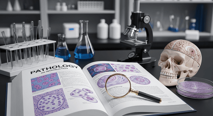
Pathology Intro: Concepts & Applications Pathology Intro: Concepts & Applications What is Pathology? Pathology is the scientific study of disease. It acts as the bridge between basic sciences (like anatomy, physiology, biochemistry, microbiology) and clinical medicine. Etymology: Derived from Greek words: Pathos = Suffering Logos = Study Pathology seeks to understand the causes (etiology), mechanisms (pathogenesis), structural alterations (morphological changes), and functional consequences (clinical manifestations) of disease. Definition Pathology is a branch of natural science that studies the etiology (cause), mechanisms (pathogenesis), and effects (morphological changes and clinical manifestations) produced by diseases in all living organisms, including humans, animals, and plants. Ancient Foundations (The Roots) Imhotep (Egypt, c. 2600 BC): Recognized as the oldest known physician/doctor in history. He transitioned medicine from purely magic to early observation. The Papyrus (Egypt, c. 1600 BC): Specifically the Edwin Smith Papyrus, it is considered the oldest study of anatomy and surgical trauma, detailing clinical observations and treatments. The Evolution of Pathological Thought 1. The Era of Religious & Supernatural Beliefs Before a rational approach was developed, disease was attributed to: Divine Punishment: A “Curse from God” or the result of sin. Magic/Supernatural: Belief in the “evil eye” or malevolent spirits. Scriptural References: Examples found in the Bible (Job 2:7—affliction with boils; Exodus 9:8-12—the plague of boils). Cultural Deities: Different regions had specific gods of disease, such as Walumbe in the Buganda kingdom (associated with death and disease). 2. The Antiquity to AD 1500: The Rational Approach This period saw the shift from mysticism to observation. Hippocrates (Greece, 460–377 BC): Known as the “Father of Medicine.” Dissociation: Permanently dissociated medicine from religious mysticism. Clinical Observation: Established the study of patient symptoms as the primary method for diagnosis. Cornelius Celsus (Rome, 53 BC–7 AD): Described the 4 Cardinal Signs of Inflammation: Rubor (redness), Calor (heat), Tumor (swelling), and Dolor (pain). Claudius Galen (130–200 AD): Postulated the Humoral Theory (Galenic Theory). He argued that illness resulted from an imbalance of four body fluids: Blood, Lymph, Black Bile (associated with the spleen), and Biliary Secretion/Yellow Bile (from the liver). 3. The Era of Gross Pathology (AD 1500 to 1800) During this time, physicians began correlating symptoms with what they saw during autopsies. Giovanni B. Morgagni (Italy, 1682–1771): The “Father of Anatomical Pathology.” Introduced Clinical Pathologic Correlation (CPC)—the practice of linking a patient’s symptoms during life to the organ changes found after death. John Hunter (Scotland, 1728–1793): Introduced the Pathology Museum as a vital tool for medical education and the systematic study of diseased specimens. R.T.H. Laennec (France, 1781–1826): Described lung diseases, including various tuberculous lesions and bronchiectasis. Described cirrhosis of the liver (still frequently called Laennec’s Cirrhosis). Invented the stethoscope, allowing for better clinical-pathological correlation during life. 4. The Era of Technology & Cellular Pathology (AD 1800 to 1950s) The invention of the microscope shifted the focus from organs to cells. Rudolf Virchow (Germany, 1821–1905): Known as the “Father of Cellular Pathology.” Proposed the Cellular Theory: Disease does not arise in organs or tissues generally, but primarily in individual cells (Omnis cellula e cellula). Established Histopathology as a formal diagnostic branch of medicine. George N. Papanicolaou (USA, 1883–1962): Known as the “Father of Exfoliative Cytology.” Developed the Pap Smear in the 1930s for the early detection of cervical cancer, proving that microscopic examination of individual cells could prevent disease. 5. Modern Pathology (1950s to the 21st Century) The focus shifted again—from the cell to the molecule and DNA. Watson and Crick (1953): Described the double-helix structure of DNA, opening the door to molecular pathology. Nowell and Hungerford (1960): Discovered the Philadelphia chromosome in Chronic Myeloid Leukemia (CML), identifying the specific translocation t(9;22). Gall and Pardue (1969): Developed In Situ Hybridization, allowing researchers to locate specific nucleic acid sequences within tissues. Kary Mullis (1983): Introduced the Polymerase Chain Reaction (PCR), a revolutionary technique that allows for the amplification of DNA, now used for diagnosing infections, genetic mutations, and cancers. Modern Diagnostic Modalities: Telepathology Telepathology is the practice of diagnostic pathology by a remote pathologist utilizing images of tissue specimens transmitted over a telecommunication network. This allows for rapid consultation and diagnosis across different geographical locations. 1. Components of Telepathology Conventional Light Microscope: The primary tool used to view the specimen. Image Capture Method: Usually a high-resolution digital camera mounted on the microscope. Telecommunications Link: A secure network (internet or satellite) to transmit data between the sending and receiving sites. Workstation: A computer at the receiving end equipped with a high-quality, medical-grade monitor for accurate interpretation. 2. Types of Telepathology Static (Store-and-Forward): Images are captured and sent as individual files. The remote pathologist views them later (passive telepathology). Dynamic (Robotic/Virtual Microscopy): This involves Virtual Pathology Slides (VPS). The remote pathologist can interact with the microscope in real-time, moving the slide or changing magnification remotely (robotic interactive telepathology). Fields and Branches of Pathology Pathology is not limited to humans; it is a universal study of disease across living systems. 1. Major Study Fields Human Pathology: Study of diseases in humans. Veterinary Pathology: Study of diseases in animals. Plant Pathology: Study of diseases in plants. Teratology: The scientific study of visible conditions/congenital malformations caused by the interruption or alteration of normal development (e.g., birth defects). Nosology: The branch of medicine that deals with the classification and description of known diseases. 2. Functional Branches Etiology: The study of the causes of disease (why it happens). Pathogenesis: The study of the mechanisms and steps of disease development (how it happens). Physiopathology (Pathophysiology): The study of the disordered physiological processes associated with disease or injury. Semiology: The study of the symptoms (subjective, felt by the patient) and signs (objective, observed by the doctor) of disease. Clinic: The practical management and treatment of the disease. Anatomic Pathology The study of morphological and structural changes in cells, tissues, and organs that underlie disease. General Pathology: Studies basic reactions of cells and tissues to abnormal stimuli that occur in all diseases (e.g., inflammation, neoplasia, cell death). Systemic Pathology: Studies diseases as they pertain to specific organs

Scroll to Top