Classification & Taxonomy of Bacteria
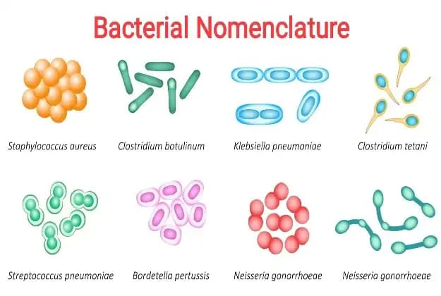
Classification & Taxonomy of Bacteria Cell Biology & Bacterial Taxonomy Why This Matters In clinical medicine, you cannot treat an invisible enemy without knowing exactly what it is. Bacterial taxonomy isn’t just memorizing names; it is the roadmap to prescribing the correct life-saving antibiotics. By understanding a bacterium’s shape, oxygen requirements, and cell wall structure, a doctor can predict exactly how a disease will progress and which drug will destroy the pathogen without harming the patient. 1. Taxonomical Hierarchies and Nomenclature Taxonomy is the science of classifying organisms. In microbiology, we classify bacteria into a strict hierarchy to understand their relationships and pathogenic behaviors. The Hierarchy of Bacteria Kingdom: Prokaryotes (organisms lacking a true nucleus). Order: The name always ends with the suffix ‘-ales’. Example: Enterobacteriales. Family: Many families exist in one order. The name always ends with the suffix ‘-eae’. Examples: Enterobacteriaceae, Pseudomonodaceae. Genus: Each family is divided into genera. Example: Within Enterobacteriaceae, you have Escherichia, Klebsiella, Enterobacter. Species: The most specific group. Example: Escherichia coli. Sub-species: Further divisions based on tiny genetic/antigenic differences (e.g., Salmonella enterica subsp. enterica). Clinical Extension: This level is vital for tracking outbreaks! For example, tracking the deadly food-poisoning strain E. coli O157:H7 distinguishes it from the harmless E. coli living normally in your gut. Rules for Bacterial Nomenclature (Naming) Medical professionals must communicate without ambiguity. Only one correct name exists, determined by the International Journal of Systematic Bacteriology (IJSB). Confusing names are rejected. Every bacterium must have a Genus and a Species name. The Genus name must start with a Capital letter. The species name must start with a small (lowercase) letter. The entire name MUST be italicized (if typing) or underlined (if handwriting). Example: Staphylococcus aureus or Escherichia coli. All names are written in Latin. 2. Classification of Medically Important Bacteria (Based on Cell Wall) This is the most critical flowchart in clinical microbiology. Bacteria are primarily classified by their cell wall structure, which dictates what antibiotics will work against them. A. Lacking Cell Wall Genera: Mycoplasma & Ureaplasma Clinical Pearl & Exam Trap The Mycoplasma Exception Because they have NO cell wall, they are naturally resistant to all beta-lactam antibiotics (like Penicillin, Amoxicillin, Cephalosporins). Why? Because beta-lactams work exclusively by destroying the cell wall (peptidoglycan). You cannot destroy a wall that doesn’t exist! Example: Mycoplasma pneumoniae causes “walking pneumonia.” You must treat it with drugs that target ribosomes (inside the cell), like Macrolides (Azithromycin) or Tetracyclines. B. Rigid Cell Wall (The vast majority of bacteria) Divided based on how they live and their shape/staining: Obligate Intracellular Bacteria: Must live inside a host cell to survive (they cannot make their own ATP—they are “energy parasites”). Genera: Chlamydia, Rickettsia, Coxiella, Ehrlichia. Filamentous Bacteria (Branching, fungus-like): Genera: Actinomyces, Nocardia, Mycobacteria. Clinical Pearl: Mycobacteria (which causes Tuberculosis) has a rigid wall heavily loaded with mycolic acid (waxy lipids), making it “Acid-Fast” instead of truly Gram-positive or negative. Regular Gram stain bounces right off this wax. We must use the Ziehl-Neelsen (Acid-Fast) stain, where TB appears as bright red snappers. Free-Living, Simple Unicellular (Gram Positive vs Gram Negative): Gram Positive (Purple/Blue Stain) Gram Negative (Pink/Red Stain) Cocci (Spheres): Staphylococcus (Clusters – looks like grapes. Common on skin). Streptococcus (Chains – like a pearl necklace. Causes strep throat). Enterococcus Peptostreptococcus Rods (Bacilli): Bacillus (Spore former – Anthrax) Clostridium (Spore former – Tetanus/Botulism) Corynebacterium (Diphtheria) Listeria (Food poisoning in pregnant women) Erysipelothrix Lactobacillus Propionibacterium Cocci: Neisseria (e.g., N. gonorrhoeae and N. meningitidis) Moraxella Acinetobacter Enteric Rods (Gut bugs): Escherichia, Klebsiella, Proteus, Salmonella, Shigella, Vibrio, Helicobacter, Campylobacter, Bacteroides. Non-Enteric Rods (Respiratory/Zoonotic): Pseudomonas, Haemophilus, Brucella, Bordetella, Legionella, Pasteurella. C. Flexible Cell Wall (Spirochetes) Corkscrew-shaped bacteria that move using axial filaments (internal flagella that twist the entire cell like a drill). Genera: Treponema (Causes Syphilis), Borrelia (Causes Lyme Disease), Leptospira. Diagnostic Note: They are so incredibly thin that they cannot be seen with a normal light microscope. Doctors must use a special “Darkfield Microscope” to see Treponema pallidum swimming in fluid from a syphilis sore. 3. Morphology: Size and Shape of Bacteria Size and Its Importance Bacterial cell size ranges from 0.1 to 5µm in diameter. Cyanobacteria: 5 × 40µm (Huge for bacteria) Escherichia coli: 1.3 × 3µm (Average) Haemophilus influenzae: 0.25 × 1.2µm (Very small) Exam Favorite Why is being small a massive advantage? The rate at which nutrients enter and waste products exit the cell is inversely proportional to cell size. This is because smaller cells have a massively larger Surface Area-to-Volume ratio. Result: The smaller the cell, the faster the metabolic rate, and the incredibly faster the growth/replication rate. This explains why a few E. coli bacteria on a piece of chicken can double their population every 20 minutes, leading to millions of bacteria and massive food poisoning overnight! Shapes of Bacteria The shape of the cell directly affects its ecology (how it survives in its environment). Coccus (Spheres): Can exist as single cocci, diplococci (pairs, e.g., Neisseria gonorrhoeae looks exactly like two kidney beans facing each other), streptococci (chains), staphylococci (grape-like clusters), tetrads (groups of 4), or sarcina (groups of 8). Bacillus (Rods): Coccobacillus (very short, plump rods that look almost like cocci), standard rods (e.g., Bacillus anthracis looks like long boxcars). Vibrio: Comma-shaped, curved rods (e.g., Vibrio cholerae, shaped like a comma to rapidly dart through thick intestinal mucus). Spirillum / Spirochete: Helical, corkscrew-shaped. Filamentous: Long, branching threads (mimicking fungal hyphae to spread through tissue). 4. Bacterial Cell Structures (Anatomy of a Prokaryote) A. Cytoplasm Structures (Inside the cell) Nuclear Region (Nucleoid): Prokaryotes do NOT have a true nucleus or a nuclear membrane. Their DNA is a single, long, circular strand that is heavily supercoiled (twisted up tight like a rubber band) to fit inside the tiny cell. Ribosomes: Small particles made of protein and rRNA, essential for translation (protein synthesis). Bacterial ribosomes are 70S in size (composed of 50S and 30S subunits). Granules (Inclusion bodies): Used to store energy (like Glycogen) or serve as structural building blocks when nutrients are plentiful. Plasmids: Extrachromosomal circular
