The Genetic Code and Gene Regulation: How Cells Read, Control and Use DNA
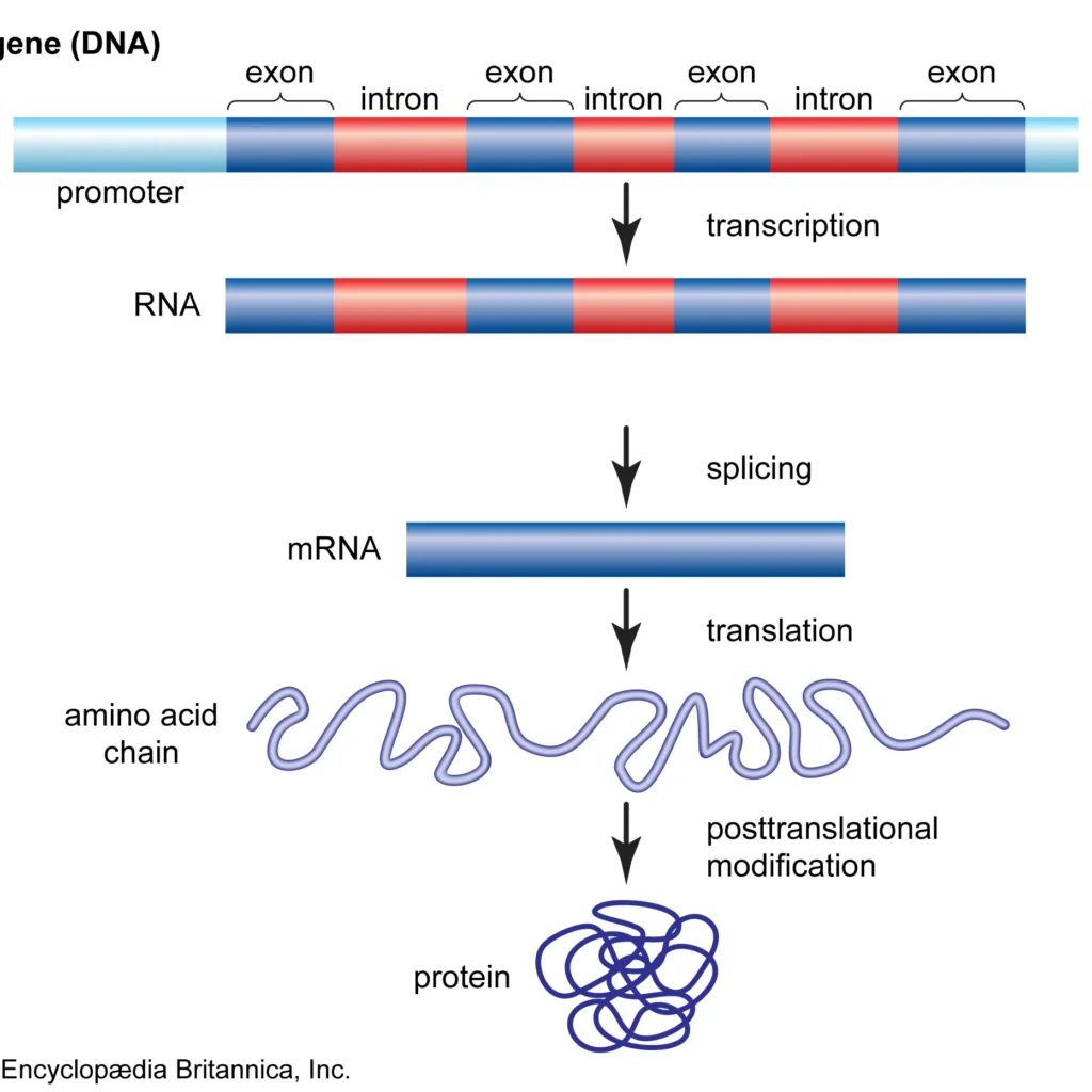
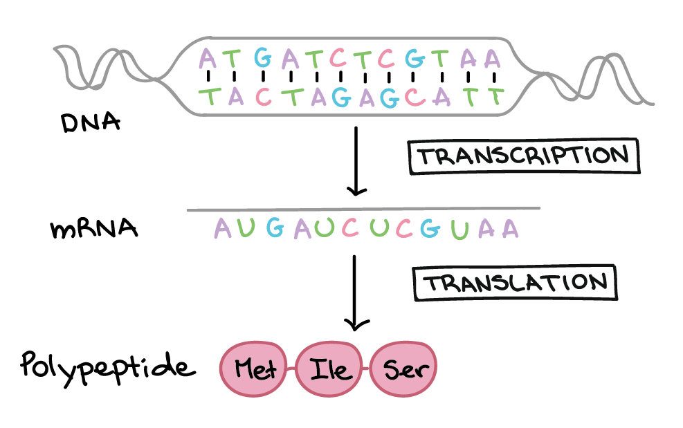
The Genetic Code and Gene Regulation: How Cells Read, Control and Use DNA The genetic code is the rule that converts a nucleotide sequence in messenger RNA into an amino-acid sequence. Gene regulation determines when, where and how strongly that information is used. Together they explain how one genome produces many specialised cells and why abnormal regulation causes developmental disease, endocrine disorders, immune disease and cancer. Why this topic matters: a patient may have a normal gene sequence but disease because the gene is not switched on, is expressed in the wrong tissue, produces unstable RNA, translates inefficiently, or makes too much protein. 1. The central dogma and its limits In most human cells, information flows from DNA to RNA to protein. Transcription copies DNA information into RNA; translation reads mRNA codons to build a polypeptide. This is not a claim that every gene produces protein—some genes produce functional RNA—and it does not mean regulation occurs at only one step. Information flow DNA sequence → transcription initiation → pre-mRNA → 5′ capping, splicing and polyadenylation → mature mRNA → ribosome reading codons → polypeptide folding and modification → functional product and phenotype. 2. What the genetic code is The code is read in non-overlapping triplets called codons. Each codon contains three mRNA bases and specifies one amino acid or a stop signal. The code is degenerate because several codons can specify the same amino acid, but it is not ambiguous: a particular codon normally has one meaning in the standard code. AUG usually acts as the start codon and also encodes methionine. UAA, UAG and UGA are stop codons. The reading frame is established at initiation; shifting it changes all downstream codons. 3. Properties of the code Property Meaning Clinical relevance Triplet Three bases form one codon Insertions/deletions not divisible by three cause frameshifts. Degenerate Several codons may specify one amino acid Some substitutions are synonymous, but may still affect splicing or expression. Nearly universal Most organisms use the same code Allows recombinant DNA and gene-expression systems. Non-overlapping Each base is read in one codon within a frame A frameshift changes the downstream message. Wobble Pairing at the third codon position is flexible Explains redundancy and some effects of tRNA abundance. 4. Transcription: making the RNA message RNA polymerase II binds a regulated promoter and synthesises a complementary RNA strand using the DNA template. Transcription has initiation, elongation and termination. Transcription factors determine which genes are accessible and help recruit or block polymerase. Regulation at transcription is powerful because it controls whether a message is made at all. A hormone-responsive promoter, for example, can rapidly increase the production of enzymes or receptors after a signal reaches the nucleus. 5. RNA processing and quality control 5′ cap: protects the transcript and helps ribosome recognition. Splicing: removes introns and joins exons. Alternative splicing: produces different transcripts from one gene. Poly-A tail: supports stability, export and translation. Nonsense-mediated decay: removes selected transcripts with premature stop codons. Errors in processing can produce disease even when the coding sequence is unchanged. This is why variant interpretation must include splice-site, deep-intronic, UTR and regulatory regions. 6. Translation: decoding mRNA at the ribosome Translation begins when the ribosome identifies the mRNA start region and assembles around the start codon. Initiator tRNA carries methionine. During elongation, tRNAs deliver amino acids, peptide bonds form, and the ribosome moves codon by codon. A stop codon recruits release factors rather than a tRNA. Translation may occur on free ribosomes or ribosomes attached to the rough endoplasmic reticulum. The destination of the new protein depends on signal sequences and determines whether it becomes cytosolic, nuclear, mitochondrial, lysosomal, secreted or membrane-bound. 7. Levels of gene regulation Chromatin level DNA packaging controls accessibility to transcription factors. Transcriptional level Promoters, enhancers, repressors and transcription factors control RNA production. RNA-processing level Capping, splicing, editing and polyadenylation determine the mature message. RNA-stability level Degradation determines how long an mRNA remains available. Translational level Ribosome recruitment and initiation determine protein production. Post-translational level Folding, modification, trafficking and degradation determine protein activity. 8. Transcription factors and regulatory elements Transcription factors are DNA-binding proteins that activate or repress genes. Activators recruit co-activators, mediator and chromatin-opening machinery. Repressors recruit co-repressors and chromatin-condensing enzymes. The same gene can respond to several factors, allowing integration of hormones, nutrients, stress, inflammation and cell identity. 9. Epigenetic regulation Epigenetic mechanisms change gene activity without changing the underlying base sequence. DNA methylation can reduce promoter accessibility. Histone acetylation often opens chromatin, while other histone marks can activate or repress depending on their location and context. Epigenetic regulation is essential in embryonic development, X-inactivation, imprinting, ageing and cancer. Clinical caution: “epigenetic” does not mean that a disease is imaginary or that a patient caused it through lifestyle. Epigenetic states are influenced by development, environment, inflammation, nutrition and disease, but causal interpretation requires evidence. 10. Non-coding RNAs Ribosomal RNA forms the structural and catalytic core of ribosomes. Transfer RNA carries amino acids. MicroRNAs bind target mRNAs and reduce translation or promote degradation. Long non-coding RNAs can influence chromatin, transcription and RNA processing. Non-coding RNA abnormalities are increasingly recognised in cancer and inherited disease. 11. Operons and eukaryotic regulation Many bacteria organise related genes into operons controlled by one promoter and regulatory region. This allows coordinated response to nutrients. Human genes are usually regulated individually through combinations of promoters, enhancers, insulators and chromatin domains, although shared regulatory networks coordinate pathways. 12. Mutations in the code versus mutations in regulation Variant location Likely first effect Example clinical reasoning Coding exon Protein sequence or termination changes Assess missense mechanism, nonsense-mediated decay or domain effect. Promoter/enhancer Amount or tissue of expression changes Normal protein sequence may be made in the wrong quantity. Splice region Abnormal mature mRNA RNA studies may clarify exon skipping or intron retention. UTR Stability, localisation or translation changes Protein may be reduced despite a normal coding sequence. Epigenetic control Gene switched off or misregulated Consider imprinting, methylation disorders or tumour silencing. 13. Gene regulation in cancer Cancer