
Antigens, Immunogens, and Immunoglobulins
Core Immunological Concepts
Key Definitions
- Antigen: Any substance capable of specifically binding to an antibody or an antigen-specific T-cell receptor (TCR). In simple terms, an antigen is a molecular target that immune receptors can recognize.
- Immunogen: Any substance that directly triggers or stimulates an active immune response. In practical terms, an immunogen can provoke an attack, while an antigen might only be recognized without necessarily provoking one on its own.
| Feature | Antigen | Immunogen |
|---|---|---|
| Primary Capability | Binds to specific immune products (antibodies or TCRs) | Actively induces an adaptive immune response |
| Relationship | All immunogens are antigens | Not all antigens are immunogenic (some require carrier proteins) |
Immunoglobulins: Basic Architecture and Chains
What Are Immunoglobulins?
Immunoglobulins (Ig), commonly termed antibodies, are specialized defensive glycoproteins synthesized by plasma cells (fully differentiated B lymphocytes) following exposure to an immunogen.
- Distribution: Circulate widely in the serum and bathe extracellular tissue fluids across mammalian systems.
- Primary Functional Role: Form the essential humoral component of adaptive immunity, delivering targeted defense throughout body fluids.
The standard basic immunoglobulin unit consists of four polypeptide chains configured in a characteristic Y-shaped monomeric layout. This structure includes two identical heavy (H) chains and two identical light (L) chains. Sturdy covalent disulfide bridges join these chains together, stabilizing the overall four-chain complex.
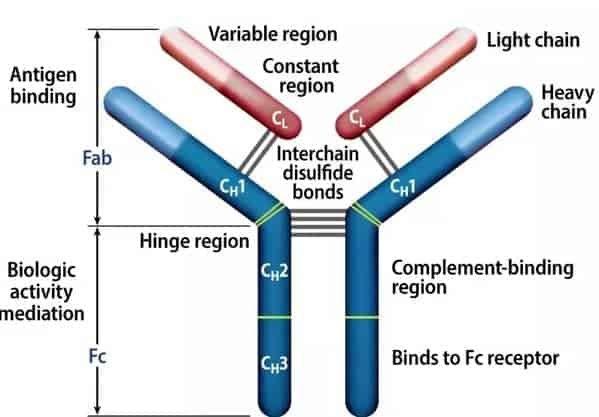
Structural Regions of an Antibody Monomer
Heavy and Light Chain Characteristics
Antibody classes are defined by distinct heavy chains, whereas light chains are shared across classes.
| Chain Category | Isotypes / Types | Molecular Characteristics | Key Structural Features |
|---|---|---|---|
| Heavy (H) Chains | γ (gamma), α (alpha), μ (mu), δ (delta), ε (epsilon) | Defines the five distinct Ig classes: IgG, IgA, IgM, IgD, and IgE | Variable length; contains one variable domain (VH) and 3 to 4 constant domains (CH) |
| Light (L) Chains | κ (kappa) and λ (lambda) | Molecular mass of approximately 23,000 Daltons | An individual antibody contains exclusively κ or λ chains, never a mixture of both |
Domains and Functional Regions
Both heavy and light chains fold into distinct three-dimensional repeating segments known as domains:
- Light Chain Domains: Contains exactly one variable domain (VL) and one constant domain (CL).
- Heavy Chain Domains: Contains one variable domain (VH) paired with either three or four constant domains:
- IgG, IgA, and IgD: Possess three constant domains (CH1, CH2, CH3) along with an accessible hinge region.
- IgM and IgE: Lack an ordinary hinge; instead, they contain an extra constant domain (CH4).
Functional Division: Variable vs. Constant Regions
- Variable Regions (VH and VL): Responsible for antigen recognition and binding specificity. Located at the amino-terminal ends of the chains, they feature three hypervariable loops called Complementarity Determining Regions (CDRs: CDR1, CDR2, CDR3). These hypervariable segments directly contact and physically secure the antigen.
- Constant Regions (CH and CL): Form the structural scaffold and drive effector mechanisms. These regions mediate complement fixation, placental crossing, and Fc receptor attachment on effector cells.
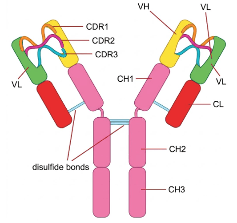
Complementarity Determining Regions (CDRs) on Heavy and Light Chains
Enzymatic Cleavage: Structure-Function Relationships
Exposing immunoglobulin monomers to proteolytic enzymes cleaves the polypeptide chains at specific sites, separating antigen-binding capacity from effector function.
| Enzyme | Cleavage Location | Resulting Fragments | Valency & Biological Activity |
|---|---|---|---|
| Papain | Above the hinge region disulfide bonds | Two Fab fragments and one Fc fragment |
Fab (Fragment antigen-binding): Univalent (valence = 1); retains antigen-binding specificity defined by VH and VL, but cannot cross-link antigens to precipitate them. Fc (Fragment crystallizable): No antigen-binding capacity; crystallizes in solution and mediates effector tasks (complement fixation, Fc receptor binding). |
| Pepsin | Below the hinge region disulfide bonds | One F(ab′)2 fragment and degraded Fc subfragments |
F(ab′)2: Bivalent (valence = 2); two Fab units remain connected by disulfide bridges, allowing both antigen binding and lattice formation (agglutination/precipitation). Fc: Digested into multiple small peptide remnants; loses crystallizability and effector mechanisms. |
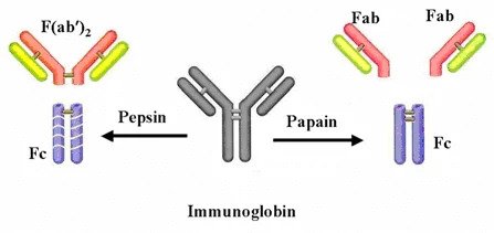
Proteolytic Cleavage of Immunoglobulin by Papain vs. Pepsin
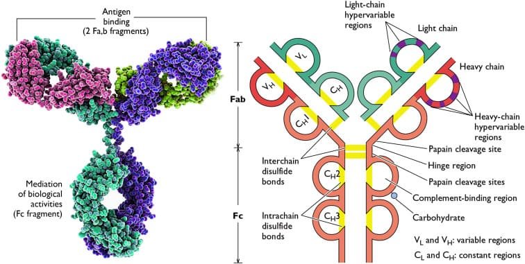
Effector and Recognition Sites Across the Antibody Molecule
Clinical Application of Fab and F(ab′)2 Fragments
Therapeutic antivenoms and monoclonal drugs (such as Digoxin Immune Fab or Abciximab) use cleaved Fab or F(ab′)2 fragments instead of intact antibodies. Because they lack the Fc stem, these fragments do not activate complement or bind non-target Fc receptors. This minimizes hypersensitivity reactions, reduces unwanted tissue damage, and allows faster systemic clearance.
The Five Mammalian Immunoglobulin Classes
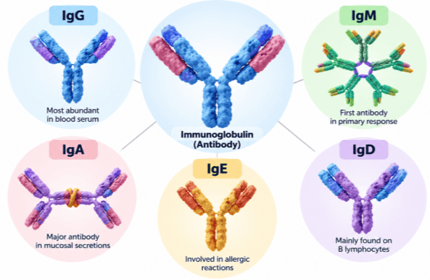
Comparative Geometries of IgG, IgE, IgD, IgA, and IgM
Immunoglobulin G (IgG)
- Structural Form: Exists as a 7S monomer. Composed of four major subclasses: IgG1, IgG2, IgG3, and IgG4. While IgG1, IgG2, and IgG4 share similar hinge arrangements, IgG3 contains an extended hinge made of multiple repeated segments and interchain disulfide links.
- Serum and Tissue Distribution: The most abundant immunoglobulin in circulating serum and the primary antibody found in extravascular spaces.
- Transplacental Passage: The only immunoglobulin class that crosses the human placenta, providing crucial passive immunity to protect the developing fetus and neonate.
- Effector Mechanisms:
- Complement Activation: Readily fixes and activates classical complement pathways (especially IgG3 and IgG1).
- Opsonization: Binds Fcγ receptors on phagocytic cells (macrophages and neutrophils), facilitating the ingestion and destruction of coated pathogens.
- ADCC (Antibody-Dependent Cell-Mediated Cytotoxicity): Binds to Natural Killer (NK) cells, triggering targeted lysis of infected or abnormal host cells.
- Staphylococcal Protein A (SPA) Binding: Reacts strongly with Protein A found on Staphylococcus aureus cell walls.
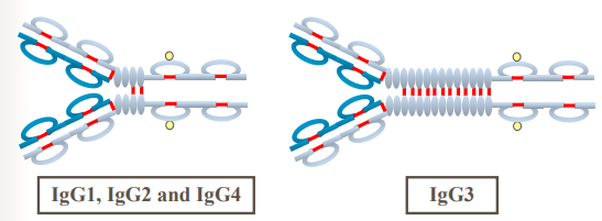
Hinge Region Variation Among IgG Subclasses
Immunoglobulin M (IgM)
- Structural Form: Secreted primarily as a large 19S pentamer built from five basic H2L2 units connected by a specialized J chain (joining chain). Heavy chains lack a classic hinge and instead contain an extra constant domain (CH4). It also includes an extended tail piece.
- Serum Abundance: Represents the third most prevalent antibody class in circulating serum.
- Developmental & Primary Response Role: The very first immunoglobulin synthesized by the developing fetus and the earliest class produced by newly differentiated B cells during a primary immune response.
- Agglutination & Lysis Efficiency: Because of its high valence (10 theoretical antigen-binding sites, typically 5 functional sites due to steric hindrance), it is the most efficient antibody for pathogen agglutination and classical complement fixation.
- Cellular Roles: Can bind to specific Fc receptors and functions on naïve B-cell surfaces as a monomeric antigen-recognition receptor.
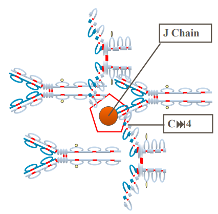
Pentameric Arrangement and J-Chain Linkage of IgM
Mechanism of Complement C1 Activation: IgM vs. IgG
The first component of the classical pathway, C1, is a large multi-subunit complex made of C1q, C1r, and C1s. For C1 to activate, two or more of the globular heads on C1q must bind to adjacent antibody Fc regions simultaneously.
- IgM Activation (Highly Efficient): A single antigen-bound pentameric IgM molecule changes its shape (from a flat planar form into a staple-like configuration), exposing multiple closely packed CH3/CH4 sites. This allows a single pentamer to easily cross-link several C1q heads and trigger complement activation.
- IgG Activation (Requires Proximity): Because monomeric IgG has only one Fc region, a single IgG cannot activate C1q alone. Two separate IgG molecules must bind to antigens right next to each other on a cell surface (within approximately 30–40 nm) so their Fc stems can bridge adjacent C1q heads.
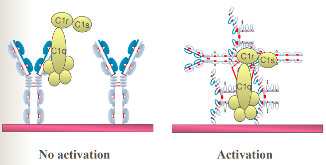
Fixation of Complement C1: Unsuccessful IgG Monomers vs. Successful IgM Pentamer
The B-Cell Antigen Receptor Complex (BCR)
On the surface of mature, non-activated B lymphocytes, antibodies operate as the primary antigen recognition system.
- Membrane-Bound Immunoglobulins: Naïve mature B cells express monomeric IgM and IgD anchored within the plasma membrane via hydrophobic transmembrane sequences.
- Signaling Invariant Heterodimers: Because membrane-bound antibody tails are too short to send intracellular signals, they pair with invariant signaling proteins called Ig-α (CD79a) and Ig-β (CD79b). These accessory proteins contain Immunoreceptor Tyrosine-based Activation Motifs (ITAMs) that transmit activation signals into the cell when an antigen binds.
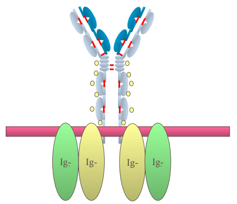
Organization of the B-Cell Antigen Receptor (BCR) and Ig-α/Ig-β Signaling Chains
Immunoglobulin A (IgA)
- Structural Forms:
- Serum IgA: Circulates primarily as an individual monomer. It is the second most abundant antibody class in serum.
- Secretory IgA (sIgA): Found in external secretions as an 11S dimer. This complex contains two H2L2 monomers linked by a J chain and protected by a polypeptide wrap called the Secretory Component (SC or Secretory Piece, SP).
- Secretion Locations: The predominant antibody in mucosal fluids, including saliva, tears, breast milk, colostrum, respiratory secretions, intestinal fluids, and urogenital tract secretions.
- Effector Functions: Blocks pathogens and toxins from attaching to mucosal surfaces. It does not activate the classical complement cascade unless artificially clustered in high concentrations, but it can bind to specific Fcα receptors on select immune cells.
- Role of the Secretory Component: Synthesized by mucosal epithelial cells (derived from the polymeric immunoglobulin receptor, pIgR). The secretory component helps transport dimeric IgA across epithelial layers and protects it from being broken down by proteolytic enzymes in the gastrointestinal tract.
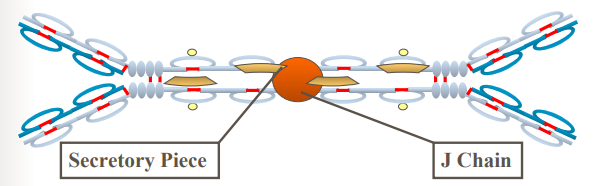
Secretory Dimeric IgA with J Chain and Secretory Piece
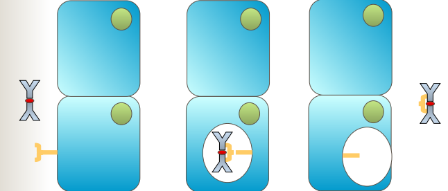
Epithelial Transcytosis and Acquisition of the Secretory Component
Immunoglobulin D (IgD)
- Structural Form: Exists as a flexible monomer containing an extended hinge and a carboxy-terminal tail piece.
- Serum Concentration: Found in very low amounts in circulation (the fourth highest serum Ig, but accounts for less than 1% of total serum antibodies).
- Primary Biological Role: Acts primarily as a membrane-bound antigen receptor on mature, naïve B cells (co-expressed alongside membrane IgM). It helps signal B-cell activation and differentiation.
- Effector Limitations: Does not bind or activate complement proteins and has no established classical effector role in circulating serum.
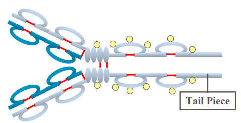
Monomeric Structure of IgD Featuring an Extended Hinge and Tail Piece
Immunoglobulin E (IgE)
- Structural Form: Exists as a monomer. Like IgM, its heavy chains lack an ordinary hinge and contain an extra constant domain (CH4).
- Serum Abundance: The least common immunoglobulin in circulating serum, present only in trace concentrations under normal conditions.
- High-Affinity Receptor Binding: Binds extremely tightly via its Fc region to high-affinity FcεRI receptors on tissue mast cells and circulating basophils. This binding occurs spontaneously in the resting state, without requiring prior antigen contact.
- Clinical Roles:
- Type I Hypersensitivity Reactions: When specific allergens cross-link IgE molecules already bound to mast cells or basophils, these cells instantly degranulate, releasing histamine, leukotrienes, and heparin. This drives acute allergic symptoms and systemic anaphylaxis.
- Defense Against Parasitic Worms: Coats helminths and recruits eosinophils via their Fcε receptors. This activates the eosinophils to release major basic protein and other enzymes, destroying the parasite's outer surface.
- Complement Inactivity: Does not fix or activate classical complement components.
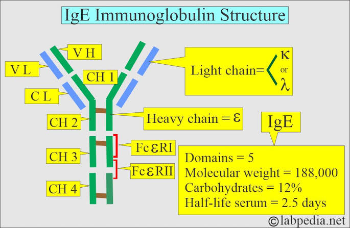
Monomeric Structure of IgE Highlighting the Extra CH4 Domain
Comprehensive Comparison of Immunoglobulin Classes
| Property | IgG | IgM | IgA | IgD | IgE |
|---|---|---|---|---|---|
| Heavy Chain | γ (gamma) | μ (mu) | α (alpha) | δ (delta) | ε (epsilon) |
| Basic Configuration | Monomer (7S) | Pentamer (19S) with J chain | Monomer (serum) / Dimer (11S, secretions) with J chain and SP | Monomer | Monomer |
| Heavy Chain Constant Domains | 3 (CH1, CH2, CH3) | 4 (has extra CH4) | 3 (CH1, CH2, CH3) | 3 (CH1, CH2, CH3) | 4 (has extra CH4) |
| Serum Abundance Ranking | 1st (most abundant) | 3rd | 2nd | 4th | 5th (least abundant) |
| Placental Transfer | Yes (only class) | No | No | No | No |
| Classical Complement Fixation | Yes (IgG3 > IgG1 > IgG2; IgG4 does not) | Yes (most potent) | No (only when aggregated) | No | No |
| Primary Biological Function | Long-term systemic immunity, opsonization, ADCC, toxin neutralization | Primary immune response, rapid agglutination, early B-cell receptor | Mucosal protection, prevents pathogen attachment in secretions | Antigen receptor on mature naïve B lymphocytes | Type I hypersensitivity, allergic reactions, defense against parasites |
Primary Biological Functions of Immunoglobulins
Antibodies protect the host through five major defensive mechanisms:
Summary of Effector Mechanisms
- Enhanced Phagocytosis (Opsonization): Antibodies coat foreign microbes through their antigen-binding sites. This exposes their Fc stems to phagocytes (macrophages and neutrophils), making the pathogens much easier to engulf and digest.
- Virus Neutralization: Antibodies bind directly to surface proteins on viruses, preventing them from docking onto host cellular receptors and blocking infection.
- Neutralization of Bacterial Toxins: Antibodies bind specifically to microbial exotoxins, changing their shape and blocking the active sites required to damage host cells.
- Complement-Mediated Lysis: When antibodies bind to foreign cell surfaces, their exposed Fc stems activate the classical complement pathway. This triggers a enzymatic cascade that forms the Membrane Attack Complex (MAC), punching holes in the cell and causing osmotic lysis.
- Antibody-Dependent Cell-Mediated Cytotoxicity (ADCC): When antibodies coat target cells (such as virus-infected or tumor cells), their exposed Fc stems bind to Fc receptors on cytotoxic killer cells (primarily Natural Killer cells). This signals the killer cells to release perforins and granzymes, destroying the target cell.
