
The Complement System
1. General Organization and Historical Discovery
Key Definitions: Fundamental Nature of Complement
- The Complement System: A primary humoral defense network made up of greater than 30 distinct proteins. The majority of these circulating factors are manufactured by hepatocytes within the liver and circulate continuously throughout blood plasma and extracellular fluids in an inactive state.
- Historical Origin: The scientific term complement was coined following pioneering experiments carried out by Jules Bordet. His work demonstrated that heat-labile substances in normal serum "complemented" or assisted specific, heat-stable antibodies in bursting and destroying foreign bacterial targets.
2. Essential Features Governing Complement Function
The system relies on four fundamental rules to defend the body while preventing accidental damage to normal, healthy host tissue:
| Principle | Molecular Mechanism | Functional Outcome |
|---|---|---|
| 1. Target-Focused Activation | Triggered directly by microbial surface components or indirectly through antibodies and lectins bound to foreign antigens. | Directs destructive immune reactions specifically onto microbial targets while sparing innocent bystander tissues. |
| 2. Sequential Zymogen Cascades | Circulating complement factors are zymogens (inactive enzyme precursors). When one protease is cleaved, it becomes active and cleaves the next zymogen in line. | Functions like the blood coagulation and kinin cascades. This chain reaction produces massive, rapid exponential enzyme amplification at the infection site. |
| 3. Covalent Membrane Anchoring | Active cleavage fragments form stable, covalent chemical bonds with microbial cell walls, bound antibodies, or apoptotic bodies. Free fragments in blood remain active for only seconds before decomposing. | Restricts full, sustained complement attack strictly to target surfaces, preventing runaway destructive reactions in circulating blood. |
| 4. Host Membrane Protection | Healthy host cells express specific surface regulatory proteins that shut down complement activation. Microbes lack these protective molecules entirely. | Protects self-tissues from autologous attack while leaving unprotected invading microorganisms fully exposed to complement destruction. |
3. Biological Outcomes: Host Protection vs. Detriment
| Category | Specific Activity | Clinical & Physiological Effect |
|---|---|---|
| Beneficial Host Responses | Opsonization | Coats microbial surfaces with complement fragments, dramatically speeding up phagocytic engulfment. |
| Leukocyte Chemotaxis | Attracts circulating neutrophils and macrophages directly toward the focus of acute infection and activates them. | |
| Target Cell Cytolysis | Forms large multiprotein transmembrane pores called the membrane attack complex (MAC), lysing gram-negative bacteria and infected cells. | |
| Immune Complex Clearance | Solubilizes circulating antigen-antibody deposits so they can be carried away by erythrocytes to the spleen and liver for destruction. | |
| Apoptotic Cell Clearance | Tags dying self-cells for clean phagocytic disposal, preventing dangerous release of intracellular autoantigens. | |
| Antibody Response Modulation | Interacts with B-cell co-receptors to amplify humoral immune responses and encourage robust antibody production. | |
| Potential Host Detriment | Anaphylatoxin Release | Excessive production of small cleavage peptides triggers systemic mast cell degranulation. This causes profound vasodilation, plasma leakage, and smooth muscle spasms, culminating in circulatory shock. |
4. Complement Nomenclature and Structural Terminology
Key Definitions: Nomenclature Rules
- Numbered Components: Factors of the classical and terminal pathway are written with a capital "C" followed by numbers 1 through 9 (e.g., C1, C2, C3, C4, C5, C6, C7, C8, C9).
- Lettered Factors: Alternative pathway components are designated by individual letters (e.g., Factor B, Factor D, Factor H, Factor I, and Properdin (Factor P)).
- Lectin Binding Elements: Mannose-binding lectin (MBL) complexes with MBL-associated serine proteases (MASP-1, MASP-2, MASP-3) and ficolins (M-ficolin, L-ficolin, H-ficolin).
- Proteolytic Fragments ("a" vs "b"): When a complement protein is cleaved by an upstream enzyme:
- The smaller peptide fragment receives the lowercase suffix "a" (e.g., C3a, C4a, C5a) and diffuses away into the surrounding fluid as a soluble signaling peptide.
- The larger active fragment receives the lowercase suffix "b" (e.g., C3b, C4b, C5b) and binds directly to the activating cell membrane.
Nomenclature Inversion of Complement Component C2
In standard classical and lectin cascade nomenclature, C2 is the sole exception to the size-letter rule:
- C2a: Represents the LARGER active enzymatic piece that stays attached to C4b to build the functional C3 convertase (C4b2a).
- C2b: Represents the SMALLER split product that diffuses away (acting as a prokinin precursor).
| Laboratory / Clinical Term | Practical Working Definition |
|---|---|
| Complement Activation | A conformational or enzymatic alteration of a complement protein that enables it to interact directly with the next factor in the cascade. |
| Complement Fixation | The consumption and binding of serum complement proteins by antigen-antibody complexes. |
| CH50 (Hemolytic Unit) Assay | A laboratory screening test measuring the dilution of patient serum required to lyse 50% of a standardized red blood cell suspension coated with anti-erythrocyte antibodies. It monitors the functional integrity of the classical and terminal pathways. |
| Complement Inactivation | The loss of hemolytic function through protein denaturation, which routinely occurs when serum samples are heated to 56°C for 30 minutes. |
| Convertase / Esterase | An assembled, multi-subunit protein complex that acts as an active protease to cleave downstream complement substrates. |
5. Classical Pathway Components: Biochemical and Physical Properties
The classical pathway depends on a precise sequence of structural assembly, enzymatic cleavage, and covalent binding:
| Protein Component | Molecular Structure & Mass | Mean Serum Level (μg/mL) | Primary Functional Mechanism |
|---|---|---|---|
| C1 (C1qr2s2 Complex) | 750 kDa complete multimolecular complex held together by Ca2+ | Not measured as a single intact unit in serum | Initiates the entire classical pathway sequence upon antibody cross-linking. |
| C1q | 460 kDa hexamer composed of 18 polypeptide chains (three pairs of distinct chains: 22, 23, and 24 kDa) arranged in a bouquet-like shape | 50–150 μg/mL | Binds to the Fc region of antibody-antigen complexes, apoptotic cell membranes, and polyanionic surfaces. |
| C1r | 85 kDa homodimer | 50 μg/mL | Autocatalytic serine protease; activated by C1q deformation, subsequently cleaving and activating C1s. |
| C1s | 85 kDa homodimer | 50 μg/mL | Active serine esterase; directly cleaves native C4 and native C2. |
| C4 | 210 kDa trimeric glycoprotein composed of 97, 75, and 33 kDa chains joined by disulfide bonds | 300–600 μg/mL | Cleaved by C1s into small C4a (anaphylatoxin) and large C4b. C4b exposes an internal thioester bond that covalently attaches to the microbial surface and binds C2 for activation. |
| C2 | 102 kDa single-chain monomer | 20 μg/mL | Binds to membrane-bound C4b in the presence of magnesium ions (Mg2+) and is cleaved by C1s into C2b and C2a. C2a functions as the catalytic serine protease core of C3 and C5 convertases. |
6. The Three Activation Cascades: Classical, Lectin, and Alternative
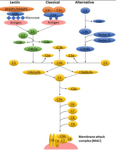
Convergence of the Three Complement Activation Pathways
| Pathway Name | Immune Classification | Initial Activating Stimulus | Assembled C3 Convertase | Assembled C5 Convertase |
|---|---|---|---|---|
| Classical Pathway | Adaptive (humoral) immunity | IgM (single pentamer) or IgG (at least two molecules side-by-side) bound to target antigens | C4b2a | C4b2a3b |
| Lectin (MBL) Pathway | Innate immunity | Mannose-binding lectin or ficolins binding to terminal mannose, fructose, or N-acetylglucosamine arrays on bacterial walls | C4b2a | C4b2a3b |
| Alternative Pathway | Innate immunity (phylogenetically oldest) | Continuous low-grade spontaneous hydrolysis (C3 tickover), protected microbial membranes, bacterial endotoxin (LPS), cobra venom factor, or nephritic factor | C3bBb (stabilized by Properdin to form C3bBbP) | C3bBb3b |
7. Step-by-Step Execution of the Classical Pathway
The classical pathway bridges the adaptive humoral antibody response with innate cell lysis:
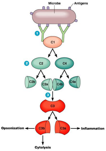
Assembly of the Classical C3 Convertase (C4b2a)
- Antibody Engagement: Soluble IgG or IgM binds to foreign antigens on a bacterial wall. This changes the shape of the antibody's Fc stem, exposing previously hidden complement docking sites.
- C1 Complex Activation: The globular heads of C1q bind to the exposed Fc segments. In the presence of calcium ions (Ca2+), this physical stress activates the serine protease C1r, which then cuts and activates its partner C1s.
- Cleavage of C4 and C2:
- Active C1s cuts C4 into a small fluid-phase fragment (C4a, an anaphylatoxin) and a large fragment (C4b). C4b quickly forms a covalent bond with the pathogen's surface next to the C1 complex.
- In the presence of magnesium ions (Mg2+), native C2 binds to C4b. C1s cleaves C2 into C2b (a small prokinin peptide that diffuses away) and C2a (a large enzymatic fragment that remains bound to C4b).
- C3 Convertase Formation: The newly joined C4b2a complex is the functional classical C3 convertase.
- Massive C3 Amplification: A single active C4b2a enzyme molecule rapidly cleaves greater than 200 molecules of C3 into C3a (inflammatory anaphylatoxin) and C3b (opsonin).
8. The Central Role of C3b in Opsonization and Immune Adherence
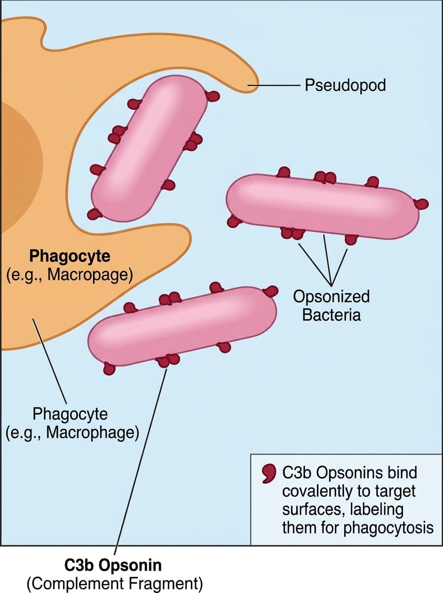
Surface Opsonization and Immune Adherence
Key Definitions: Opsonization Dynamics
- Opsonin: A specialized immune molecule that binds simultaneously to microbial surface antigens and specific surface receptors on phagocytes, acting like a bridge.
- Opsonization Enhancement: Coating a bacterial capsule with C3b increases the rate and efficiency of neutrophil and macrophage phagocytosis by up to 1,000-fold compared to uncoated targets.
- Immune Adherence: The high-affinity physical binding between particles coated with C3b or C4b and the Complement Receptor 1 (CR1 / CD35) found on leukocytes and red blood cells.
9. Assembly of the C5 Convertase and the Membrane Attack Complex (MAC)
Once the bacterial surface is heavily coated with C3b, the cascade shifts from opsonization to direct membrane destruction:
| Assembly Phase | Molecular Event | Downstream Biological Effect |
|---|---|---|
| 1. C5 Convertase Assembly | Excess C3b binds directly to the preexisting C4b2a enzyme to build the trimolecular complex C4b2a3b. | Switches the enzyme's binding pocket so it now binds and cleaves native C5 instead of C3. |
| 2. Cleavage of C5 | C4b2a3b splits C5 into two products:
|
C5a triggers systemic inflammation, while membrane-bound C5b serves as the foundation for the terminal pore complex. |
| 3. Terminal Pore Assembly (C5b–C9) |
|
Forms the functional Membrane Attack Complex (MAC) with an open central transmembrane pore measuring roughly 10 nm in diameter. |
| 4. Osmotic Lysis (Cytolysis) | The open MAC channel permits free, unregulated movement of ions and fluid: intracellular cytoplasmic solutes escape while extracellular water rushes in down its osmotic gradient. | The target cell swells rapidly, loses membrane integrity, and undergoes explosive osmotic cytolysis. |
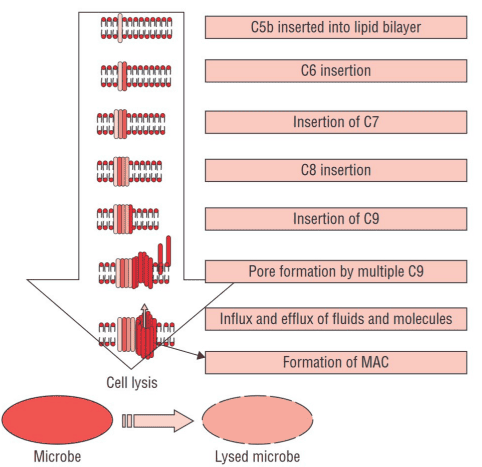
Assembly and Insertion of the Membrane Attack Complex (MAC)
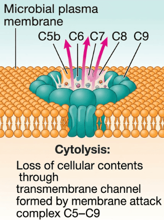
Cytolytic Mechanism of the Assembled Transmembrane Channel
10. Inflammatory Mediators: Anaphylatoxins and Cellular Interactions
Complement cleavage produces diffusible peptide fragments that coordinate acute local and systemic inflammatory responses:
| Split Product | Primary Functional Classification | Specific Target Cells and Biological Actions |
|---|---|---|
| C5a | Most powerful anaphylatoxin & chemokine | Binds to specific G-protein-coupled receptors on mast cells, basophils, and neutrophils. Stimulates massive histamine release, triggers vascular leakage, induces smooth muscle contraction, and provides the body's strongest chemical gradient to guide leukocyte migration. |
| C3a | Intermediate anaphylatoxin | Stimulates mast cells and basophils to degranulate. Causes endothelial cell contraction, increased capillary permeability, and smooth muscle bronchospasm. |
| C4a | Weakest anaphylatoxin | Promotes modest smooth muscle contraction and mild local fluid extravasation. |
| C2b (Prokinin) | Vasoactive kinin precursor | Cleaved by serum plasmin into an active kinin peptide that dramatically increases vascular permeability, producing marked tissue edema. |
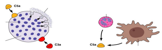
Inflammatory Signaling via Anaphylatoxins (C3a and C5a)
| Cell Type | Granule Molecule Class | Specific Clinical Examples | Biological Function |
|---|---|---|---|
| Neutrophil | Proteases | Elastase, Collagenase | Breaks down extracellular matrix to remodel damaged tissue and allow immune cell transit. |
| Antimicrobial proteins | Defensins, Lysozyme | Inflicts direct, lethal damage on microbial cell membranes and cell walls. | |
| Protease inhibitors | α1-antitrypsin | Controls and limits runaway protease activity, protecting nearby healthy tissues. | |
| Vasoactive amines | Histamine | Promotes vasodilation and local inflammatory recruitment. | |
| Eosinophil | Cationic proteins | Eosinophil peroxidase (EPO) | Drives the generation of reactive oxygen species (ROS) to kill helminths and large targets. |
| Ribonucleases | Major basic protein (MBP) | Induces direct cytotoxicity, causes vasodilation, and stimulates basophil degranulation. | |
| Cytokines | ECP, EDN | Antiviral defenses and tissue-destructive enzymatic actions. | |
| Chemokines | IL-4, IL-10, IL-13, TNF-α, RANTES, MIP-1α | Modulates adaptive immune responses and attracts circulating leukocytes to the inflammatory focus. | |
| Basophil / Mast Cell | Cytokines | IL-4, IL-13 | Directs B-cell class-switching toward IgE and drives Th2-type immune responses. |
| Lipid mediators | Leukotrienes | Sustained regulation of vascular permeability and bronchial constriction. | |
| Vasoactive amines | Histamine | Causes rapid capillary endothelial contraction, fluid extravasation, and smooth muscle activation. |
11. The Lectin (Mannose-Binding Lectin) Pathway
The lectin pathway operates entirely independent of antibodies, recognizing unique sugar patterns on microbial surfaces:
Key Definitions: Lectin Recognition Complex
- Lectins: Circulating pattern-recognition proteins that bind specifically to carbohydrate targets displayed on microbial cell walls.
- Mannose-Binding Lectin (MBL): A large serum collectin structurally homologous to C1q. MBL binds specifically to nonreduced mannose, fructose, and N-acetylglucosamine residues found on bacterial and fungal cell walls (mannans).
- MASP Proteases: MBL-associated serine proteases (MASP-1, MASP-2, MASP-3) that act as the functional equivalents of C1r and C1s. When MBL binds carbohydrates, MASP-2 cleaves native C4 and C2 to form the identical C3 convertase (C4b2a) used by the classical pathway.
| Lectin Pathway Protein | Structural Composition | Serum Level (μg/mL) | Physiological Function |
|---|---|---|---|
| Mannose-Binding Lectin (MBL) | Helical trimer of 32 kDa chains; forms dimers up to hexamers of this triple helix | 1–8 μg/mL | Agglutinin, opsonin, activates complement by binding microbial carbohydrates. |
| M-ficolin (ficolin-1) | Helical trimer of 34 kDa chains; tetramer of this triple helix | Undetectable in normal serum (primarily cell-associated) | Secretory agglutinin and opsonin; fixes complement on target microbes. |
| L-ficolin (ficolin-2) | Helical trimer of 34 kDa chains; tetramer of this triple helix | 1–7 μg/mL | Systemic agglutinin and opsonin; triggers lectin pathway activation. |
| H-ficolin (ficolin-3) | Helical trimer of 34 kDa chains; tetramer of this triple helix | 6–83 μg/mL | Abundant serum opsonin; recognizes acetylated carbohydrates to fix complement. |
| MASP-1 | 90 kDa homodimer; structural homology to C1r/C1s | 2–13 μg/mL | Forms active complex with MASP-2 and collectins/ficolins; cleaves and activates MASP-3. |
| MASP-2 | 110 kDa homodimer; structural homology to C1r/C1s | 2–13 μg/mL | Primary catalytic enzyme of the lectin pathway; directly cleaves both C4 and C2 to form C4b2a. |
| MASP-3 | 76 kDa homodimer; structural homology to C1r/C1s | 0.02–1.0 μg/mL | Regulatory protease; associates with collectins/ficolins and works alongside MASP-1. |
12. The Alternative Pathway: Constant Tickover and Amplification
Described by Louis Pillemer in 1954, the alternative pathway serves as an antibody-independent, continuous surveillance system:
Key Definitions: C3 Tickover Mechanism
- C3 Tickover: The spontaneous, continuous non-enzymatic hydrolysis of circulating native C3 into fluid-phase C3(H2O) or C3b at a baseline rate of 1% to 2% of total plasma C3 per hour.
- Unstable Thioester Bond: Native C3 contains a buried, reactive internal thioester bond. Spontaneous reaction with water breaks this bond, enabling the molecule to bind covalently to nearby sugars or proteins on foreign surfaces.
- Sialic Acid Protection: Healthy mammalian cell membranes are rich in sialic acid. Sialic acid promotes the binding of inhibitory Factor H over Factor B, rapidly inactivating bound C3b. Microbes lack sialic acid, which allows the alternative pathway to proceed unimpeded on their surfaces.
| Step | Molecular Event | Chemical Requirements & Product |
|---|---|---|
| 1. Factor B Binding | C3b attached to a foreign surface binds circulating Factor B to create a reversible surface complex (C3bB). | Requires magnesium ions (Mg2+). Mg2+ is the only ion required for the functional activation of the alternative pathway. |
| 2. Factor D Cleavage | The binding of C3b to Factor B exposes an enzymatic cleavage site on Factor B, allowing plasma protease Factor D to cleave Factor B into Ba and Bb. | Ba diffuses into the surrounding fluid, while Bb stays bound to C3b, generating the active C3 convertase: C3bBb. |
| 3. Properdin Stabilization | Unbound C3bBb has an extremely short half-life (roughly 90 seconds). Circulating Properdin (Factor P) binds to the C3bBb complex on the microbial membrane. | Forms the stabilized alternative C3 convertase: C3bBbP, extending its functional half-life 6- to 10-fold. |
| 4. Amplification Loop | C3bBbP rapidly cleaves native C3 into massive numbers of C3a and C3b fragments. Each newly generated C3b can recruit more Factor B and Factor D, creating a self-fueling cycle. | Generates up to 2 × 10⁶ molecules of C3b within 5 minutes. |
| 5. C5 Convertase Assembly | An additional C3b fragment binds directly to the C3bBb convertase to form the alternative C5 convertase: C3bBb3b. | Cleaves native C5 into C5a (anaphylatoxin) and C5b (MAC initiator), feeding into the common terminal pathway. |
13. Complement Regulation: Protecting Host Cells from Bystander Lysis
Because activated complement enzymes can destroy host tissue, fluid-phase and membrane-bound inhibitors regulate every stage of the cascade:
| Regulatory Protein | Target Component(s) | Precise Biochemical Mechanism of Action |
|---|---|---|
| Antibody Availability | C1 complex initiation | Unbound circulating IgG and IgM hide their C1q docking sites. The classical pathway cannot initiate until antibodies undergo a conformational change after binding target antigens. |
| C1 Inhibitor (C1-INH / Serpin) | C1r, C1s, and intact C1qrs | Covalently binds and permanently blocks C1r and C1s esterases, forcing them to dissociate from C1q. This dismantles the active C1 complex and detaches it from antigen-antibody pairs. |
| Decay-Accelerating Factor (DAF / CD55) | C4b2a (Classical/Lectin) and C3bBb (Alternative) | A GPI-anchored membrane protein expressed on host cells. It binds membrane-bound C4b and C3b, displacing C2a and Factor Bb respectively. This prevents the formation of both C3 convertases and spares host cells from autologous damage. |
| Factor H | C3b (Alternative pathway) | Soluble plasma regulator that binds host surface sialic acid and membrane-bound C3b. It displaces Factor Bb from the alternative convertase and acts as an essential cofactor for Factor I-mediated cleavage of C3b. |
| Factor I | C3b and C4b | Active circulating serine protease. Cleaves membrane-bound C3b into inactive iC3b (and further to C3dg) and cleaves C4b into inactive fragments. Requires cofactors (Factor H, C4-BP, MCP, or CR1) to function. |
| C4-Binding Protein (C4-BP) | C4b (Classical/Lectin) | Soluble plasma protein that accelerates the decay of classical C3 convertase by displacing C2a from C4b. It also acts as an obligate cofactor for Factor I to permanently degrade C4b. |
| C3a-Inactivator (C3a-INA / Carboxypeptidase B) | C3a, C4a, C5a | Removes the C-terminal arginine residue from circulating anaphylatoxins, converting them into des-Arg forms. This dramatically reduces their inflammatory and spasmogenic potency. |
| Protein S (Vitronectin) / CD59 (Protectin) | Terminal C5b-7, C5b-8, and C9 | Vitronectin binds soluble C5b-7 in the fluid phase to prevent membrane insertion. CD59 on host cells binds C5b-8 and prevents C9 molecules from polymerizing into a lytic MAC pore. |
Deficiencies of Complement Regulators in Clinical Medicine
1. C1-Inhibitor Deficiency (Hereditary Angioedema): Without C1-INH, uninhibited activation of the early classical cascade and the kallikrein-kinin system leads to excessive production of vasoactive kinins (bradykinin and C2b-prokinin). Patients experience recurrent, life-threatening episodes of non-pitting subcutaneous and mucosal edema, particularly affecting the larynx and intestines.
2. Paroxysmal Nocturnal Hemoglobinuria (PNH): An acquired mutation in the PIGA gene disrupts the production of GPI anchors, leaving erythrocytes deficient in membrane-bound DAF (CD55) and Protectin (CD59). Without these shields, circulating red cells are vulnerable to spontaneous alternative pathway activation and MAC-mediated intravascular hemolysis, presenting with episodic dark morning urine, anemia, and thrombosis.
