
Comprehensive Immunology: Host Defense, Cellular Communication, Phagocytosis, and Adaptive Immunity
Key Definitions: Foundations of Immunology
- Immunology: The biological and medical science dedicated to exploring the immune system. This system consists of an intricate, highly cooperative network of specialized cells, organ tissues, and soluble circulating factors that shield the human body from external infectious agents and internal cellular damage.
- Pathogens: Infectious biological agents that can bring about illness, tissue injury, or functional impairment. Major categories include:
- Viruses: Obligate intracellular invaders such as Human Immunodeficiency Virus (HIV) and Influenza virus.
- Bacteria: Extracellular and intracellular microbes such as Staphylococcus and Mycobacterium tuberculosis.
- Fungi: Opportunistic or true pathogenic fungi such as Candida albicans.
- Parasites: Complex single-celled or multicellular organisms, including Plasmodium species that cause malaria.
1. The Dual Architecture of Host Defense: Innate versus Adaptive Immunity
The human immune system organizes its protective responses across two cooperative functional arms: the immediate, non-tailored innate immune system and the highly specialized, delayed adaptive immune system.
| Feature | Innate Immunity (First Line) | Adaptive Immunity (Specialized Line) |
|---|---|---|
| Speed of Response | Rapid; operates within minutes to hours after exposure. | Delayed; requires several days to develop and mature during primary exposure. |
| Target Specificity | Non-specific; responds against common microbial signatures identically across different invaders. | Highly specific; precision-tailored to unique individual antigens. |
| Immunological Memory | Absent; repeat challenge generates the exact same baseline response intensity. | Present; creates long-lasting memory cells producing faster and stronger secondary responses. |
| Primary Elements | Physical barriers (skin, mucosa), chemical shields (gastric acid, lysozymes), phagocytes (macrophages, neutrophils), natural killer cells, complement cascade, and cytokines. | B lymphocytes (humoral immunity via antibodies) and T lymphocytes (cell-mediated immunity via CD4+ helper and CD8+ cytotoxic T cells). |
Primary versus Secondary Immune Responses and Vaccine Biology
The first time the host encounters a foreign antigen, the adaptive arm reacts through a slow primary response. The patient often develops clinical symptoms and signs of disease while the system works to generate defensive clones. Once the pathogen is cleared, specialized long-lived memory B and T lymphocytes remain behind.
Upon subsequent exposure to the exact same antigen, these memory cells launch a rapid, high-magnitude secondary response. Pathogens are routinely cleared before clinical symptoms appear. This memory principle is the scientific foundation of vaccination.
2. Hematopoiesis, Stem Cells, and Leukocyte Classification
Immune cells constantly travel throughout the body using blood and lymphatic vessels. While these cells reside in normal circulating blood, their absolute counts rise rapidly during infection.
The primary reservoir and birthplace for these cells is the bone marrow. All mature blood elements arise from immature precursors known as hematopoietic stem cells (HSCs) through the regulated process of hematopoiesis.
Key Definitions: Major Formed Elements of Blood
- Erythrocytes (Red Blood Cells): Hemoglobin-containing cells focused primarily on carrying oxygen from lungs to peripheral tissues.
- Platelets (Thrombocytes): Anucleated cytoplasmic fragments derived directly from giant bone marrow cells called megakaryocytes; essential for forming blood clots and maintaining vascular integrity.
- Leukocytes (White Blood Cells): Nucleated cells dedicated to immune defense. These are categorized into three main functional groups: granulocytes, mononuclear phagocytes, and lymphocytes.
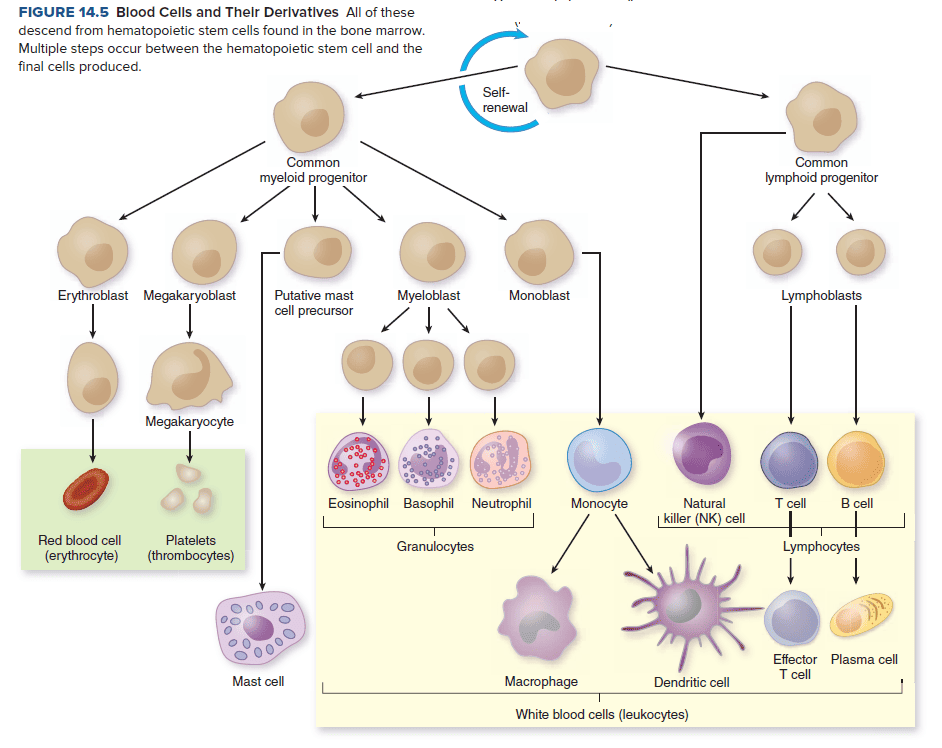
Blood Cells and Their Derivatives
| Cell Class | Cell Type (% Circulating Leukocytes) | Primary Tissue Location | Core Functional Role |
|---|---|---|---|
| Granulocytes (Contain fluid-filled, chemically active cytoplasmic granules) |
Neutrophils (55% to 65%) Also termed PMNs, polymorphonuclear leukocytes, or polys |
Mainly circulating in blood; sparse in healthy tissues, but migrate quickly in large numbers during inflammation. Bone marrow holds large reserve pools. | Professional phagocytes functioning as the body's rapid response team. They arrive first at tissue damage sites, exhibit very high killing capacity, and have a short functional lifespan of 1 to 2 days. |
| Eosinophils (2% to 4%) | Low numbers in circulation; most enter mucosal tissues and reside near local secretions. | Involved in allergic reactions and defense against parasitic worms. Granules contain antimicrobial compounds and the enzyme histaminase (which breaks down histamine). | |
| Basophils (0% to 1%) & Mast Cells | Basophils circulate in blood; mast cells reside within peripheral connective tissues near vessels and mucosal boundaries. | Key players in allergic reactions and acute inflammation. Their granules release vasoactive mediators like histamine to boost capillary permeability. (Note: while they share similar appearance and functions, they represent distinct cell lineages). | |
| Mononuclear Phagocyte System (MPS) | Monocytes (3% to 8%) | Circulate within blood vessels following release from bone marrow. | Precursor cells that travel through blood and enter injured or infected tissues, where they differentiate into active tissue macrophages or dendritic cells. |
| Macrophages | Present in almost all tissues (abundant in spleen, lungs, liver, lymph nodes, and peritoneal cavity). Named according to their home tissue. | Professional tissue sentinels and scavengers. They clear daily cell debris, phagocytose pathogens, produce cytokines, regenerate their lysosomes to sustain killing, and live for weeks to months. | |
| Dendritic Cells | Originate in tissues, then travel to secondary lymphoid organs (lymph nodes, spleen, appendix, tonsils). | Highly motile, phagocytic cells during early life. They capture antigens in peripheral tissues and migrate to lymphoid organs to present them to lymphocytes. | |
| Lymphocytes | B Cells, T Cells, and Natural Killer (NK) Cells (25% to 35%) | Abundant in secondary lymphoid organs (lymph nodes, spleen, mucosal-associated lymphoid tissue) and constantly recirculate through blood and lymph. | Primary mediators of adaptive immunity. B cells differentiate into antibody-producing plasma cells; T cells direct responses and destroy infected host cells; NK cells eliminate virus-infected and malignant cells. |
3. Primary and Secondary Lymphoid Organs
| Organ Category | Anatomical Sites Included | Physiological Role |
|---|---|---|
| Primary (Central) Lymphoid Organs |
|
Sites where lymphocytes are generated from stem cells and mature into immunocompetent cells without prior foreign antigen stimulation. |
| Secondary (Peripheral) Lymphoid Organs |
|
Sites where mature lymphocytes encounter trapped antigens and antigen-presenting cells to launch active adaptive immune responses. |
4. Cellular Communication: Receptors, Cytokines, and Adhesion Molecules
Leukocytes must communicate clearly with one another and with local endothelial tissues to coordinate defense responses. They exchange information using three main tools:
Key Definitions: Cellular Communication Tools
- Surface Receptors: Transmembrane proteins spanning the cell's outer lipid bilayer. They connect the extracellular environment with internal signaling pathways. The specific molecule that binds a receptor is called a ligand. Binding changes the receptor's internal shape, alerting the cell and driving responses like directional movement (chemotaxis) or gene activation.
- Adhesion Molecules: Specialized surface proteins that let immune cells attach to other cells. For example, during tissue injury, endothelial cells display adhesion molecules that grab flowing leukocytes from the bloodstream.
- Cytokines: Low-molecular-weight proteins released by cells to serve as chemical messengers. Cytokines made by lymphocytes are called lymphokines. They bind specific cytokine receptors on target cells to regulate cell growth, differentiation, movement, and programmed death. Cytokines act at very low concentrations, have short lifespans, and can work locally, regionally, or throughout the entire body.
| Cytokine Class | Representative Factor | Cellular Source | Physiological & Immune Action |
|---|---|---|---|
| Chemokines | Chemokines (chemotactic cytokines) | Various tissue and immune cells | Guide directional movement (chemotaxis) of leukocytes toward sites of infection or tissue damage. |
| Colony-Stimulating Factors (CSFs) | CSFs | Various immune and stromal cells | Promote bone marrow stem cells to grow and differentiate into mature leukocytes. |
| Interferons (IFNs) | Interferon alpha (IFN-α) | Leukocytes | Blocks viral replication in neighboring cells (antiviral defense). |
| Interferon beta (IFN-β) | Fibroblasts | Provides antiviral protection in non-immune connective tissues. | |
| Interferon gamma (IFN-γ) | T lymphocytes and Natural Killer (NK) cells | Strongly activates macrophages and directs adaptive immune pathways. | |
| Interleukins (ILs) | IL-1 | Macrophages and epithelial cells | Stimulates T cells and macrophages; triggers inflammatory responses and fever. |
| IL-2 | T cells | Drives T-cell growth and clonal expansion. | |
| IL-4 | T cells and mast cells | Encourages B-cell antibody production and class switching. | |
| IL-6 | T lymphocytes and macrophages | Promotes T- and B-cell growth; drives acute inflammation and systemic fever. | |
| Tumor Necrosis Factor (TNF) | TNF-α | Macrophages, T cells, and Natural Killer cells | Drives local inflammation, kills certain tumor cells, triggers liver acute-phase proteins, and coordinates immune responses. |
5. Pattern Recognition: Toll-Like Receptors (TLRs)
Sentinels must spot microbial invaders early. They rely on Toll-like receptors (TLRs), a family of more than 10 distinct pattern-recognition receptors. Each TLR detects a specific microbial molecule or shared pathogen pattern.
Key TLR Ligands and Cellular Locations
- Extracellular / Cell Surface Membrane TLRs: Monitor the space outside the cell.
- TLR-4: Binds lipopolysaccharide (LPS) from Gram-negative bacterial outer membranes.
- TLR-2: Detects peptidoglycan found in bacterial cell walls.
- TLR-5: Detects flagellin, the primary structural protein of bacterial flagella.
- Intracellular / Endosomal Membrane TLRs: Face the lumen of endosomes and phagosomes to detect ingested viral and bacterial nucleic acids.
- Detect double-stranded RNA (dsRNA) from replicating viruses.
- Detect single-stranded RNA (ssRNA) from RNA viruses.
- Detect unmethylated bacterial DNA patterns.
- Signaling Pathway: When a ligand binds directly or indirectly to a TLR, it sends an intracellular signal into the host cell nucleus. This alters gene expression:
- Stimulates macrophages and monocytes to release chemokines that recruit more phagocytes.
- Triggers blood vessel endothelial cells to make pro-inflammatory cytokines and surface adhesion molecules.
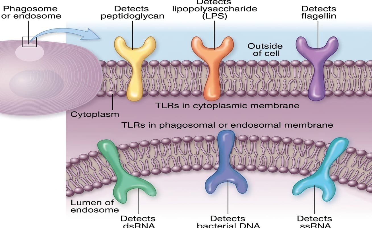
Toll-Like Receptors (TLRs) in Cell and Endosomal Membranes
6. The Complement Cascade: Triggers, Pathways, and Biological Outcomes
The complement system consists of nine major circulating proteins, designated C1 through C9. These proteins circulate in an inactive form within blood and tissue fluids. When triggered by foreign substances, they activate in a rapid, sequential enzymatic cascade.
| Complement Pathway | Triggering Stimulus | Initial Biochemical Assembly |
|---|---|---|
| Alternative Pathway | Spontaneous activation; complement fragment C3b binds directly to foreign microbial surfaces. | C3b deposits directly on the foreign cell wall without needing antibodies. |
| Lectin Pathway | Soluble host pattern-recognition protein called mannose-binding lectin (MBL) binds mannose sugars on microbial invaders. | MBL attaches to repeating carbohydrates on bacterial or fungal surfaces. |
| Classical Pathway | Specific antibodies bind to foreign antigens, forming immune complexes. | Antibody stems expose binding sites that recruit and activate the C1 complex. |
Central Convergence: Formation of C3 and C5 Convertase
All three activation pathways converge at a shared enzymatic step: the formation of C3 convertase.
- Cleavage of C3: C3 convertase splits circulating C3 into two active pieces: C3a (small fragment) and C3b (large fragment).
- Formation of C5 Convertase: The newly made C3b attaches to C3 convertase to form C5 convertase, which splits C5 into C5a and C5b.
| Biological Outcome | Key Complement Mediators | Detailed Physiological Mechanism |
|---|---|---|
| 1. Induction of Inflammation | C3a and C5a (Often termed anaphylatoxins) |
These fragments bind receptors on blood vessel endothelial cells and mast cells, increasing vascular permeability. C5a also serves as a potent chemoattractant that draws flowing phagocytes toward the activation site. C3a and C5a also stimulate phagocyte metabolism and increase their surface C3b receptors. |
| 2. Opsonization | C3b | C3b coats foreign microbial surfaces, acting as a molecular tag (opsonin). Phagocytes carry specific surface C3b receptors that let them easily grip and ingest the coated microbe. |
| 3. Cell Lysis via MAC | C5b, C6, C7, C8, and multiple C9 molecules | Fragment C5b binds the target membrane and recruits C6, C7, and C8. This complex drives the assembly of multiple C9 units into a doughnut-shaped pore called the Membrane Attack Complex (MAC). These pores disrupt membrane integrity, leading to cell lysis. |
Bacterial Sensitivity to the Membrane Attack Complex (MAC)
Gram-Negative Bacteria: These organisms have a thin peptidoglycan layer and an exposed outer lipid bilayer, making them highly susceptible to MAC insertion and lysis.
Gram-Positive Bacteria: These microbes are resistant to MAC lysis. Their thick, multi-layered peptidoglycan cell wall physically blocks complement proteins from reaching and inserting into the inner cytoplasmic membrane.
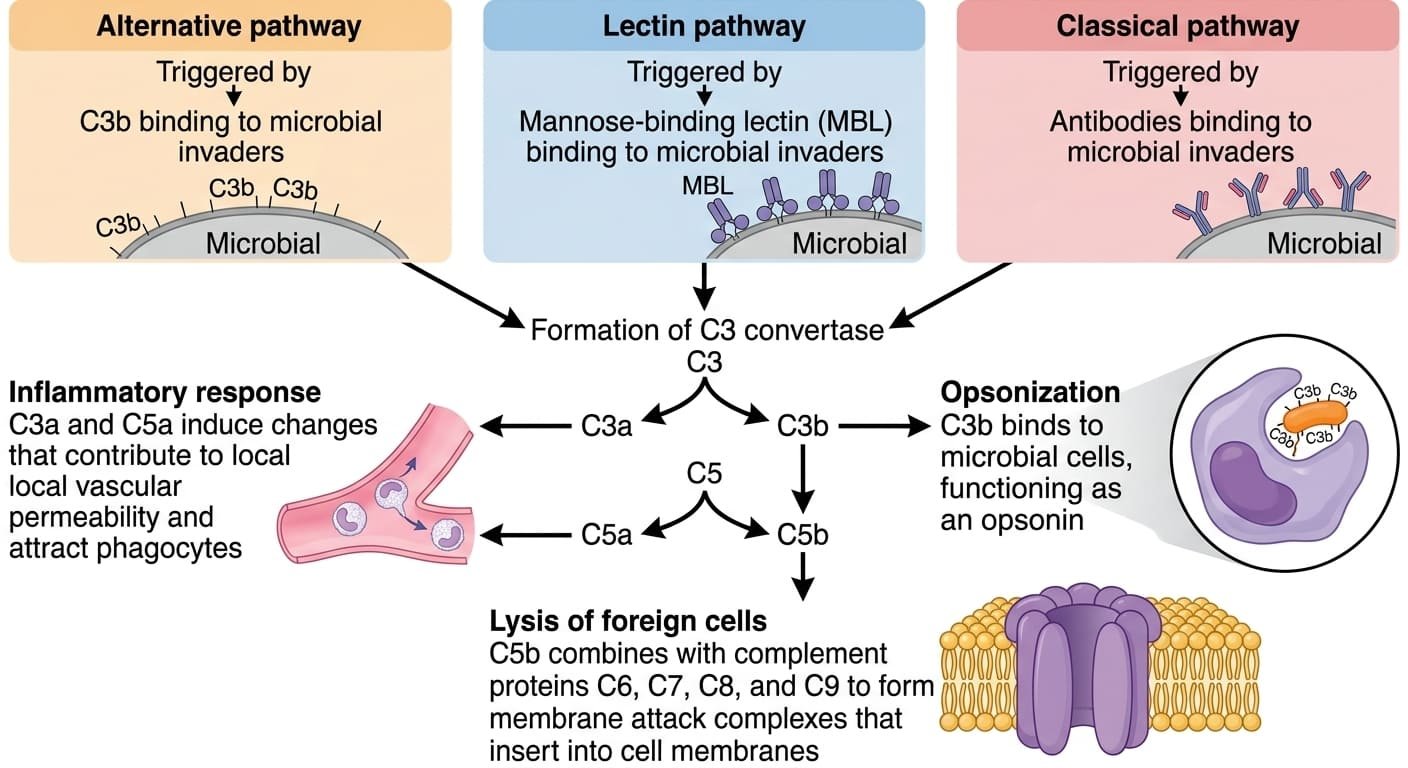
The Complement Activation Pathways and Terminal Biological Outcomes
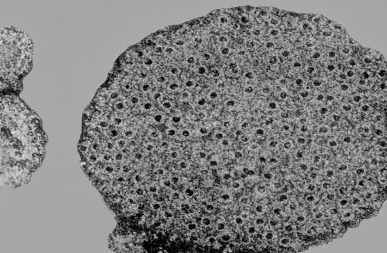
Membrane Attack Complexes (MACs) Formed Within a Target Cell Membrane
7. The Inflammatory Response: Mechanisms, Vascular Changes, and Phagocyte Recruitment
Inflammation is a coordinated response to tissue damage or microbial invasion. Injuries caused by penetrating objects or microbial toxins trigger local defenses designed to isolate the injury, eliminate invaders, and restore tissue function.
The Classic Signs of Inflammation
- Redness (Rubor) & Heat (Calor): Pro-inflammatory chemicals cause local small blood vessels to dilate. This increases local blood flow to the damaged area, while blood flow within capillaries slows.
- Swelling (Tumor): Vessel dilation pulls apart the tight junctions between endothelial cells. Fluid rich in antibodies, complement proteins, and transferrin leaks out of vessels into the surrounding tissue.
- Pain (Dolor): Caused by increased tissue fluid pressure and the direct stimulation of sensory nerve endings by local chemical mediators.
- Loss of Function (Functio Laesa): Can occur when swelling, tissue disruption, and pain prevent normal physiological movement and function.
| Initiating Event | Molecular Mediators Involved | Downstream Inflammatory Effect |
|---|---|---|
| 1. Microbial Molecular Products | Microbial flagellin, LPS, and bacterial DNA binding macrophage TLRs | Macrophages release pro-inflammatory cytokines such as TNF-α. TNF-α stimulates hepatocytes in the liver to synthesize acute-phase proteins, which enhance complement activation and phagocytosis. |
| 2. Microbial Surface Activation of Complement | Generation of complement split products C3a and C5a | Directly trigger mast cell degranulation, releasing histamine and pro-inflammatory cytokines that increase vascular permeability. |
| 3. Physical Tissue Injury | Activation of enzymatic cascades:
|
Prevents blood loss, blocks microbial spread through the body, and triggers localized pain and vascular widening. |
8. The Multistep Leukocyte Extravasation Cascade
Phagocytes travel quickly through normal blood vessels. To reach infected tissues, they complete a coordinated sequence of recruitment steps:
Step-by-Step Extravasation and Migration
- Endothelial Activation & Rolling: Pro-inflammatory cytokines trigger local vessel endothelial cells to make surface adhesion molecules. Circulating phagocytes loosely bind these molecules, slowing down and rolling along the vessel wall until they stop.
- Tight Adherence: Phagocytes synthesize complementary surface adhesion molecules that anchor them securely to the activated endothelial lining.
- Diapedesis (Transmigration): Phagocytes squeeze between adjacent endothelial cells through widened junctions in the dilated vessel wall to enter the surrounding tissue space.
- Chemotactic Migration: Once inside the tissue, phagocytes move toward the injury site along a chemical concentration gradient of chemoattractants (such as C5a, chemokines, and damaged cell phospholipids).
- Sequential Influx:
- Neutrophils (PMNs): The first cells to arrive from the bloodstream, dominating the early inflammatory site.
- Monocytes & Lymphocytes: Arrive later. Monocytes mature in the tissue into large, active macrophages that engulf pathogens and dead neutrophils.
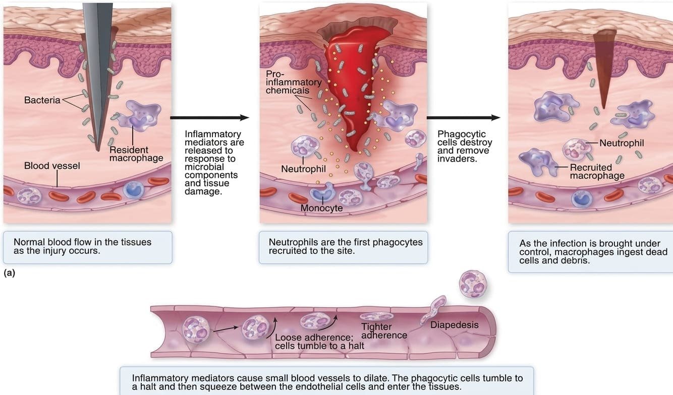
The Acute Inflammatory Response and Leukocyte Diapedesis
9. Resolution, Chronic Inflammation, and Systemic Complications
| Stage / Outcome | Dominant Cells & Structures | Pathological Process & Clinical Features |
|---|---|---|
| Acute Inflammation & Pus Formation | Neutrophils (PMNs), fibrin clots, and tissue debris | Short-term, localized response starting right after injury. Clotting factors seal the wound and trap pathogens. As short-lived neutrophils die in large numbers, their remains mix with cellular debris to form pus. A localized collection of pus forms a boil or abscess. During normal healing, new capillaries grow into the site, damaged tissue is replaced, and a scar forms. |
| Chronic Inflammation & Granulomas | Macrophages, Multinucleated Giant Cells, T-helper cells | Develops when acute inflammation fails to clear an infection. This response can persist for months or years. If activated macrophages cannot digest the pathogen, many macrophages fuse together to form multinucleated giant cells. Macrophages, giant cells, and helper T cells then form tight clusters called granulomas. Granulomas wall off pathogens to keep them from spreading. This illustrates cooperation between innate and adaptive defenses, and is a hallmark of diseases like tuberculosis and histoplasmosis. |
| Host Tissue Damage | Degranulating phagocytes releasing reactive chemicals | Activated phagocytes can release toxic oxygen radicals and digestive enzymes into surrounding tissues. This response can become life-threatening in enclosed, delicate spaces like the meninges of the brain and spinal cord. |
| Septic Shock | Systemic monocytes, macrophages, and endothelial cells | Occurs when Gram-negative bacteria enter the bloodstream and release massive amounts of endotoxin (lipopolysaccharide / LPS). This triggers widespread cytokine release, leading to profound systemic vasodilation, a precipitous drop in blood pressure, widespread capillary microclotting, hypoperfusion, and extensive tissue necrosis. |
10. Cellular Specialization: Macrophages versus Neutrophils
| Comparative Parameter | Tissue Macrophage | Circulating Neutrophil (PMN) |
|---|---|---|
| Normal Physiological Role | Tissue sentinel and scavenger; phagocytoses debris and dead cells; monitors for danger; calls for reinforcements. | Circulating rapid responder; sparse in healthy tissues; rapidly recruited to damage sites. |
| Arrival Timeline | Already resident within tissues; additional cells recruited after the initial neutrophil wave. | First leukocyte recruited from the blood to arrive at damaged or infected tissue. |
| Lifespan & Lysosome Renewal | Long-lived (weeks to months); continuously regenerates lysosomes to maintain killing activity. | Short-lived (1 to 2 days); dies after degranulating and phagocytosing targets. |
| Intrinsic Killing Power | Moderate at baseline; killing capacity increases significantly when activated. | Naturally high baseline killing power, exceeding that of resting and activated macrophages. |
| Activation & Tissue Toxicity | Stimulated by T-helper cell signals (like IFN-γ) to become activated macrophages. They produce large amounts of oxygen radicals and nitric oxide. Because these chemicals damage host tissues, macrophages cannot safely remain activated indefinitely. | Releases enzymes from cytoplasmic granules and uses reactive oxygen bursts to rapidly destroy microbes. |
11. The Six Sequential Steps of Phagocytosis
Phagocytosis is the process by which specialized cells engulf, kill, and digest foreign microorganisms and cellular debris. Understanding each step is clinically important because many pathogens have developed ways to block specific stages of this pathway.
Step-by-Step Mechanism of Phagocytosis
- Chemotaxis: Phagocytes migrate toward the site of damage or infection along a gradient of chemical attractants. These attractants include complement fragment C5a, microbial components, and phospholipids released from injured mammalian cell membranes.
- Recognition and Attachment: Phagocytes use specialized surface receptors to bind targets directly or indirectly:
- Direct Binding: Receptors bind microbial molecules directly. For example, phagocyte mannose receptors bind mannose sugars on yeast and bacterial walls. Surface scavenger receptors bind charged molecules on foreign targets.
- Indirect Binding (Opsonization): Phagocytes attach to targets that have been coated with opsonins. Common opsonins include complement fragment C3b and specific classes of antibodies. Phagocytes carry dedicated surface receptors that bind these opsonin molecules.
- Engulfment: Binding triggers the phagocyte's internal cytoskeleton to rearrange. The cell extends membrane-bound, arm-like projections called pseudopods around the microbe. These pseudopods fuse to enclose the microbe inside a membrane-bound vacuole called a phagosome. (Note: engulfment alone does not kill the microbe).
- Fusion with Lysosome: The phagosome travels along cytoskeletal tracks deep into the cell. It fuses with a digestive vesicle called a lysosome (or a neutrophil granule) to create a single shared chamber: the phagolysosome. Lysosomes contribute digestive enzymes, including lysozymes and proteases.
- Destruction and Digestion: Microbes are destroyed using two coordinated metabolic mechanisms:
- Respiratory Burst / Oxidative Killing: Oxygen uptake spikes as sugars are oxidized through the tricarboxylic acid (TCA) cycle. This produces toxic reactive oxygen species, including superoxide, hydrogen peroxide, hydroxyl radicals, and singlet oxygen.
- Fermentation and Acidification: As available oxygen is consumed, metabolism shifts to fermentation. This generates lactic acid, lowering the internal pH. Acid-activated lysosomal enzymes then break down the bacterial peptidoglycan cell wall and internal components.
- Exocytosis: Following digestion, the membrane-bound vesicle carries leftover debris to the cell's outer surface. It fuses with the plasma membrane, releasing the digested remnants out of the phagocyte.
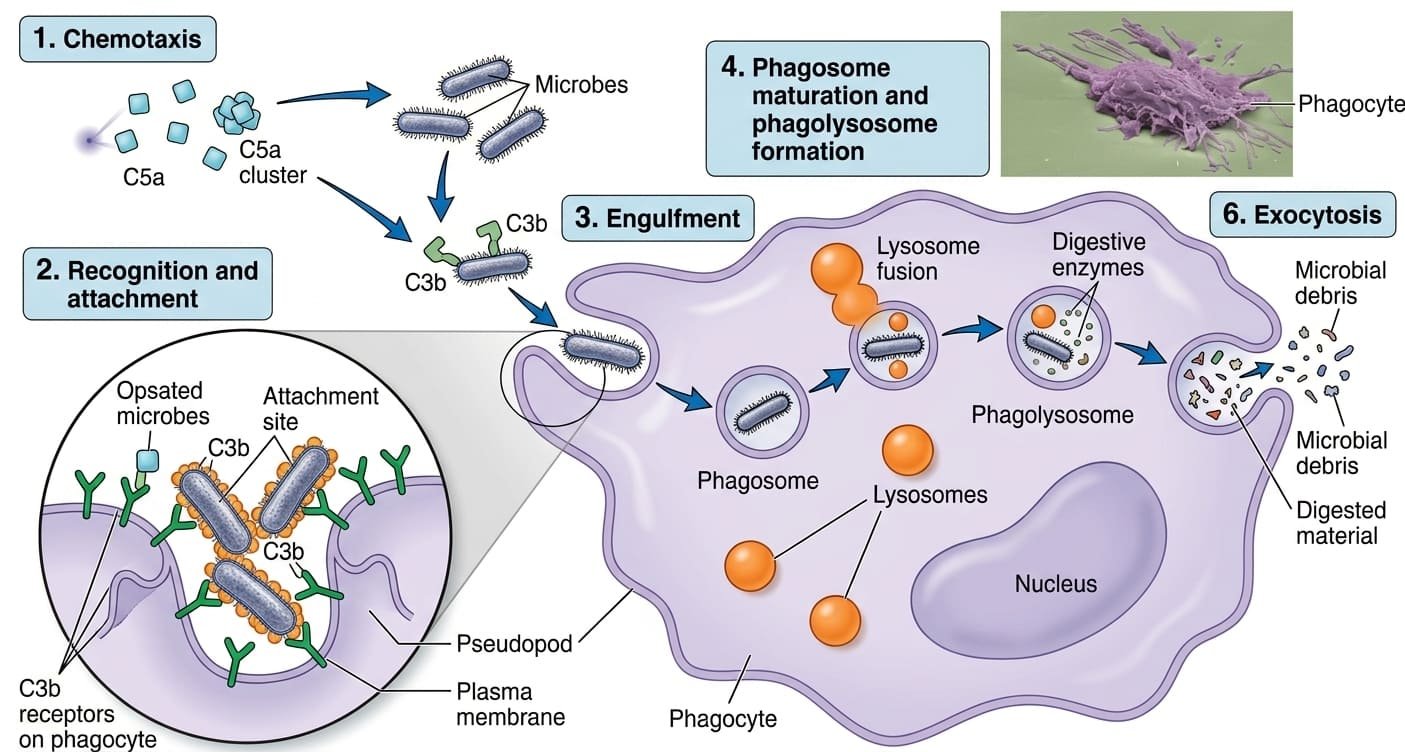
The Sequential Stages of Phagocytosis and Microbial Killing
12. Adaptive Immunity: Humoral Defense and B Lymphocytes
Humoral immunity is carried out by B lymphocytes (B cells), which develop and mature in the bone marrow. This arm protects mainly against extracellular pathogens, such as bacteria and toxins in the blood and lymph.
B-Cell Activation and Clonal Differentiation
- B-Cell Receptor (BCR): B cells display numerous BCRs on their outer membrane. A BCR is a membrane-bound version of the specific antibody that the cell is programmed to produce.
- Antigen Recognition: When a BCR binds its target antigen, the B cell takes in the signal. However, before the B cell can multiply, it typically requires a "second opinion" activation signal from an antigen-specific helper T cell that confirms the target is dangerous.
- Differentiation: Once activated, the B cell proliferates and differentiates into two cell types:
- Plasma Cells: Specialized antibody factories that synthesize and secrete large quantities of soluble, antigen-specific antibodies into blood and lymph.
- Memory B Cells: Long-lived cells that persist in the body for years, enabling a faster, stronger antibody response if the same antigen is encountered again.
13. Antibody Structure and Functional Protection Mechanisms
Antibodies (immunoglobulins) are Y-shaped proteins that bind specific foreign antigens.
| Structural Component | Molecular Characteristics | Functional Activity |
|---|---|---|
| Two Identical Arms (Fab Regions) | Formed by variable amino acid sequences that differ widely among antibody clones. | Provide high antigen-binding specificity. The unique tip shapes let each antibody bind exclusively to its matching target antigen. |
| Stem Region (Fc Region) | Constant amino acid sequence shared within each immunoglobulin class. | Acts as a "red flag" tag. When the arms bind an antigen, the stem recruits and activates other immune components (such as complement proteins and phagocyte Fc receptors) to destroy the target. |
Direct versus Indirect Mechanisms of Antibody Protection
- Direct Mechanism (Neutralization): Antibodies bind directly to toxins or surface spikes on viruses and bacteria. By coating the invader, they physically block it from attaching to host cell receptors, preventing infection or toxin entry.
- Indirect Mechanisms:
- Opsonization: The antibody stem sticks outward from the coated pathogen, giving phagocytes a firm grip via their surface Fc receptors.
- Complement System Activation: The bound antibody stems expose binding sites for C1, launching the classical complement cascade to promote inflammation, further opsonization, and MAC lysis.
14. Adaptive Immunity: Cell-Mediated Defense and T Lymphocytes
Cell-mediated immunity is driven by T lymphocytes (T cells), which develop in the bone marrow and mature within the thymus. This arm targets intracellular pathogens (such as viruses and intracellular bacteria) as well as aberrant host cancer cells. (Note: T cells do not secrete antibodies).
T-Cell Receptor (TCR) Recognition Rules
- Antigen Presentation Required: Unlike B cells, T cells cannot bind free-floating antigens. A T-cell receptor (TCR) only recognizes an antigen if it is processed into short peptide fragments and displayed on the surface of another host cell.
- Two-Signal Activation: T cells require verification from an antigen-presenting cell—especially dendritic cells—confirming that the displayed antigen is dangerous before they begin multiplying.
- Clonal Expansion: Once activated, T cells proliferate into antigen-specific effector T cells and long-lived memory T cells that respond rapidly upon reinfection.
| T-Cell Subpopulation | Core Functional Role | Effector Mechanisms and Actions |
|---|---|---|
| Helper T Cells (CD4+) | Often called the "Generals" of the immune system. | Coordinate broad immune responses by releasing cytokines. They do not directly kill infected cells. Instead, their cytokines activate B cells, stimulate cytotoxic T cells, and activate macrophages to enhance their killing capacity. |
| Cytotoxic T Cells (CD8+) | Often called the "Assassins" of the immune system. | Directly bind and destroy host cells that harbor intracellular viruses, intracellular bacteria, or cancerous mutations. |
15. Intracellular Surveillance, Apoptosis, and Autoimmunity
Because cytotoxic T cells kill host cells, the body uses strict quality control mechanisms to target only infected or abnormal cells while sparing healthy tissue.
Surveillance Mechanisms and Prevention of Autoimmunity
- Internal Quality Inspection: Host cells routinely display short peptide fragments of their cytoplasmic proteins on their surface membranes. Effector cytotoxic T cells inspect these displayed fragments.
- Normal vs. Foreign Peptides: If the cell displays normal self-peptides, inspecting T cells leave it unharmed. If it displays foreign peptides (such as viral proteins), the cytotoxic T cell binds the cell and delivers death packages.
- Induction of Apoptosis: The cytotoxic T cell triggers the target cell to undergo apoptosis (programmed cell suicide). This process stops the virus from using host machinery to assemble new virions. In addition, the dying cell releases unassembled, non-infectious viral parts that recruit phagocytes and stimulate antibody production.
- Autoimmune Disease: If these screening mechanisms break down and the immune system attacks healthy host cells indiscriminately, autoimmune disease develops.
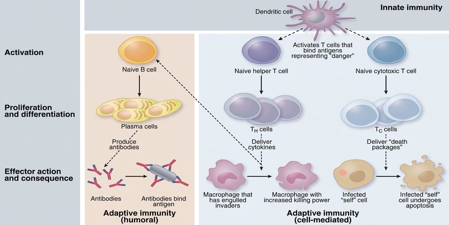
Overview of Adaptive Immune Branches: Humoral Immunity versus Cell-Mediated Immunity
16. Clinical Applications and Pathological Correlations
| Clinical Discipline | Representative Conditions / Therapies | Core Immunological Connection |
|---|---|---|
| Vaccinology | Preventive and therapeutic vaccines | Introduces harmless antigens to stimulate primary adaptive immunity, generating protective memory B and T cells without causing full disease. |
| Allergy & Asthma | Hypersensitivity reactions, allergic asthma, anaphylaxis | Occurs when the immune system mounts inappropriate, exaggerated responses against harmless environmental antigens, driven by IgE, basophils, eosinophils, and mast cells. |
| Autoimmunity | Type 1 Diabetes Mellitus, Rheumatoid Arthritis, Multiple Sclerosis (MS) | Develops when self-tolerance fails and adaptive lymphocytes attack healthy host tissues. |
| Immunodeficiency | Acquired: HIV/AIDS; Congenital: Severe Combined Immunodeficiency (SCID) | Weakened immune components leave the patient vulnerable to frequent, severe opportunistic infections. |
| Cancer Immunotherapy | CAR-T cell therapy, immune checkpoint inhibitors | Harnesses and reprograms the patient's own cytotoxic T cells to overcome tumor suppression and destroy cancer cells. |
| Transplantation Medicine | Solid organ and bone marrow grafting | Uses targeted immunosuppression to prevent recipient T cells and antibodies from recognizing donor tissue antigens and rejecting the graft. |
