Pathological Foundations of Genetic and Epigenetic Disease
1. Fundamental Concepts of Molecular Genetics and Inheritance
Key Definitions in Genetic Pathology
- Gene: The elementary unit of hereditary transmission, consisting of a designated sequence of deoxyribonucleic acid situated at a precise chromosomal address termed a locus. Genes exert their physiological influence by serving as templates that direct the synthesis of functional polypeptides and cellular proteins.
- Genetic Disorder: Any pathological deviation in cellular physiology or anatomical structure originating from an intrinsic molecular aberration within the nuclear or mitochondrial genome. The scale of disruption encompasses microscopic single-base substitutions to macrostructural chromosomal gains, losses, or complex rearrangements.
- Hereditary Disorder: Pathological conditions derived directly from parental germline genetic material, carried within gametes, transmitted transgenerationally, and therefore displaying an inherently familial distribution.
- Congenital Disorder: Any clinical anomaly or malformation physically present at the time of birth, irrespective of whether the underlying etiology is genetic or environmental.
Human disease results from an intricate continuum of genetic alterations, epigenetic modulations, and external environmental exposures. While classical genetics focuses on permanent, physical modifications of the linear nucleotide sequence, epigenetics encompasses chemical alterations to chromatin and associated regulatory ribonucleic acids that dictate transcriptional activity without modifying the underlying genomic code.
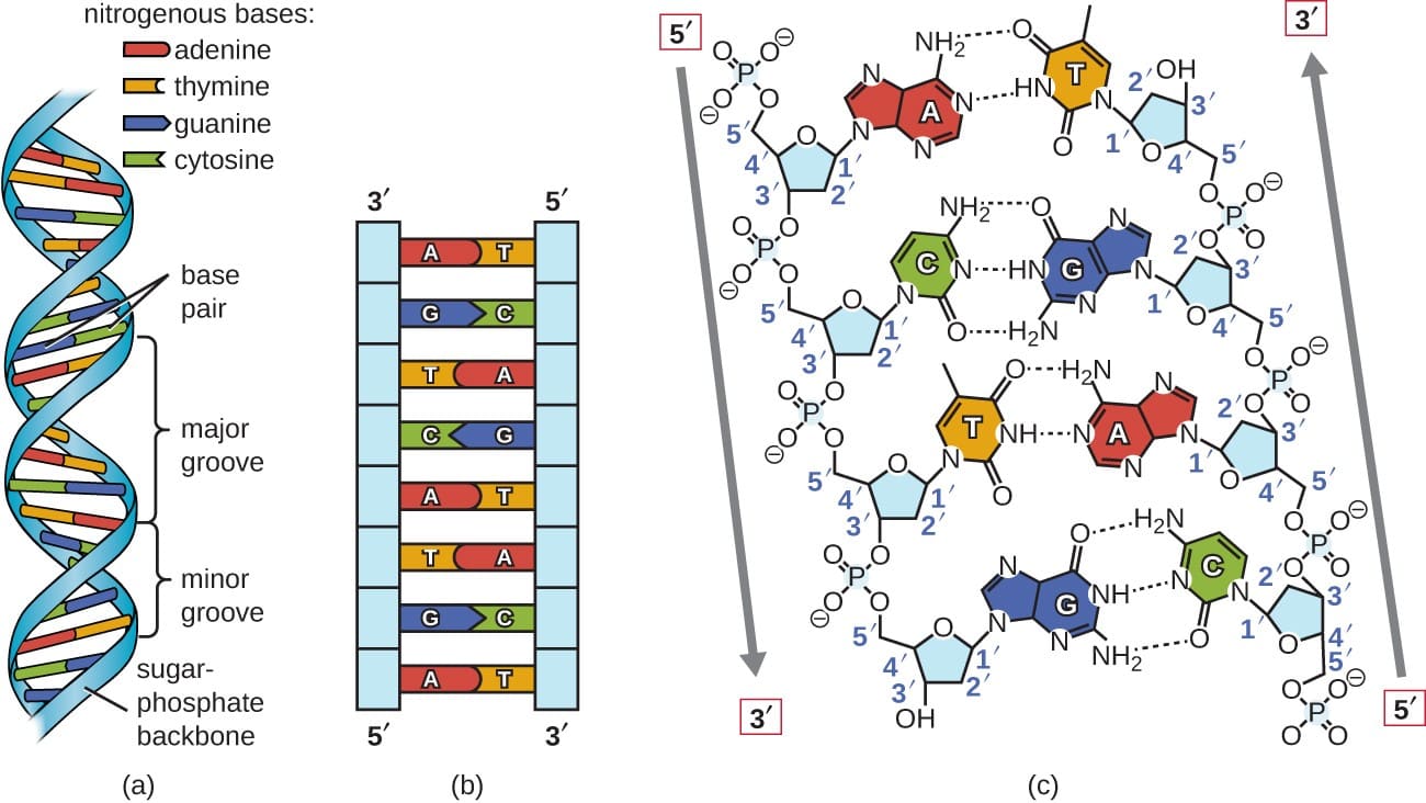
Structural Organization and Base Pairing of the Deoxyribonucleic Acid Double Helix
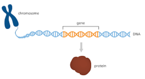
Relationship Between Chromosomal Loci, Linear DNA Sequences, and Polypeptide Synthesis
2. Clinical Differentiation: Hereditary Versus Congenital Pathologies
A crucial principle in diagnostic pathology is recognizing that not all genetic disorders are congenital, and not all congenital conditions are genetic in origin:
| Classification | Etiological Mechanism | Familial Transmission | Representative Clinical Examples |
|---|---|---|---|
| Hereditary (Familial) Genetic Disorders | Heritable alterations in parental germ cells transmitted to offspring via gametes. | Yes; vertical transmission across generational pedigrees. | Cystic fibrosis, Marfan syndrome, Sickle cell anemia, Huntington disease. |
| Acquired (Somatic) Genetic Disorders | Postzygotic mutagenesis confined exclusively to somatic tissue lineages. | No; cannot be transferred to progeny. | Sporadic neoplastic malignancies (e.g., driver mutations in solid organ carcinomas). |
| Congenital Non-Genetic Conditions | Intrauterine teratogenic insults, maternal-fetal infections, or physical trauma during gestation. | No; limited strictly to the exposed fetus. | Congenital syphilis (Treponema pallidum infection), Congenital rubella syndrome, Fetal alcohol spectrum disorders. |
| Delayed-Manifestation Genetic Diseases | Constitutional germline gene defects present at conception that remain asymptomatic for decades until adult onset. | Yes; heritable despite delayed phenotypic expression. | Huntington disease (chorea begins in 4th–5th decade), Autosomal dominant polycystic kidney disease (ADPKD). |
3. Molecular Mechanics of Genomic Aberrations
Structural and functional genomic abnormalities originate from two primary pathways:
- Chromosomal Non-Disjunction: The failure of paired homologous chromosomes or sister chromatids to disjoin appropriately during meiotic or mitotic nuclear division, generating quantitative aneuploidy (monosomy or trisomy).
- DNA Sequence Mutations: Permanent biochemical modifications altering the linear nucleotide order within the genome.
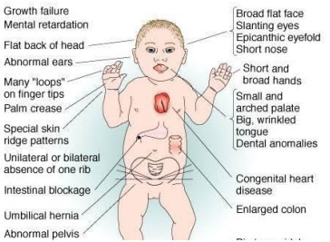
Phenotypic Manifestations of Aneuploidy Induced by Chromosomal Non-Disjunction (Trisomy 21)
Classification of Gene Mutations
| Mutation Category | Biochemical Mechanism | Impact on Translational Product | Archetypal Pathology |
|---|---|---|---|
| Point Mutation: Missense | Single base substitution resulting in an altered codon that specifies an incorrect amino acid residue. | Production of a full-length protein possessing altered physical properties, folding kinetics, or enzymatic capacity. | Sickle Cell Disease: Point mutation in β-globin gene substituting thymine for adenine (GAG → GTG), replacing glutamic acid with valine at position 6. |
| Point Mutation: Nonsense | Single base substitution converting an amino acid-coding codon into a premature stop codon (e.g., UAA, UAG, UGA). | Premature termination of ribosomal translation, producing truncated polypeptides subject to rapid proteasomal or nonsense-mediated decay. | β0-Thalassemia: Premature stop codons disrupting synthesis of the adult β-globin polypeptide chain entirely. |
| Frameshift Mutation | Insertion or deletion of a number of nucleotides that is not a multiple of three (i.e., 1 or 2 base pairs). | Total realignment of the downstream triplet reading frame, yielding an aberrant downstream amino acid sequence terminating in a premature stop codon. | Tay-Sachs Disease: A four-base-pair insertion in exon 11 of the HEXA gene causing catastrophic frame disruption in the α-subunit of β-hexosaminidase A. |
| Trinucleotide Repeat Expansion | Amplification of unstable tandem 3-base repeating units exceeding the physiological threshold during DNA replication. | Toxic gain-of-function protein aggregates, structural mRNA entrapment, or gene promoter hypermethylation and transcriptional silencing. | Fragile X Syndrome: Amplification of CGG repeats (250–4000 repeats vs. ~29 normal) in the 5'-untranslated region of the FMR1 gene. Huntington Disease: Expansion of CAG repeats (>35 vs. 11–34 normal) in the HTT gene. |
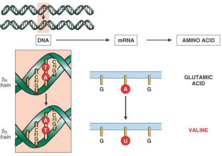
Biochemical Mechanism of a Missense Point Mutation Illustrated via the Sickle β-Globin Gene
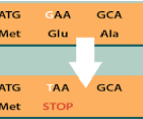
Translational Halting Induced by a Point Mutation Generating a Premature Termination Codon
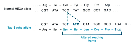
Frameshift Alteration Driven by Base Insertion in the Human HEXA Gene
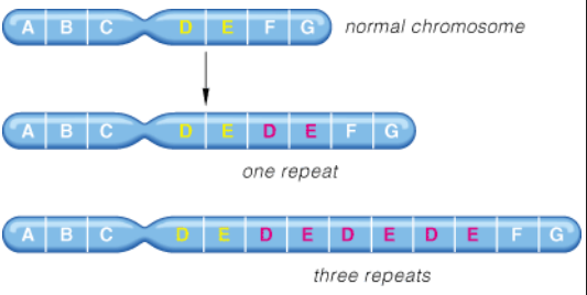
Dynamic Meiotic Expansion of Unstable Triplet Repeat Sequences
Pliotropy, Heterogeneity, and Anticipation
Pleiotropy: A phenomenon wherein a single pathogenic gene mutation culminates in multiple, seemingly unrelated phenotypic and anatomical consequences across distinct organ systems. Clinical Example: In Marfan syndrome, a solitary mutation in the fibrillin-1 (FBN1) gene causes defects spanning the skeletal system (arachnodactyly, pectus excavatum, tall stature), ocular system (ectopia lentis), and cardiovascular system (cystic medial necrosis, aortic root dilatation, catastrophic aortic dissection).
Genetic Heterogeneity: A circumstance where multiple distinct mutations, occurring either at different loci within the same gene (allelic heterogeneity) or across completely different genes (locus heterogeneity), produce an identical clinical disease phenotype. Clinical Example: Retinitis pigmentosa can be precipitated by defects across dozens of independent genetic loci.
Genetic Anticipation: A clinical feature characteristic of trinucleotide repeat disorders wherein the pathology manifests at progressively earlier ages and with escalating clinical severity in successive generations. In Huntington disease, profound CAG expansion occurs preferentially during paternal spermatogenesis, leading to juvenile-onset presentations when passed through the male germline.
4. Mendelian Patterns of Monogenic Disease
Monogenic (single-gene) conditions adhere to classical Mendelian transmission dynamics, categorized into three distinct modes: Autosomal Dominant, Autosomal Recessive, and X-Linked inheritance.
4.1 Autosomal Dominant Disorders
- Genotypic Definition: Clinical pathology is fully manifested in the heterozygous state; inheriting a solitary mutant allele from either parent is sufficient to trigger phenotypic expression.
- Pedigree Characteristics: Vertical transmission is evident; every affected proband typically possesses at least one affected parent. Both sexes are afflicted in equal proportions, and male-to-male transmission is readily observed.
- Recurrence Risk: When an affected heterozygote mates with an unaffected homozygote, each conception carries a 50% (1 in 2) probability of inheriting the disease allele.
- De Novo (New) Mutations: A patient may present with an autosomal dominant disease despite having clinically and genetically normal parents. This arises from a new spontaneous mutational event during gametogenesis in the parental egg or sperm. The patient's siblings remain at baseline population risk, while the patient's future offspring will face the standard 50% recurrence risk.
- Modifiers of Dominant Phenotypes:
- Reduced Penetrance: An individual inherits the mutant pathogenic allele but remains completely devoid of clinical manifestations (all-or-none statistical measure).
- Variable Expressivity: The pathogenic trait is expressed across all carriers of the mutant gene, but the severity and organ-specific profile diverge markedly between individuals.
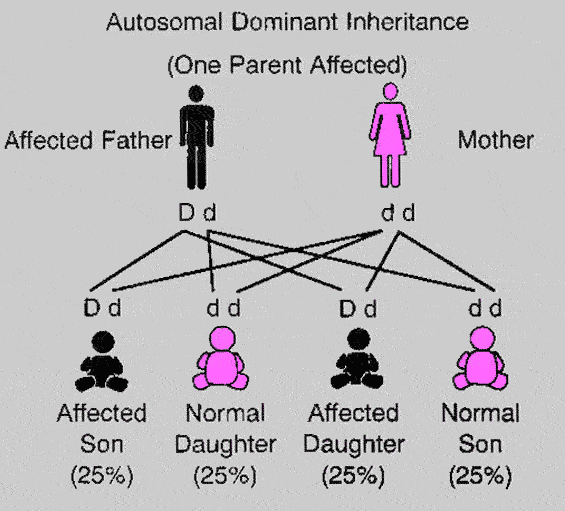
Classic Inheritance Pedigree of Autosomal Dominant Transmission
Hallmark Autosomal Dominant Syndromes
Marfan Syndrome: Arises from mutations within the FBN1 gene on chromosome 15q21, which encodes the glycoprotein fibrillin-1, an essential scaffold for extracellular elastin deposition. Defective microfibrils destabilize structural matrix elements and trigger excess bioavailability of active Transforming Growth Factor-β (TGF-β). Clinical signs include disproportionate skeletal elongation (arachnodactyly, pectus excavatum, dolichostenomelia), joint hypermobility, upward/outward dislocation of the ocular lens (ectopia lentis), and cardiovascular fragility (mitral valve prolapse, ascending aortic aneurysms, and fatal aortic dissection).
Familial Hypercholesterolemia (FH): Initiated by loss-of-function mutations in the LDLR gene (encoding the low-density lipoprotein receptor), or less frequently via gain-of-function lesions in PCSK9 or mutations in APOB. Impaired receptor-mediated hepatic clearance of circulating intermediate-density lipoprotein (IDL) and LDL leads to profound hypercholesterolemia. Diagnostic physical signs include:
- Tendon Xanthomas: Firm nodular cholesterol aggregates situated on extensor tendons (particularly the Achilles tendon and extensor tendons of the hands; highly specific for FH).
- Xanthelasmas: Soft, yellow, lipid-laden plaques deposited along the inner canthi and periorbital subcutaneous tissue of the eyelids.
- Corneal Arcus (Arcus Senilis): Opaque grayish-white stromal lipid ring circumscribing the corneal periphery (highly specific when appearing in patients <45 years of age).
- Accelerated systemic atherosclerosis precipitating premature coronary artery disease, myocardial infarction, and stroke at an early age.
Autosomal Dominant Polycystic Kidney Disease (ADPKD): Precipitated by pathogenic mutations in PKD1 (encoding polycystin-1, representing ~85% of cases) or PKD2 (encoding polycystin-2, ~15%). Polycystins are localized to the primary cilia of renal tubular epithelia, governing cellular mechanosensation, cell-matrix communication, and intracellular calcium signaling. Defective signaling induces uncontrolled tubular epithelial proliferation, fluid hypersecretion, and progressive bilateral enlargement of kidneys replaced by thousands of fluid-filled cysts. Symptoms include severe flank pain, secondary hypertension, microscopic/gross hematuria, and inevitable progression to end-stage renal disease (ESRD) requiring chronic hemodialysis or renal allograft transplantation.
Hereditary Spherocytosis: Caused by diverse molecular lesions affecting red blood cell membrane skeletal scaffold proteins: ankyrin (ANK1), α-spectrin (SPTA1), β-spectrin (SPTB), band 3 anion transport protein (SLC4A1), and protein 4.2 (EPB42). Destabilization of the erythrocyte vertical cytoskeletal linkages provokes continuous membrane microvesiculation and loss of surface-area-to-volume ratio, forcing the cell into an inflexible spherical shape (spherocyte). These mechanically rigid cells undergo premature sequestration and extravascular lysis within splenic cords, producing a classic triad of chronic hemolytic anemia, fluctuating hyperbilirubinemic jaundice, and splenomegaly. Management involves folic acid supplementation, blood transfusions, and therapeutic splenectomy.
Familial Adenomatous Polyposis (FAP): Driven by a heterozygous constitutional germline mutation in the adenomatous polyposis coli (APC) tumor suppressor gene on chromosome 5q21. Under physiological conditions, APC facilitates the proteasomal degradation of β-catenin; loss of APC permits constitutive nuclear translocation of β-catenin and permanent activation of the WNT proliferative signaling pathway. Affected individuals develop hundreds to thousands of adenomatous polyps carpeting the mucosal surface of the colorectal tract starting during second/third decades of life, carrying a 100% inevitable risk of progression to invasive colorectal adenocarcinoma if prophylactic total proctocolectomy is not performed.
Huntington Disease (HD): An autosomal dominant neurodegenerative movement disorder provoked by a pathogenic CAG trinucleotide repeat expansion within the HTT gene on chromosome 4p16.3, encoding an elongated polyglutamine tract in the huntingtin protein. Wild-type alleles harbor 11 to 34 copies, whereas disease manifests when repeat copy numbers expand to ≥36 (often into the hundreds). The abnormal protein undergoes misfolding and accumulates as toxic intranuclear aggregates, precipitating selective, progressive neuronal apoptosis in the caudate nucleus and putamen (striatum), alongside cortical atrophy. Clinically, patients manifest choreiform involuntary movements, psychiatric disturbances, executive cognitive decline, and progressive dementia.
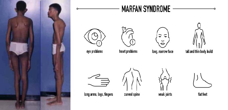
Phenotypic Spectrum and Structural Abnormalities Characterizing Marfan Syndrome
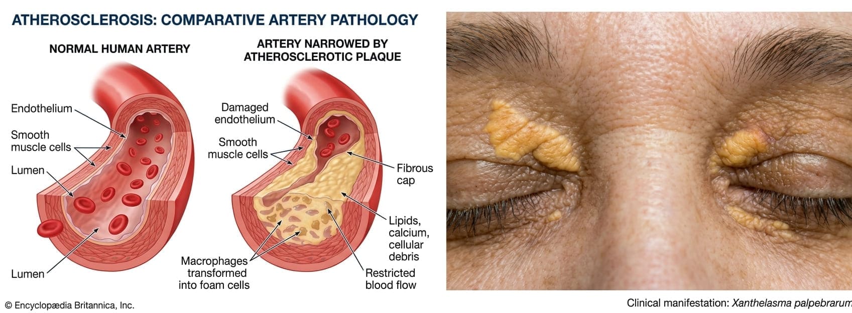
Pathological Vascular Alterations and Ophthalmic Lipid Deposition in Severe Hypercholesterolemia
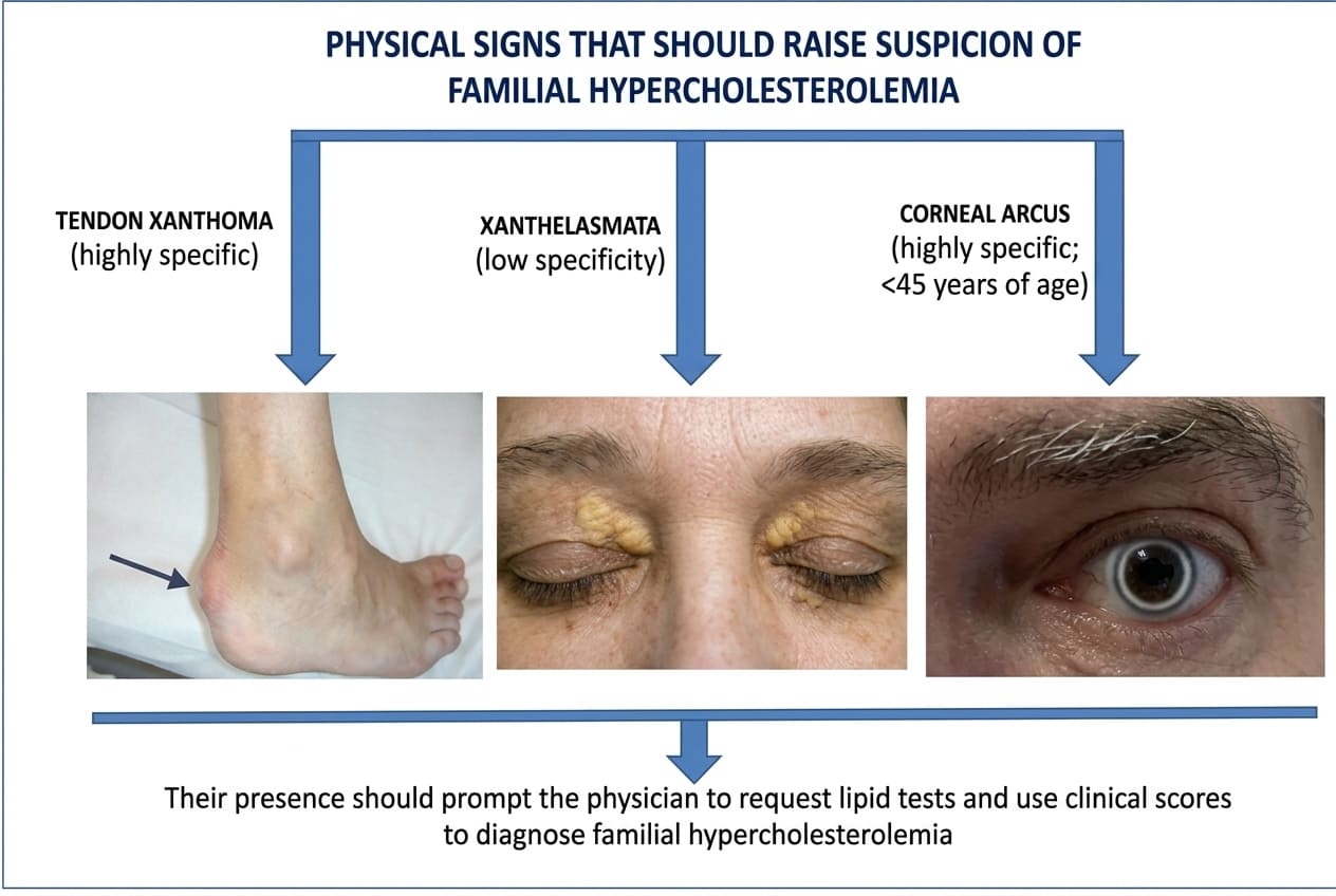
Pathognomonic Cutaneous and Ocular Stigmata Prompting Clinical Evaluation for Familial Hypercholesterolemia
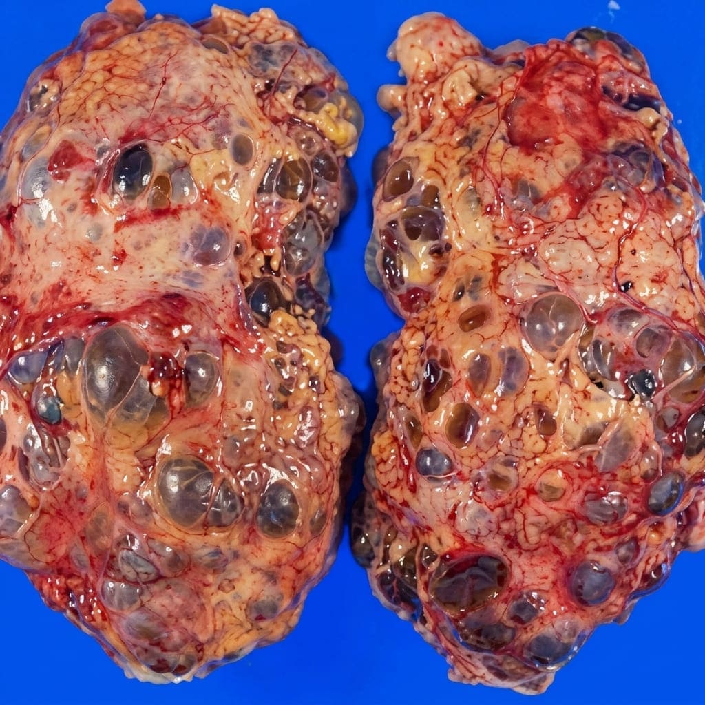
Gross Macroscopic Renal Architecture in Advanced Autosomal Dominant Polycystic Kidney Disease
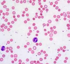
Microscopic Morphology of Peripheral Spherocytes Lacking Central Pallor in Hereditary Spherocytosis
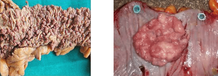
Diffuse Colonic Polyposis Progressing to Invasive Mucinous Carcinoma in APC Gene Mutation
4.2 Autosomal Recessive Disorders
- Epidemiological Scale: Constitute the largest unified category of Mendelian disorders in clinical practice.
- Molecular Basis: Pathology is expressed only in individuals who are homozygous or compound heterozygous for deleterious alleles; pathogenic mutations must disrupt function in both alleles of the gene.
- Pedigree and Parental Carrier Dynamics: Parents of an affected proband are obligate heterozygous carriers who possess one normal functioning allele and one mutated copy; because the physiological output of the remaining wild-type allele compensates, carriers remain entirely asymptomatic. Consanguinity markedly increases the likelihood of mating between two carriers of identical rare recessive mutations.
- Recurrence Probability: A union between two heterozygous carriers produces offspring with a 25% (1 in 4) chance of being affected, a 50% chance of being an asymptomatic carrier, and a 25% chance of being a homozygous normal non-carrier.
- Sex Distribution and Clinical Onset: Affects males and females in equal distributions because the affected loci reside on autosomal chromosomes. Unlike dominant diseases, autosomal recessive conditions frequently present with an early, uniform childhood onset and typically involve inborn errors of metabolism or structural enzymatic deficiencies.
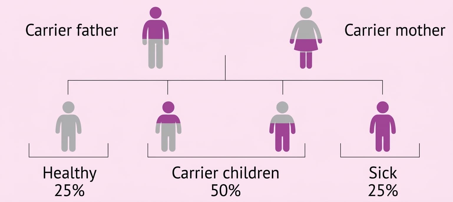
Mendelian Segregation Ratios Resulting from Mating Between Two Heterozygous Recessive Carriers
Hallmark Autosomal Recessive Syndromes
Cystic Fibrosis (CF): A severe, multi-organ disorder resulting from autosomal recessive mutations within the CFTR (Cystic Fibrosis Transmembrane Conductance Regulator) gene, mapped and cloned on chromosome 7q31.2 in 1989. CFTR is an ATP-binding cassette transporter operating as an epithelial, cyclic AMP-regulated chloride channel that governs transcellular chloride and sodium transport across mucosal surfaces.
- Respiratory & Exocrine System: Loss of functional CFTR leads to failure of chloride secretion and compensatory hyperabsorption of sodium and water via epithelial sodium channels (ENaC), severely dehydrating luminal fluid layers and precipitating the secretion of abnormally thick, viscous, tenacious mucus. This inspissated mucus plugs the pulmonary bronchial tree, precipitating recurrent, intractable endobronchial infections (e.g., Pseudomonas aeruginosa), progressive bronchiectasis, and respiratory failure. In the pancreas, ductal obstruction causes acinar atrophy, exocrine pancreatic insufficiency, and malabsorption.
- Hepatobiliary Pathophysiology: Defective CFTR in biliary cholangiocytes causes hyperviscous, inspissated bile that plugs intrahepatic bile ducts. Chronic focal biliary obstruction triggers progressive portal inflammation, periductal fibrosis, and development of biliary cirrhosis characterized by macroscopic nodular remodeling (macronodules measuring >3 mm). End-stage liver disease represents the third most frequent cause of mortality in cystic fibrosis patients, with orthotopic liver transplantation serving as the sole curative intervention for hepatic failure.
Sickle Cell Disease (SCD): Caused by a homozygous missense mutation in the β-globin gene (HBB), generating abnormal hemoglobin S (HbS). Upon deoxygenation, HbS molecules undergo polymer assembly into rigid, crystalline filaments that distort erythrocytes into rigid, sickle-shaped configurations. These fragile cells suffer intravascular and extravascular hemolysis (chronic hemolytic anemia, scleral icterus, cholelithiasis) and become mechanically lodged in the microvasculature, inducing widespread vaso-occlusion, tissue ischemia, and excruciating pain crises. Highly prevalent among ancestral populations of African, Mediterranean, Middle Eastern, and Indian descent. Clinical hallmarks begin in early infancy with dactylitis (painful microvascular swelling of the hands and feet), splenic autoinfarction, and susceptibility to encapsulated bacterial infections. Management incorporates oral folic acid, prophylactic penicillin administration, immunizations, hydroxyurea, and emerging CRISPR-Cas9-mediated gene therapies.
Tay-Sachs Disease: A catastrophic neurodegenerative lysosomal storage disorder caused by loss of function in the HEXA gene on chromosome 15q23, resulting in complete deficiency of the enzyme β-hexosaminidase A. Unable to catabolize GM2 gangliosides within lysosomes, lipid compounds accumulate toxically inside central nervous system neurons, inducing cellular swelling, dendritic vacuolization, and progressive neuronal death. Infants present after several months of normal development with rapid developmental regression, motor weakness, hyperacusis (startle response), spasticity, generalized seizures, macroscopic blindness accompanied by pathognomonic ophthalmoscopic cherry-red macular spots, and fatal outcomes typically by age 4 to 5. Carrier identification and prenatal diagnostic amniocentesis remain central preventive tools.
The Thalassemias: A heterogeneous constellation of hemoglobin synthesis disorders caused by mutations that quantitatively downregulate or abolish the production of α- or β-globin chains.
- Thalassemia Trait (Thalassemia Minor): Heterozygous state wherein an individual carries a solitary defective globin allele. Subjects present with microcytic, hypochromic red cell indices and mild asymptomatic anemia that rarely requires intervention, but carrier detection is vital to prevent unions at risk of conceiving offspring with severe Thalassemia Major (Cooley's anemia or Hemoglobin Barts hydrops fetalis).
Hereditary Hemochromatosis: An autosomal recessive inborn error of iron metabolism precipitated primarily by pathogenic mutations within the HFE gene (predominantly C282Y and H63D missense substitutions) on chromosome 6p21. Disruptions in HFE downregulate the synthesis of hepcidin, the master iron-regulatory hormone secreted by hepatocytes. Low hepcidin allows unrestricted ferroportin-mediated dietary iron absorption across the duodenal enterocyte basolateral membrane, accumulating total body iron stores to 20–50 grams (normal: ~3–4 grams). Over decades, excess iron catalyzes Fenton reactions and free radical formation, depositing as hemosiderin within parenchyma of the liver (micronodular cirrhosis, hepatocellular carcinoma risk), heart (dilated or restrictive cardiomyopathy, arrhythmias), endocrine pancreas (bronze diabetes mellitus), joints (destructive arthropathy), and skin (hyperpigmentation). Management centers on regular therapeutic phlebotomy to deplete iron stores, coupled with dietary iron and alcohol restriction.
Phenylketonuria (PKU): Caused by a constitutional homozygous deficiency in the hepatic enzyme phenylalanine hydroxylase (PAH). Inability to convert dietary phenylalanine into tyrosine forces toxic accumulation of phenylalanine and alternate metabolites (phenylpyruvate, phenyllactate) within the developing brain, triggering profound irreversible mental retardation, microcephaly, hypopigmentation (due to impaired melanin synthesis), and a mousy body odor. Managed through strict lifelong dietary limitation of dietary phenylalanine initiated immediately after neonatal screening.
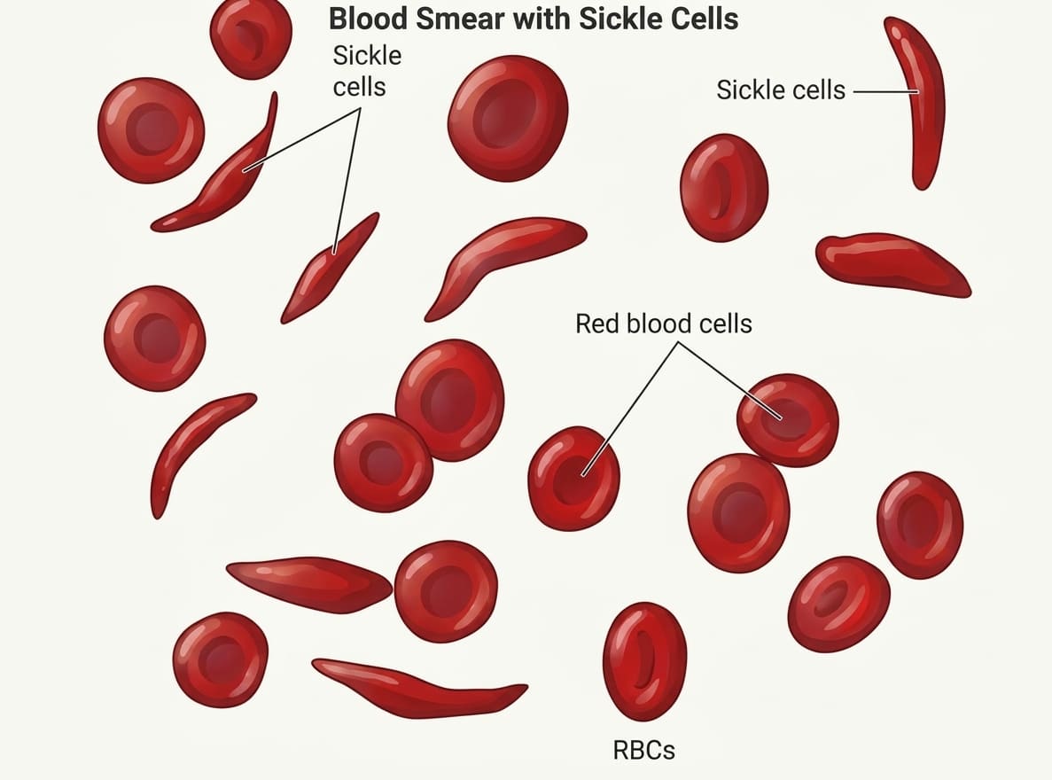
Erythrocyte Morphological Alterations and Vaso-Occlusive Sickled Cells
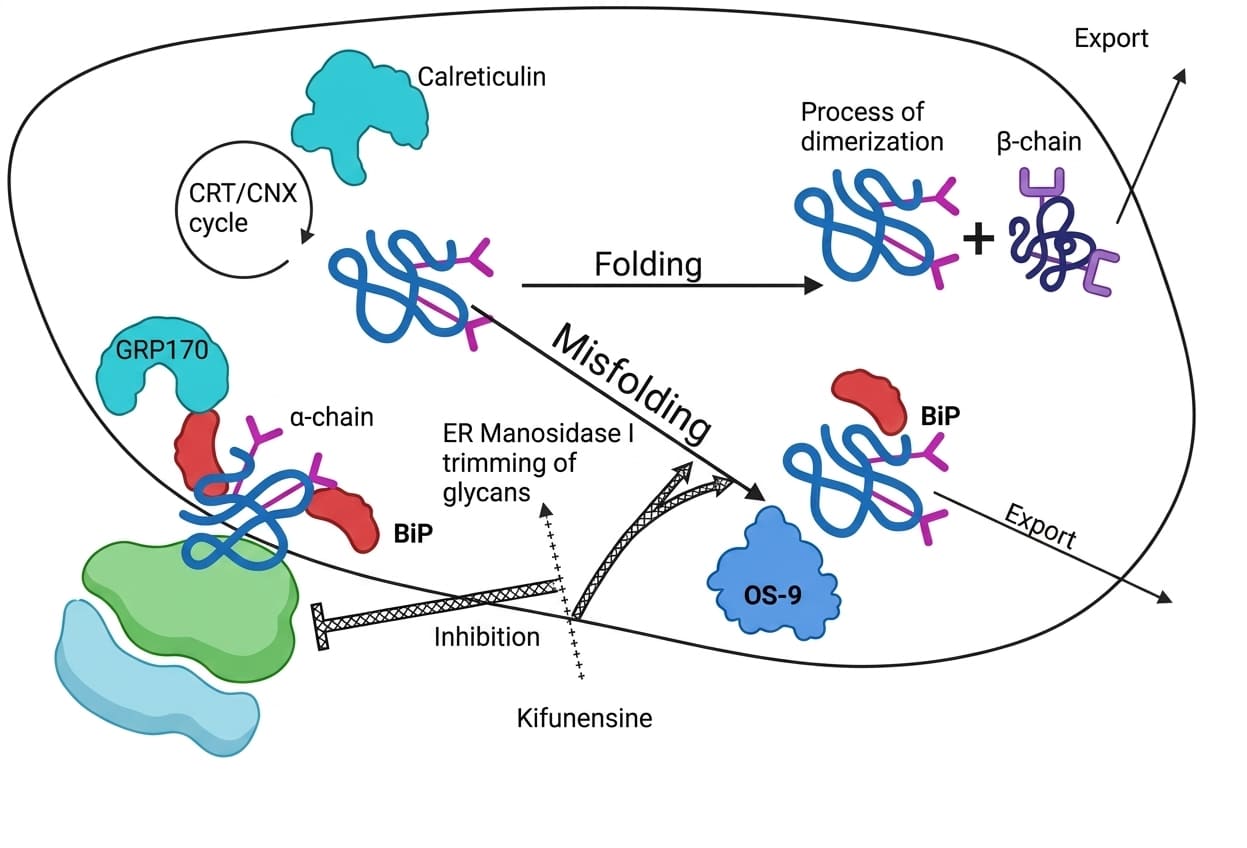
Endoplasmic Reticulum Quality Control, Misfolding, and Degradation of Mutant Polypeptides in Tay-Sachs Disease
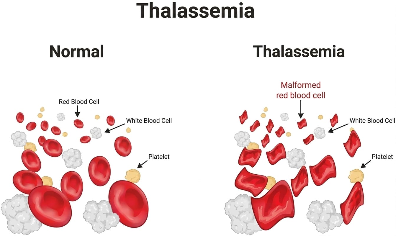
Comparative Morphological Evaluation of Normal Versus Microcytic Hypochromic Thalassemic Red Cells
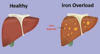
Macroscopic Hepatic Hemosiderosis and Cirrhotic Transformation in Hereditary Hemochromatosis
4.3 X-Linked Recessive Transmission
- Chromosomal Mechanism: Dictated by mutations in genetic loci located exclusively on the X chromosome. Because biological males inherit a single maternal X chromosome paired with a paternal Y chromosome, they are hemizygous for all X-linked loci.
- Gender Predilection: The clinical disease phenotype is expressed overwhelmingly in hemizygous males, who lack a second homologous allele to mask the deleterious mutation. Affected males cannot transmit the mutated X chromosome to their sons (absence of male-to-male transmission), but obligately transmit it to 100% of their daughters, who become heterozygous carriers.
- Female Carrier Dynamics & Lyonization: Females possessing one mutant allele are usually asymptomatic carriers due to the presence of a normal paired allele. However, heterozygous females may clinically manifest variable disease symptoms under two specific genetic circumstances:
- Skewed (Unfavorable) X-Inactivation (Lyonization): Epigenetic condensation of the normal wild-type maternal or paternal X chromosome occurs preferentially during early embryonic blastocyst development, leaving the mutant-bearing X chromosome active in the majority of somatic tissues.
- Compound Homozygosity: The exceedingly rare scenario where a carrier female mates with an affected hemizygous male, inheriting mutant alleles from both parents.
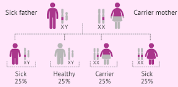
Pedigree Dynamics and Offspring Segregation in X-Linked Recessive Inheritance
Hallmark X-Linked Recessive Syndromes
Red-Green Color Blindness (Daltonism): An X-linked recessive sensory deficit caused by mutations or unequal crossover deletions within the opsin genes (OPN1LW and OPN1MW) located at chromosomal locus Xq28, which encode the photopigment apoproteins in retinal red- and green-sensitive cone photoreceptor cells. As a direct consequence of hemizygosity, the trait affects approximately 9% of males, but only 1% of females in the general population. Clinical diagnosis involves Ishihara pseudoisochromatic plate testing, with management focusing on specialized chromatic-filtering optical lenses.
Hemophilia A: A life-threatening bleeding diathesis caused by mutations (inversions, point mutations, nonsense codons) in the F8 gene at locus Xq28, resulting in a quantitative deficiency or absolute absence of procoagulant Factor VIII within the intrinsic coagulation pathway. Diminished Factor VIII impairs tenase complex assembly and activation of Factor X, preventing normal thrombin generation and fibrin clot stabilization. Severity correlates with residual functional factor levels: severe disease (<1% activity) presents in infancy with spontaneous hematomas, easy ecchymosis, intractable post-surgical hemorrhages, recurrent debilitating joint bleeding (hemarthrosis causing joint ankylosis and crippling arthropathy), and fatal intracranial hemorrhages. Managed via recombinant Factor VIII replacement therapy and non-factor bispecific antibodies (e.g., emicizumab).
Duchenne Muscular Dystrophy (DMD): A severe, progressive, lethal muscle-wasting disease of childhood driven by loss-of-function frameshift mutations or large deletions in the massive DMD gene situated at locus Xp21.2 (spanning 2.4 megabases). The gene encodes dystrophin, a structural cytoskeletal protein that links internal subsarcolemmal actin filaments to the extracellular laminin matrix via the transmembrane dystroglycan-sarcoglycan glycoprotein complex, functioning as a vital shock-absorber during mechanical muscle contraction. Absence of dystrophin compromises myofiber sarcolemmal integrity, allowing toxic extracellular calcium influx, hyperactivation of intracellular proteases, myofiber necrosis, chronic inflammation, and ultimate replacement of skeletal muscle by dense fibrofatty tissue. Boys display early delayed gross motor milestones, clumsy frequent falling, a waddling gait, pseudohypertrophy of the calf muscles (fat and connective tissue deposition replacing atrophied muscle), and the classic Gowers sign (using the arms to push against the legs and 'climb up' the body to achieve an upright posture from the floor). Progressive weakness forces wheelchair dependence by early adolescence, with death occurring in the second or third decade from progressive dilated cardiomyopathy or diaphragmatic respiratory failure.
Fragile X Syndrome: The second most common genetic etiology of intellectual impairment after Down syndrome, caused by massive expansion of a dynamic unstable CGG trinucleotide repeat in the 5'-untranslated promoter region of the FMR1 gene at Xq27.3. When the CGG copy number exceeds 200–230 repeats (full mutation; normal copy number is ~29, expanding to 250–4000 repeats), the expansion triggers extensive promoter hypermethylation, recruiting histone deacetylases that condense chromatin and abolish production of Fragile X Mental Retardation Protein (FMRP). FMRP is an RNA-binding translation regulator in dendritic spines; its absence arrests synaptic pruning and neuroplasticity. Clinical features include moderate to severe intellectual disability, macroorchidism (enlarged testes evident post-puberty), facial dysmorphisms (long narrow face, large prominent ears, prominent jaw), joint hypermobility, high-arched palate, mitral valve prolapse, and behavioral spectrum disorders (autistic features, severe social anxiety, hand-flapping).
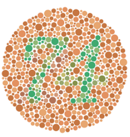
Ishihara Pseudoisochromatic Color Vision Plate for Detecting X-Linked Cone Photoreceptor Dysfunctions
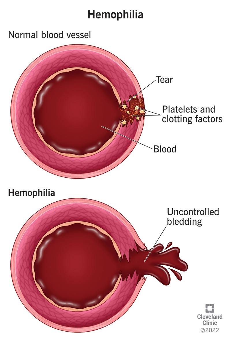
Disruption of Intrinsic Clotting Factor Cascades and Uncontrolled Bleeding in Hemophilia
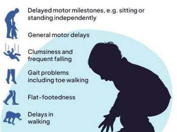
Motor Milestones Regression and Compensatory Gowers Sign in Duchenne Muscular Dystrophy
5. Master Reference Table of Classical Mendelian Pathologies
| Mendelian Inheritance Class | Disease Entity | Abnormal Polypeptide / Protein Product | Native Physiological Role |
|---|---|---|---|
| Autosomal Dominant | Familial Hypercholesterolemia | Low-Density Lipoprotein Receptor (LDLR) | Receptor-mediated endocytosis and hepatic clearance of LDL cholesterol |
| Marfan Syndrome | Fibrillin-1 | Extracellular matrix microfibrillar structural scaffolding and TGF-β sequestration | |
| Ehlers-Danlos Syndrome (select variants) | Collagen (Type I, III, or V chains) | Extracellular matrix mechanical tensile strength in skin, joints, and vascular walls | |
| Hereditary Spherocytosis | Spectrin (α/β), Ankyrin, or Protein 4.1 / 4.2 | Erythrocyte cytoskeleton stabilization, deformability, and membrane anchoring | |
| Neurofibromatosis Type 1 (NF-1) | Neurofibromin-1 | GTPase-activating protein (GAP) acting as a negative regulator of RAS proto-oncogene signaling | |
| Adult Polycystic Kidney Disease (ADPKD) | Polycystin-1 (PKD1) or Polycystin-2 (PKD2) | Ciliary mechanosensory calcium channels regulating tubular cell proliferation and differentiation | |
| Autosomal Recessive | Cystic Fibrosis (CF) | CFTR Transmembrane Regulator | cAMP-dependent epithelial chloride and bicarbonate channel |
| Phenylketonuria (PKU) | Phenylalanine Hydroxylase (PAH) | Hepatic catalytic conversion of phenylalanine into tyrosine | |
| Tay-Sachs Disease | β-Hexosaminidase A (α-subunit) | Lysosomal degradation and cleavage of GM2 gangliosides in central nervous system neurons | |
| α- and β-Thalassemias | Hemoglobin α- or β-Globin Subunits | Heterotetrameric adult hemoglobin (HbA: α2β2) assembly for oxygen transport | |
| Sickle Cell Anemia | Hemoglobin β-Globin (HbS Variant) | Reversible systemic oxygen delivery without polymerization or membrane sickling | |
| X-Linked Recessive | Hemophilia A | Coagulation Factor VIII | Essential cofactor for Factor IXa-mediated activation of Factor X in the intrinsic pathway |
| Duchenne Muscular Dystrophy | Dystrophin | Sarcolemmal-cytoskeletal linking protein dissipating contractile mechanical stress | |
| Fragile X Syndrome | Fragile X Mental Retardation Protein (FMRP) | RNA-binding translational suppressor modulating synaptic structure and plasticity |
6. Epigenetic Modifications in Human Pathology
Epigenetics Defined
The scientific discipline investigating heritable, reversible changes in gene expression and transcriptional competence that occur without altering the underlying linear sequence of adenine, cytosine, guanine, and thymine nucleotides. Epigenetic mechanisms establish and stabilize distinct cellular phenotypes across differentiation, development, and environmental stress.
Major Molecular Epigenetic Mechanisms
1. DNA Methylation: The enzymatic addition of a methyl group (-CH3) from S-adenosylmethionine (SAM) onto the carbon-5 position of cytosine residues, forming 5-methylcytosine. This reaction is catalyzed by DNA methyltransferases (DNMTs) and occurs almost exclusively in symmetrical dinucleotide clusters termed CpG islands, which populate the promoter and first exon regions of approximately 60% of human genes. Hypermethylation of promoter CpG islands physically impedes the binding of basic transcription factors and recruits methyl-CpG-binding domain (MBD) proteins, which assemble corepressor complexes, causing stable transcriptional silencing of the downstream gene.
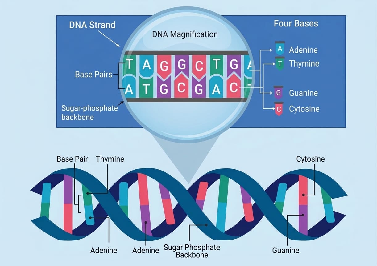
Covalent Methylation of Cytosine Residues within Genomic CpG Islands
2. Histone Tail Post-Translational Modifications: Genomic DNA is spooled around octamers of basic core histone proteins (two units each of H2A, H2B, H3, and H4) to form fundamental repeating units called nucleosomes. The amino-terminal tails projecting from these histone cores undergo complex covalent modifications that alter chromatin topology:
- Histone Acetylation: Catalyzed by histone acetyltransferases (HATs), transfers acetyl groups to positively charged lysine residues, neutralizing their basic charge. This relaxes the electrostatic attraction between histones and the negatively charged DNA phosphate backbone, uncoiling condensed heterochromatin into transcriptionally permissive, open euchromatin.
- Histone Deacetylation & Methylation: Mediated by histone deacetylases (HDACs) and histone methyltransferases (HMTs), restores positive charges or recruits repressive complexes, condensing nucleosomes tightly into transcriptionally silent heterochromatin. Other regulatory marks include histone phosphorylation and ubiquitination.
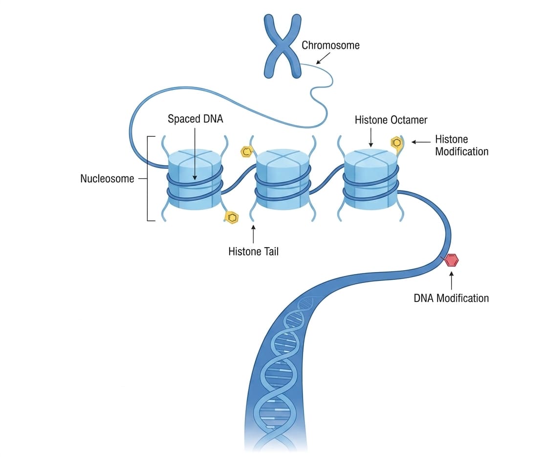
Chromatin Architectural Transitions Mediated by Post-Translational Histone Modifications
3. Non-Coding Regulatory Ribonucleic Acids (ncRNAs): Functional RNA molecules that are transcribed from genomic DNA but do not undergo translation into polypeptides. They function as epigenetic regulators of gene expression post-transcriptionally:
- MicroRNAs (miRNAs): Short, single-stranded non-coding RNAs (~21–25 nucleotides) that assemble into the RNA-Induced Silencing Complex (RISC) and bind via imperfect base-pairing to complementary 3'-untranslated regions (3'-UTRs) of specific messenger RNAs (mRNAs), triggering translational repression or target mRNA enzymatic degradation.
- Long Non-Coding RNAs (lncRNAs): Transcripts exceeding 200 nucleotides in length that act as structural molecular scaffolds, enhancers, or guides that recruit chromatin-modifying enzymes (such as polycomb repressive complexes) directly to specific target gene loci.
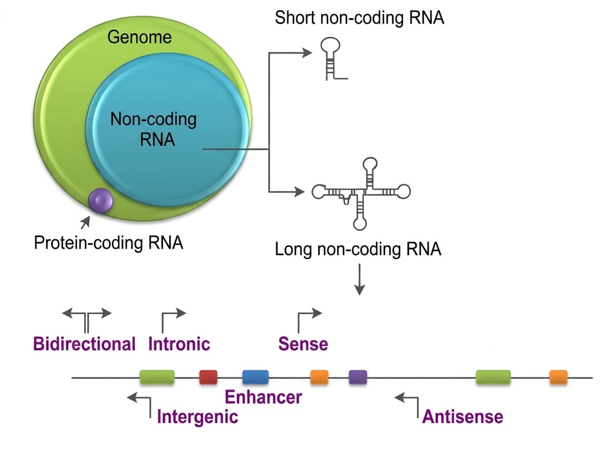
Classification, Biogenesis, and Transcriptional-Translational Interference Mediated by Non-Coding RNAs
7. Epigenetics in Clinical Disease Pathogenesis
Epigenetic Pathophysiology Across Major Organ Systems
1. Oncogenesis and Cancer Progression:
- Tumor Suppressor Hypermethylation: Aberrant, localized promoter hypermethylation silences critical cell-cycle regulators and repair genes without any change in their base sequence. Prominent examples include transcriptional silencing of CDKN2A (p16INK4a) (cyclin-dependent kinase inhibitor) and BRCA1 (homologous recombination DNA repair).
- Global DNA Hypomethylation: Widespread hypomethylation of repetitive intergenic elements and transposons induces genomic instability, chromosomal translocations, and the aberrant activation of proto-oncogenes.
- Disordered Histone Marks: Overexpression of histone methyltransferases or histone deacetylases represses differentiation programs, permitting uncontrolled neoplastic proliferation.
2. Neurodegenerative Diseases: Aberrant global and locus-specific DNA methylation dynamics within central nervous system tissue contribute to cognitive decline in Alzheimer's disease and Parkinsonism. Transcriptional dysregulation of genes governing synaptic neuroplasticity, brain-derived neurotrophic factor (BDNF) synthesis, and tau microtubule phosphorylation accelerates neuroinflammation and progressive synaptic loss.
3. Cardiovascular Diseases: Endothelial cells and vascular smooth muscle cells (VSMCs) exposed to chronic hemodynamic shearing stress, oxidized LDL, and systemic inflammation undergo pathological epigenetic remodeling. Promoter alterations in genes governing endothelial nitric oxide synthase (eNOS) and vascular matrix metalloproteinases promote chronic endothelial dysfunction, arterial stiffness, persistent essential hypertension, and accelerated fibrofatty atheromatous plaque formation.
4. Autoimmune Diseases: Epigenetic dysregulation in T- and B-lymphocyte populations disrupts immunological self-tolerance. In Systemic Lupus Erythematosus (SLE), defective DNA methyltransferase-1 (DNMT1) activity causes abnormal, widespread DNA hypomethylation in CD4+ T-cells. This promotes constitutive overexpression of autoreactive costimulatory molecules (such as CD70 and CD11a), driving spontaneous autoimmune attacks against host nuclear antigens.
5. Metabolic Disorders: Nutritional states and lifestyle factors induce lasting epigenetic modifications in hepatic tissue, pancreatic β-cells, and adipose depots. Altered promoter methylation and chromatin remodeling across genes controlling insulin receptor signaling, glucose transporter (GLUT4) translocation, and adipogenesis promote chronic peripheral insulin resistance, contributing directly to Type 2 Diabetes Mellitus and Morbid Obesity.
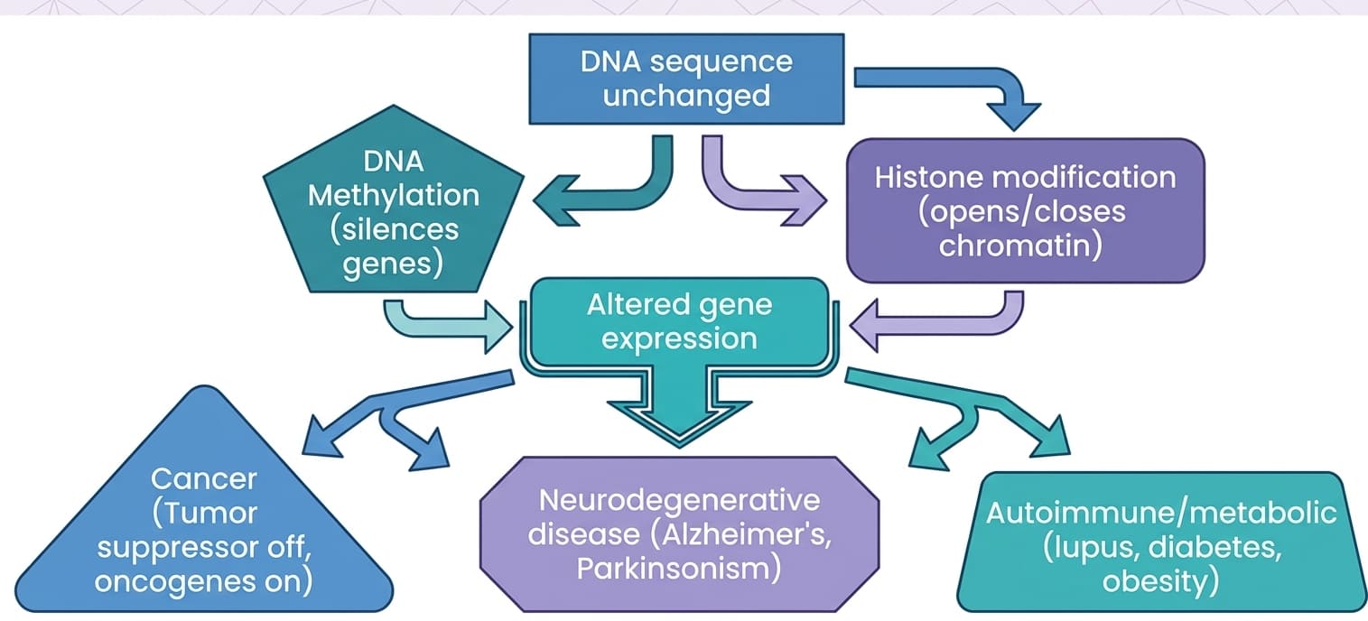
Mechanistic Pathway Linking Intact DNA Sequences to Epigenetic Reprogramming and Systemic Disease States
8. Interplay Between Genetics, Epigenetics, and Environmental Modulators
Genetics and epigenetics function not as isolated systems, but in continuous bidirectional communication:
- Genetic Mutations Disrupting Epigenetic Machinery: Primary genetic alterations (point mutations, frameshifts) frequently afflict genes that encode key chromatin-modifying enzymes. For example, mutations in DNMT3A, histone methyltransferases (e.g., EZH2), or chromatin remodelers (e.g., ARID1A) compromise the molecular machinery of epigenetics, causing widespread secondary disruptions in global gene expression.
- Epigenetic Inheritance: Certain chromatin modifications established in ancestral germlines escape post-fertilization epigenetic reprogramming and are transmitted transgenerationally, influencing disease susceptibility in subsequent generations.
- Epigenetics as an Environmental Mediator: Epigenetic marks provide an adaptive molecular interface linking environmental exposures to alterations in gene expression. Environmental stressors—such as maternal/childhood malnutrition, persistent psychological stress, endocrine-disrupting chemicals, cigarette smoke, and toxic heavy metals—catalyze stable changes in DNA methylation and histone marks that elevate chronic disease susceptibility throughout an individual's lifetime.
9. Genetic Polymorphisms and Polygenic Disease: Pathogenesis of Obesity
Polymorphism Defined
A stable genomic sequence variation existing at a specific locus across individuals within a biological population, occurring at an allele frequency of at least 1%. Polymorphisms represent non-pathogenic normal diversity, adaptive variations, or subtle quantitative modifiers of disease susceptibility, distinct from rare, highly penetrant pathogenic mutations.
Spectrum of Polymorphism: From Organismal Morphs to DNA Single-Nucleotide Variations
Organismal & Morphological Polymorphisms: Natural populations maintain distinct phenotypes ("morphs") within a single interbreeding species:
- Peppered Moth (Biston betularia): Industrial melanism displaying dark (melanic) and light (typica) wing color variations driven by selective predatory pressures.
- Gouldian Finches (Erythrura gouldiae): Stable chromatic head polymorphisms (red-headed, black-headed, and yellow-headed morphs) existing concurrently in wild populations.
- Ant Colony Social Castes: Morphological divergence between reproductive queens, sterile workers, and soldiers within the same genetic background.
- Jaguars (Panthera onca): Coexistence of standard rosetted coats alongside melanic ("black panther") variants.
Human DNA Polymorphisms: Molecular variations that yield measurable biochemical or anatomical traits:
- Human ABO Blood Group System: Allelic variations (IA, IB, i) in the gene encoding glycosyltransferase enzymes that add terminal sugar moieties to cell-surface H-antigens on erythrocytes.
- Melanocortin 1 Receptor (MC1R) Polymorphisms: Single-nucleotide changes altering receptor signaling that dictate phenotypic pigmentation, skin phototype, and red hair color.
- Lactase Persistence: Polymorphic regulatory variants upstream of the lactase gene (LCT) that permit continuous lactase expression and dietary lactose tolerance throughout adulthood.
- Single-Nucleotide Polymorphisms (SNPs): Widespread single-base substitutions across the genome (e.g., GCGA/CGCT transitioning to GTGA/CACT) serving as genetic markers for polygenic traits and complex disease risk.

Phenotypic Morphological Polymorphism Illustrated by Head Plumage Variation in Gouldian Finches
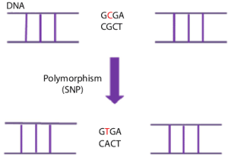
Structural Schematic of a Single-Nucleotide Polymorphism (SNP)
9.1 Polygenic and Endocrine Pathogenesis of Obesity
Obesity represents a quintessential multifactorial medical condition resulting from continuous interactions between an obesogenic environment and an individual's underlying genetic architecture. Constitutional inheritance strongly influences human body mass index (BMI), adiposity distribution, basal metabolic rate, and central appetite regulation.
Neuroendocrine and Metabolic Dysregulation in Obesity
Central caloric homeostasis and satiety are governed by reciprocal neuroendocrine loops linking peripheral tissues with the hypothalamic arcuate nucleus:
- Leptin: An adipokine synthesized and secreted by subcutaneous adipocytes in direct proportion to total body lipid mass. Leptin circulates to the brain and binds the leptin receptor (LEPR) on POMC/CART neurons in the hypothalamic arcuate nucleus, stimulating anorexigenic signaling that suppresses hunger, promotes satiety, and enhances systemic energy expenditure. In common polygenic obesity, hyperleptinemia fails to curb appetite due to central leptin resistance. Rare monogenic mutations causing complete deficiency of leptin or its receptor produce severe, early-onset morbid obesity and hyperphagia.
- Ghrelin: An orexigenic peptide hormone secreted by neuroendocrine cells of the gastric fundus during fasting states. Ghrelin crosses the blood-brain barrier and activates AgRP/NPY neurons within the arcuate nucleus, stimulating hunger and prompting meal initiation. Circulating levels rise preprandially and are suppressed following nutrient ingestion.
- Polymorphic Susceptibility: Genome-wide association studies (GWAS) have identified numerous SNPs across genes regulating appetite, energy expenditure, lipid storage, and insulin sensitivity (e.g., polymorphisms within the FTO, MC4R, and PPARG loci). These variants shift homeostatic energy balance toward increased caloric consumption and preferential lipid storage, predisposing individuals to excessive weight gain and metabolic syndrome.
10. Unified Conceptual Summary
- Dual Etiological Framework: Human diseases stem from permanent physical changes in genomic DNA sequences (spontaneous or inherited mutations) or from reversible, non-sequence chemical modifications (epigenetic dysregulation).
- Translational Consequences of Mutations: Structural genomic mutations cause cellular dysfunction through missense amino acid substitutions (Sickle cell anemia), premature nonsense termination codons (β0-thalassemia), frameshift reading realignments (Tay-Sachs disease), or unstable dynamic trinucleotide repeat expansions (Huntington disease, Fragile X syndrome).
- Mendelian Segregation: Monogenic disorders follow classical inheritance patterns: autosomal dominant (50% risk per pregnancy, structural proteins, delayed onset, variable expressivity), autosomal recessive (25% risk, metabolic enzymes, uniform early onset), or X-linked recessive (overwhelming hemizygous male predilection, female carrier transmission).
- Epigenetic Mechanisms: Gene expression is dynamically regulated through DNA methylation at CpG islands (transcriptional silencing), covalent post-translational histone tail modifications (acetylation opening chromatin vs. deacetylation/methylation condensing chromatin), and non-coding RNAs (miRNAs/lncRNAs inducing translational repression or degradation).
- Multifactorial Integration: Common chronic diseases—such as neoplasia, autoimmune disorders, neurodegeneration, atherosclerosis, and obesity—result from complex interactions between underlying genetic polymorphisms, altered epigenetic marks, and environmental exposures that together dictate disease susceptibility and clinical severity.