Comprehensive Pathology: Mechanisms and Manifestations of Chronic Inflammation
Key Definitions
- Inflammation: Derived from the Latin inflammatio ("to ignite" or "set on fire"), this represents an essential vascularized tissue defense mechanism that detects, isolates, and neutralizes noxious threats—including biological pathogens, necrotic debris, and toxic chemical irritants. Crucially, inflammation is not synonymous with infection; an infectious pathogen can trigger inflammation, but aseptic physical or immune-mediated tissue injury incites identical inflammatory cascades without viable microbes.
- Chronic Inflammation: An indolent, protracted immune reaction extending over weeks, months, or years. It is defined by the ongoing, simultaneous occurrence of active cellular injury, mononuclear leukocyte infiltration, and concurrent attempts at healing via connective tissue replacement (fibrogenesis) and vascular sprouting (angiogenesis).
1. Pathophysiologic Divergence: Acute Versus Chronic Inflammation
The transition from an immediate vascular response to a chronic process reflects a fundamental shift in effector leukocyte recruitment, signaling mediators, and clinical consequences. While acute episodes rely on rapid vascular permeability shifts and microvascular neutrophil emigrations designed for rapid resolution, unresolved stimuli lead to tissue reorganization dominated by mononuclear phagocytes.
| Pathologic Feature | Acute Inflammation | Chronic Inflammation |
|---|---|---|
| Temporal Profile | Abrupt onset; short-lived (hours to several days) | Protracted, indolent course (weeks, months, to lifelong) |
| Dominant Leukocytes | Polymorphonuclear neutrophils (PMNs) | Mononuclear cells: Macrophages, lymphocytes, plasma cells |
| Microvascular Changes | Prominent vasodilation, high plasma exudation, inflammatory edema | Variable/minimal exudate; neoangiogenesis with perivascular fibrosis |
| Tissue Consequences | Complete resolution, suppurative necrosis (abscess), or early scarring | Progressive tissue destruction, collagenous fibrosis, organ distortion |
2. Etiologic Triggers of Chronic Inflammation
Chronic inflammation develops when host defense mechanisms fail to eradicate an inciting agent or when abnormal adaptive immune responses sustain tissue injury. Clinical causes fall into four major pathophysiological categories:
- Infections with Intracellular or Non-Digestible Pathogens: Microorganisms that exhibit high resistance to phagocytic enzymatic breakdown or intracellular destruction—such as Mycobacterium tuberculosis, Treponema pallidum (syphilis), Mycobacterium leprae, fungi, and persistent viruses (e.g., Hepatitis B and Hepatitis C).
- Hypersensitivity and Immune-Mediated Inflammatory Disorders: Inappropriate, uncontrolled activation of the immune system:
- Autoimmune pathologies: Self-antigens evoke persistent cellular and humoral responses, including rheumatoid arthritis (RA), systemic lupus erythematosus (SLE), and Hashimoto's thyroiditis.
- Allergic and mucosal conditions: Chronic exposure to environmental antigens, as observed in bronchial asthma and Crohn's inflammatory bowel disease.
- Exogenous and Endogenous Non-Degradable Materials: Inability of phagocytes to digest foreign or metabolic particulates:
- Inorganic environmental dusts: Inhaled crystalline silica (silicosis), beryllium, or asbestos fibers.
- Iatrogenic / trauma implants: Retained organic splinters, ballistic fragments, surgical sutures, or long-term medicinal implants (such as subcutaneous levonorgestrel contraceptive capsules [Norplant]).
- Endogenous toxic aggregates: Hypercholesterolemic lipid deposition inciting inflammatory plaques in atherosclerosis.
- Unresolved Acute Inflammation and Chronic Mechanical Irritation: Repeated insults or anatomical barriers that impair physiological healing:
- Cavitation or unresolved necrosis resulting in a lung abscess following lobar bacterial pneumonia.
- Chronic bacterial osteomyelitis secondary to an open bone fracture, characterized by an ongoing discharging fistulous or sinus tract.
- Recurrent ascending urinary tract obstruction causing chronic obstructive pyelonephritis.
- Recurrent mechanical injury and chemical irritation from cholelithiasis (gallstones), promoting chronic cholecystitis, or acid-peptic erosion in gastric ulcers.
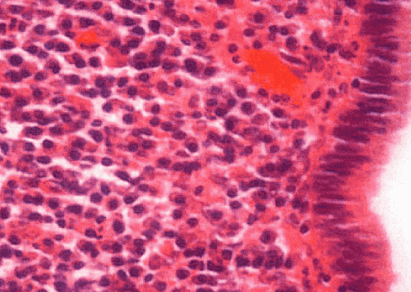
Histopathology of Chronic Mucosal Inflammation: Chronic Cervicitis
3. Cellular Cast of Chronic Inflammation
The defining histomorphologic hallmark of chronic inflammation is the persistent infiltration of mononuclear leukocytes combined with active tissue destruction and structural fibroblastic replacement.
| Cell Type | Lineage & Tissue Names | Functional Role & Mediators |
|---|---|---|
| Macrophages | Derived from bone marrow monoblasts → circulating monocytes → tissue macrophages:
|
Central coordinators of chronic inflammation. They phagocytose antigens, present antigenic peptides via MHC class II to T cells, release inflammatory cytokines (IL-1, TNF-α, IL-6), and drive tissue repair via growth factors. |
| Lymphocytes | Adaptive immune effectors: CD4+ Helper T cells, CD8+ Cytotoxic T cells, and B cells | Drive cell-mediated responses via cytokine release (IFN-γ, IL-2), recruit macrophages, and induce cytotoxic cell death. |
| Plasma Cells | Terminally differentiated B lymphocytes with "cartwheel" or "clock-face" nuclear chromatin | Synthesize and secrete targeted immunoglobulins directed against persistent local foreign antigens or altered self-epitopes. |
| Eosinophils | Granulocytes with bilobed nuclei and brick-red acidophilic granules | Recruited via eotaxin and regulated by IgE; release major basic protein to kill helminthic parasites and mediate allergic tissue remodeling. |
| Mast Cells | Sentinel connective tissue cells armed with high-affinity FcεRI receptors | Bind the Fc portion of IgE antibodies; release histamine, leukotrienes, and cytokines during both acute flares and persistent fibro-inflammatory disorders. |
| Fibroblasts | Mesenchymal stromal cells | Respond to macrophage-derived growth factors (PDGF, TGF-β, FGF); synthesize extracellular matrix and collagen to deposit scars (fibrosis). |
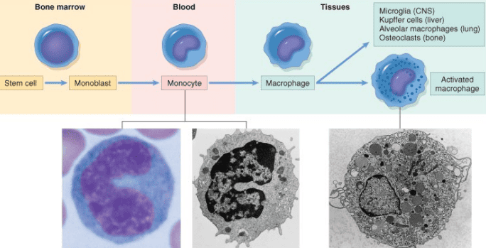
Lineage and Anatomical Differentiation of the Mononuclear Phagocyte System
4. Macrophage Dynamics, Signaling Networks, and Activation
Within several hours of an inflammatory insult, circulating monocytes migrate through endothelial junctions into the interstitium, guided by chemokines. In chronic lesions, macrophages remain elevated in tissue via three distinct mechanisms:
- Continuous Monocyte Recruitment: The most significant source of tissue macrophages; sustained adhesion molecules (selectins, integrins) and chemotactic signals draw circulating precursors into the affected site.
- In Situ Local Mitotic Proliferation: Interstitial macrophages retain the capacity to undergo local cell division upon stimulation by macrophage colony-stimulating factors.
- Immobilization and Longevity: Cytokines (e.g., macrophage migration inhibitory factor [MIF]) and oxidized lipids prevent these cells from migrating away, anchoring them in the lesion.
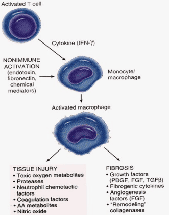
Dual Effector Arms of Macrophage Activation: Tissue Injury Versus Fibroplasia
Summary of Molecular Mediators
- Pro-inflammatory Cytokines: IL-1, IL-6, and TNF-α upregulate endothelial adhesion molecules, induce acute-phase proteins, and maintain immune cell recruitment.
- Macrophage-Activating Lymphokines: Interferon-gamma (IFN-γ) released by antigen-primed T helper cells increases phagocytic capacity, microbial killing, and epithelioid cell transformation.
- Fibrogenic and Angiogenic Growth Factors: Platelet-Derived Growth Factor (PDGF), Transforming Growth Factor-beta (TGF-β), and Vascular Endothelial Growth Factor (VEGF) trigger fibroblast proliferation, matrix production, and endothelial capillary budding.
- Degradative and Toxic Products: Reactive oxygen species (ROS), nitric oxide (NO), and matrix metalloproteinases (MMPs) break down parenchymal tissue, perpetuating injury when host regulation fails.
5. Granulomatous Inflammation: Architecture and Pathogenesis
Key Definitions
- Granulomatous Inflammation: A specialized histological pattern of chronic inflammation triggered by poorly digestible or persistent antigens, defined by compact aggregations of transformed, epithelioid macrophages frequently accompanied by multinucleated giant cells and a collar of lymphocytes and fibroblasts.
- Granuloma: A microscopic, circumscribed nodule (typically ~1 mm in diameter) composed primarily of epithelioid histiocytes with an outer lymphocytic rim. The term combines granule (small particle-like lesion) with -oma (indicating an inflammatory mass rather than a true neoplasm).
Granuloma Versus Granulation Tissue
A critical pathology distinction exists between these two similarly named entities:
- Granuloma: An organized immune assembly of epithelioid histiocytes, giant cells, and lymphocytes designed to sequester indigestible agents.
- Granulation Tissue: The hallmark of physiological wound healing and early scar formation, composed of proliferating thin-walled capillaries (angiogenesis), edematous loose stroma, and active fibroblasts.
Immunopathogenesis of the Granuloma
Granuloma morphogenesis represents a Type IV cell-mediated (delayed-type) hypersensitivity response. While protective in containing dissemination, it can induce severe secondary tissue damage via the following four-stage sequence:
- Antigen Ingestion and Phagocytic Failure: Circulating monocytes and resident macrophages engulf the inciting agent. Because the substance resists enzymatic degradation (e.g., mycobacterial wax/mycolic acids, non-absorbable silk or talc), phagolysosomes fail to clear it.
- Antigen Presentation to CD4+ T Cells: Macrophages process and present peptide fragments on MHC Class II molecules to naive CD4+ T helper lymphocytes, driving their differentiation into antigen-specific Th1 cells.
- Cytokine Cascade and Macrophage Differentiation:
- Activated CD4+ T cells release IL-2, driving autocrine and paracrine clonal T-lymphocyte expansion.
- T cells secrete IFN-γ, which activates macrophages, increasing bactericidal activity and converting them into pale, secretory epithelioid cells.
- TNF-α secretion stimulates microvascular endothelia to produce prostaglandins and recruits additional leukocytes.
- Fusion and Fibrous Encapsulation: Sustained cytokine stimulation induces adjacent epithelioid cells to fuse, creating multinucleated giant cells. Concurrently, macrophage-derived TGF-β and PDGF stimulate peripheral fibroblasts to synthesize a collagenous envelope, walling off the inflammatory focus.
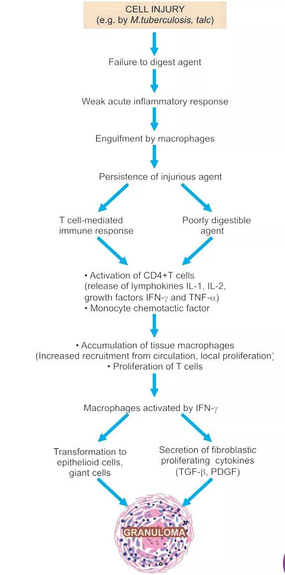
Cascade of Granuloma Formation in Delayed-Type Hypersensitivity
6. Granuloma Composition and Structural Morphology
Under light microscopy, a mature granuloma shows five concentric structural zones:
- Epithelioid Cells: Transformed macrophages that resemble epithelial cells. They feature elongated slipper-shaped or oval nuclei with pale, finely vesicular chromatin. Their cytoplasm is abundant and eosinophilic with indistinct, interdigitating cell borders. Unlike standard macrophages, they have diminished phagocytic capacity and function primarily as secretory cells.
- Multinucleated Giant Cells: Formed through the cytoplasmic fusion of multiple epithelioid cells (rather than incomplete cytokinesis), containing 20 or more nuclei within a shared cytoplasm:
- Langhans Giant Cells: Nuclei arrange in a horseshoe, peripheral ring, or bipolar cluster along the outer edge of the cytoplasm; characteristic of tuberculosis and immune granulomas.
- Foreign Body Giant Cells: Nuclei are scattered haphazardly throughout the center of the cell, typical of reactions to exogenous fibers, talc, or sutures.
- Lymphoid Rim: A peripheral collar of lymphocytes—primarily CD4+ Th1 memory T cells and occasional plasma cells—that continually release cytokines to maintain the granuloma.
- Central Necrosis: Often present in infectious granulomas. In tuberculosis, this manifests as caseous necrosis—grossly appearing as crumbly, white, cheese-like material and microscopically showing structureless, granular, eosinophilic debris with complete loss of cellular architecture.
- Fibrous Capsule: Peripheral fibroblasts and collagen fibers deposited around the rim of long-standing lesions as a reparative barrier.
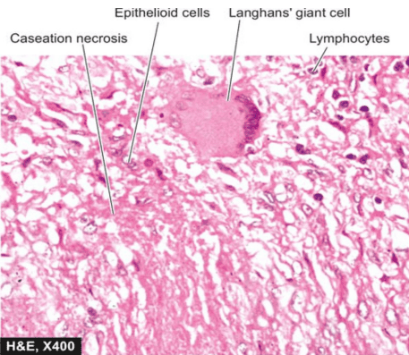
Histopathology of a Tuberculous Tubercle: Central Caseation and Langhans Giant Cells
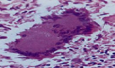
Foreign Body Giant Cell: Random Central Nuclear Distribution
7. Granuloma Classification and Clinical Spectra
Granulomas are broadly classified based on whether they involve an adaptive immune response:
| Granuloma Category | Immunologic Mechanism | Diagnostic Hallmarks & Clinical Examples |
|---|---|---|
| Immune Granulomas | Driven by a Type IV cell-mediated response to persistent, insoluble immunogenic antigens. | Epithelioid cells, Langhans giant cells, dense lymphocytic rim:
|
| Foreign Body Granulomas | Triggered by inert non-immunogenic particulates; lacks a significant T-cell-mediated response. | Foreign material occupies the center of the granuloma, rimmed directly by epithelioid cells and foreign body giant cells; absence of necrosis:
|
| Sarcoid Granulomas | Systemic inflammatory disease of unknown etiology; suspected dysregulated CD4+ Th1 cell response to uncharacterized environmental antigens. | Non-caseating (naked) granulomas with minimal lymphocytic cuffs. Typically involves bilateral hilar/mediastinal lymphadenopathy, pulmonary parenchyma, skin, eyes, salivary glands, liver, and spleen. |
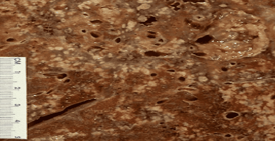
Histopathology of Pulmonary Granulomatous Infiltration (Low-Power Field)
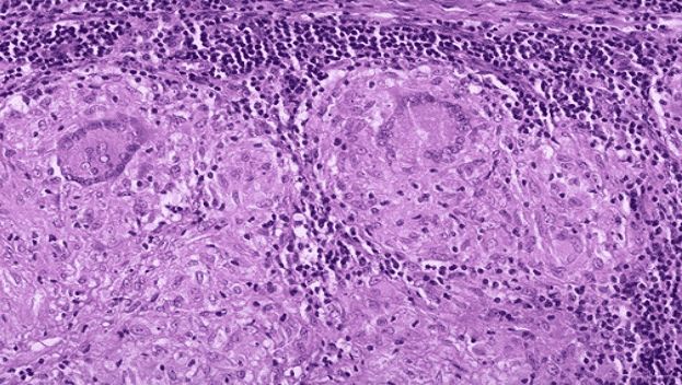
High-Power Histology: Epithelioid Histiocyte Aggregation and Multinucleated Giant Cells
8. Tuberculosis: The Prototype of Granulomatous Infection
Tuberculosis (TB), caused by Mycobacterium tuberculosis hominis, represents the classic clinical example of granulomatous inflammation. The bacterium is a slender, slightly curved, non-motile rod measuring approximately 0.5 μm by 3 μm. It is neutral or poorly staining on standard Gram stain due to a thick, waxy, lipid-rich cell wall.
Transmission Routes
- Airborne Inhalation: The most common route; occurs through inhalation of aerosolized droplet nuclei coughed out by patients with active, open cavitary pulmonary TB.
- Ingestion: Primary tonsillar or intestinal mucosal infection via swallowing infected sputum or drinking unpasteurized milk contaminated with Mycobacterium bovis.
- Direct Inoculation: Rare cutaneous inoculation occurring through accidental exposure (e.g., in pathologists or morticians handling infected postmortem tissue).
- Transplacental Route: Congenital tuberculosis acquired hematogenously via umbilical circulation from an infected mother; rare.
Dissemination Pathways
- Local Extension: Alveolar macrophages carry ingested bacilli into adjacent interstitial spaces and neighboring pulmonary segments.
- Lymphatic Spread: Mycobacteria drain through lymphatic channels to regional nodes. The combination of a peripheral parenchymal subpleural lesion and enlarged hilar lymph nodes, bridged by inflamed lymphatic vessels, forms the classical Ghon (Primary) Complex.
- Hematogenous Dissemination (Miliary TB): Drainage of bacilli into the thoracic duct or direct erosion into a pulmonary venule seeds organisms through the systemic circulation, creating 1–2 mm, uniform, yellow-white lesions resembling millet seeds across the lungs, liver, spleen, kidneys, adrenal glands, and bone marrow.
- Spread Across Natural Anatomical Passages:
- Pleural cavity erosion → tuberculous pleurisy and pleural effusion.
- Transbronchial passage into contralateral lobes or larynx → tuberculous laryngitis.
- Swallowed sputum passing through the digestive tract → ileocecal tuberculosis.
- Fallopian tube salpingitis descending into the peritoneal space → tuberculous peritonitis.
- Renal parenchymal papillary necrosis shedding into the ureter → ureteral strictures and bladder trigone ulceration.
Gross Pathology of the Lung: Disseminated Miliary Tuberculosis
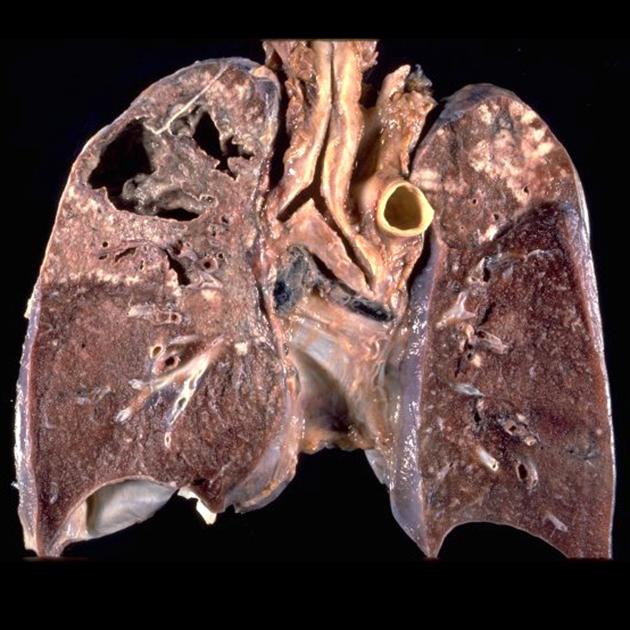
Gross Pathology of Advanced Pulmonary Tuberculosis: Apical Fibrocaseous Cavitation
9. Laboratory Identification and Diagnostic Methods for Mycobacteria
Accurate diagnosis of mycobacterial infections requires combining microscopy, culture isolation, and molecular assays:
| Diagnostic Modality | Mechanism & Protocol | Clinical Performance, Speed, & Limitations |
|---|---|---|
| Acid-Fast Staining (Ziehl-Neelsen [ZN]) | Heated carbol fuchsin penetrates the cell wall's lipid-rich mycolic acids. Bacilli resist decolorization by acid-alcohol. Decolorized with 20% sulfuric acid (in contrast to M. leprae, which is less acid-fast and decolorized with 5% H2SO4). Counterstained with methylene blue. | Rapid initial detection. Bacilli appear as bright red/magenta beaded rods against a light blue background. False positives occur with other partially acid-fast organisms: Nocardia, Rhodococcus, Legionella micdadei, and coccidian oocysts (Cryptosporidium, Isospora / Cystoisospora). |
| Fluorochrome Staining | Fluorescent dyes auramine-O and rhodamine bind cell wall mycolic acids; examined under ultraviolet illumination. Mycobacteria also display natural autofluorescence. | Higher sensitivity than ZN staining; allows rapid screening of clinical smears under low magnification, reducing technician fatigue. |
| Solid Media Culture (Lowenstein-Jensen) | Egg-based solid medium containing malachite green to inhibit contaminating commensal flora. | Traditional gold standard for definitive isolation; highly specific. Very slow: takes 8 to 12 weeks for visible buff-colored, crumbly colonies to emerge. |
| Rapid Automated Culture (e.g., BACTEC) | Automated liquid broth systems using radiolabeled (14C) or fluorometric sensors to monitor mycobacterial metabolic CO2 output. | Shortens confirmation time to 2 to 3 weeks, facilitating faster drug susceptibility testing. |
| Chromatography (HPLC) | High-Performance Liquid Chromatography analyzes specific species profiles of extracted cell-wall mycolic acids. | Provides rapid, definitive species confirmation once initial growth appears in liquid culture. |
| Molecular Assays (NAAT / PCR) | Nucleic acid amplification (e.g., GeneXpert MTB/RIF) targets conserved mycobacterial DNA sequences (such as IS6110 and rpoB). | Results available in hours with high sensitivity and specificity. Essential for distinguishing M. tuberculosis from non-tuberculous mycobacteria and detecting rifampin resistance mutations. |
| Animal Inoculation | Subcutaneous inoculation of clinical specimens into guinea pigs to observe typical caseating disease. | Historical method; largely obsolete due to the availability of rapid automated cultures and molecular assays. |
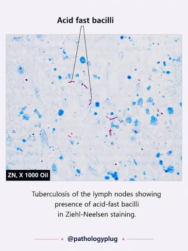
Microscopic Demonstration of Mycobacterium tuberculosis: Acid-Fast Bacilli in Lymph Node (ZN, x1000 Oil)
10. Non-Tuberculous Mycobacteria (NTM)
Non-tuberculous mycobacteria (NTM), also known as atypical or environmental mycobacteria, comprise all species outside the M. tuberculosis complex and M. leprae. They are widespread in soil and water systems. Key differences from classical tuberculosis include:
- Absence of Person-to-Person Transmission: Humans acquire infections via direct environmental exposure (inhalation, water exposure, or trauma).
- Reduced Virulence: Generally less virulent in immunocompetent hosts, but causes severe opportunistic disease in patients with AIDS or pre-existing lung damage.
- Drug Resistance: Intrinsically resistant to standard first-line anti-tubercular regimens (isoniazid, rifampin, pyrazinamide, ethambutol); requires targeted combinations including macrolides and fluoroquinolones.
- Histologic Spectrum: Lesions range from non-caseating granulomas to loose aggregates of foamy macrophages or acute neutrophilic microabscesses.
Runyon Classification of Non-Tuberculous Mycobacteria
| Growth Category | Pigmentation Characteristics | Growth Rate & Representative Species |
|---|---|---|
| Rapid Growers | Variable pigmentation | Develop visible solid media colonies in less than 7 days. Less virulent; cause skin abscesses and catheter infections. Examples: M. fortuitum, M. chelonae, M. abscessus. |
| Slow: Photochromogens | Produce deep yellow/orange carotenoid pigments only upon exposure to light; colonies remain non-pigmented in the dark. | Require 2 to 3 weeks to grow. Examples: M. kansasii (cavitary pulmonary disease), M. marinum (fish tank granuloma). |
| Slow: Scotochromogens | Produce yellow-to-orange pigmentation continuously, whether grown in the dark or light. | Require 2 to 3 weeks to grow. Example: M. scrofulaceum (cervical lymphadenitis in young children). |
| Slow: Non-Chromogens | Non-pigmented; form pale, buff-colored, or tan colonies regardless of light exposure. | Require 2 to 3 weeks to grow. Examples: Mycobacterium avium-intracellulare (MAC) complex, M. ulcerans (Buruli ulcer). |
Five Recognized Clinical Patterns of NTM Disease
- Chronic Pulmonary Disease: Indolent, cavitary, or bronchiectatic respiratory infections caused by M. kansasii or M. avium-intracellulare (MAC), frequently mimicking classical tuberculosis in patients with underlying chronic obstructive pulmonary disease (COPD) or bronchiectasis.
- Superficial Lymphadenitis: Unilateral submandibular or cervical lymph node enlargement ("scrofula") in pediatric patients (typically 1 to 5 years of age), primarily caused by M. avium-intracellulare or M. scrofulaceum.
- Ulcerative Cutaneous and Soft Tissue Lesions: Indolent necrotizing dermal ulcers, nodules, and verrucous plaques, notably Buruli ulcer caused by M. ulcerans (driven by the cytotoxic macrolide toxin mycolactone) and "swimming pool/fish tank granuloma" caused by M. marinum following minor aquatic trauma.
- Soft Tissue and Surgical Wound Abscesses: Localized, painful subcutaneous abscesses and post-procedural wound breakdowns caused by rapidly growing species, including M. fortuitum, M. chelonae, and M. abscessus, often following contaminated surgical equipment or cosmetic procedures.
- Disseminated Bacteremia: Life-threatening, widely disseminated systemic infections with high bacteremic loads, seen in severely immunocompromised patients (particularly advanced HIV/AIDS with CD4+ T-cell counts < 50 cells/μL), overwhelmingly caused by M. avium-intracellulare (MAC).
11. Diagnostic Strategy for Granulomatous Inflammation
Evaluating granulomatous lesions requires a systematic approach using histology, special stains, microbiology, and serology:
- Histomorphologic Evaluation:
- Caseating granulomas with Langhans giant cells strongly indicate tuberculosis.
- Non-caseating ("naked") granulomas in a multi-organ distribution suggest sarcoidosis.
- Stellate necrotizing granulomas with central microabscesses point to cat-scratch disease (Bartonella henselae) or lymphogranuloma venereum (Chlamydia trachomatis).
- Special Stains and Optical Microscopy:
- Acid-Fast Stains (Ziehl-Neelsen, Fite-Faraco): Identify M. tuberculosis (strongly acid-fast) and M. leprae (partially acid-fast).
- Silver Stains (Grocott's Methenamine Silver [GMS], Periodic Acid-Schiff [PAS]): Highlight fungal cell walls (e.g., budding yeasts of Histoplasma or Blastomyces).
- Polarized Light Microscopy: Detects birefringent crystalline foreign materials (e.g., surgical talc, silica, synthetic suture fragments) inside foreign body giant cells.
- Microbiological Isolation: Inoculation onto specialized media (Lowenstein-Jensen slants, BACTEC automated liquid broths, fungal Sabouraud dextrose agar) remains essential to recover viable pathogens and determine antibiotic susceptibility profiles.
- Autoimmune Serology and Immune Biomarkers: When infectious stains and cultures are negative, serological testing helps exclude underlying connective tissue and autoimmune diseases:
- Rheumatoid factor (RF) and anti-cyclic citrullinated peptide (anti-CCP) for rheumatoid arthritis.
- Antinuclear antibodies (ANA) and anti-dsDNA for systemic lupus erythematosus.
- Serum angiotensin-converting enzyme (ACE) levels to support a clinical diagnosis of sarcoidosis.
12. Clinical Review Assessment
Sample Examination Question
Which of the following alterations is NOT considered a histological hallmark of chronic inflammation?
- A. Interstitial infiltration by antibody-secreting plasma cells
- B. Neoangiogenesis with proliferation of small capillary channels
- C. Connective tissue deposition and fibroplasia
- D. Proliferation of visceral smooth muscle fibers
- E. Dense interstitial accumulation of CD4+ and CD8+ lymphocytes
Explanation: The classical histological triad of chronic inflammation consists of (1) mononuclear leukocyte infiltration (macrophages, lymphocytes, plasma cells), (2) parenchymal tissue destruction, and (3) healing through fibrovascular connective tissue proliferation (fibroblasts depositing collagen) and angiogenesis (sprouting endothelial cells). Visceral smooth muscle proliferation is not a general histopathologic hallmark of chronic inflammation.
13. Advanced Pathophysiologic Discussion Topics
Take-Home Analytical Concepts
- Chronic Inflammation as a Driver of Malignancy: Long-standing inflammatory microenvironments increase cancer risk through continuous oxidative stress:
- Activated macrophages and neutrophils release reactive oxygen and nitrogen species (ROS/RNS) that induce direct oxidative DNA damage, point mutations, and chromosomal breaks.
- Concurrent secretion of mitogenic cytokines and growth factors (e.g., TNF-α, IL-6, EGF) stimulates persistent compensatory cellular proliferation in an attempt to regenerate damaged tissue.
- Proliferation in the setting of damaged DNA increases the likelihood of oncogene activation or tumor suppressor inactivation (e.g., TP53). Classical examples include gastric adenocarcinoma in chronic Helicobacter pylori gastritis, hepatocellular carcinoma following chronic Hepatitis B or C cirrhosis, and colorectal adenocarcinoma in longstanding ulcerative colitis.
- Macrophage Plasticity and Polarization Dynamics:
- Classically Activated Macrophages (M1 Phenotype): Induced by microbial endotoxins (LPS) and Th1 cytokines (IFN-γ). M1 macrophages produce abundant pro-inflammatory cytokines (IL-1, IL-12, IL-23, TNF-α), inducible nitric oxide synthase (iNOS), and ROS to execute high-capacity microbicidal killing and tissue destruction.
- Alternatively Activated Macrophages (M2 Phenotype): Triggered by Th2 cytokines (IL-4, IL-13). M2 macrophages suppress inflammation by secreting IL-10 and arginase, while releasing growth factors (TGF-β, PDGF, VEGF) that drive collagen deposition, angiogenesis, and tissue fibrosis. In chronic inflammation, an uncoordinated transition or persistent dual activation drives both parenchymal destruction and pathological organ scarring.
- Therapeutic Targets in Chronic Inflammatory Diseases: Modern pharmacological interventions target the cytokine and cellular networks sustaining the chronic response:
- TNF-α Inhibitors: Monoclonal antibodies (infliximab, adalimumab) and soluble receptor fusion proteins (etanercept) neutralize TNF-α, serving as foundational treatments for rheumatoid arthritis, Crohn's disease, and plaque psoriasis.
- Interleukin Receptor Antagonists: Tocilizumab (anti-IL-6 receptor) and anakinra (recombinant IL-1 receptor antagonist) dampen systemic acute-phase reactions.
- Immune Cell Depletion & Costimulation Blockers: Rituximab (anti-CD20) depletes antibody-producing B lymphocytes; abatacept blocks CD28-CD80/86 costimulatory pathways required for T-cell activation.
- Anti-Fibrotic Signaling Inhibitors: Small molecules targeting TGF-β and tyrosine kinase receptors (e.g., nintedanib, pirfenidone) slow down fibroblast proliferation in idiopathic pulmonary fibrosis.