Female Reproduction
Female Reproduction
Female reproductive functions can be broadly divided into two major phases:
Preparation for conception and pregnancy
- Development and maturation of ova
- Ovulation
- Preparation of the reproductive tract for fertilization and implantation
Pregnancy and post-pregnancy reproductive functions
- Maintenance of pregnancy
- Parturition (childbirth)
- Lactation after delivery
Vulva: Mons pubis, labia minora, labia majora and vestibule are referred to collectively as the vulva.
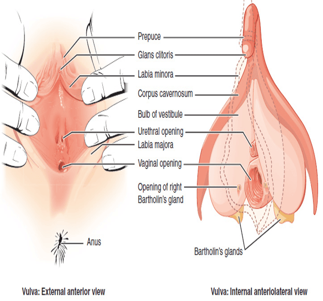
Vulva: External anterior view and internal anteriolateral view
Anatomy of the Female Reproductive System
Female reproductive system consists of:
Ovaries
- Female gonads and primary sex organs
- Produce ova (oocytes) and female sex hormones, mainly estrogens and progesterone
Uterine (Fallopian) tubes
- Transport the ovum toward the uterus
- Usual site of fertilization is the ampulla
Uterus
- Site of implantation and development of the embryo and fetus
Vagina
- Receives the penis and semen during intercourse
- Serves as the birth canal and passage for menstrual flow
External genitalia (Vulva)
- Mons pubis
- Labia majora
- Labia minora
- Clitoris
- Vestibular bulbs
- Greater vestibular (Bartholin) glands
Internal vs. External Organs
Note: Vulva refers collectively to the external female genital structures, while the ovaries, uterine tubes, uterus, and vagina form the main internal reproductive organs.
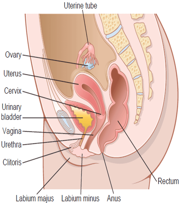
Anatomy of the Female Reproductive System
Ovaries
- Paired female gonads, approximately the size and shape of large almonds, located in the pelvic cavity and supported by ligaments.
- The fimbriae of the uterine tubes lie close to the ovaries and help capture the ovulated oocyte.
- After ovulation, the oocyte is transported through the uterine tube toward the uterus by ciliary action and smooth-muscle contractions.
Functions of the Ovaries
- Gametogenic function: Production and maturation of female gametes through oogenesis.
- Endocrine function: Secretion of estrogens, progesterone, inhibin, relaxin and small amounts of androgens.
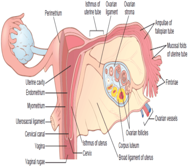
Internal structures of the uterus, ovary, and a uterine tube
Accessory sex organs of the female reproductive organs
1. Vagina
- Receives the penis during coitus and sperm deposition and transport unfertilized ova and menstrual products to the exterior. It’s the birth canal during parturition.
2. Fallopian Tubes
- Transport the oocyte from the ovary toward the uterus and are the usual site of fertilization, especially the ampulla.
Main Parts
| Part of Fallopian Tube | Anatomical Description | Functional Significance |
|---|---|---|
| Infundibulum | Funnel-shaped distal end with finger-like projections called fimbriae | Help capture the ovulated oocyte from the peritoneal space |
| Ampulla | Wider middle portion of the tube | Common and usual site of fertilization |
| Isthmus | Narrow medial portion connected directly to the uterus | Transports pre-implantation embryo into the uterine cavity |
| Intramural (uterine) part | Segment that passes through the uterine wall | Opens into the uterine cavity |
Layers of the Fallopian Tube
- Serosa: outer covering.
- Smooth muscle layer: produces peristaltic contractions.
- Mucosa: contains secretory cells (nourish the gamete/embryo) and ciliated epithelial cells (propel the oocyte toward the uterus).
Transport of the Oocyte
Movement toward the uterus is aided by:
- Ciliary beating
- Smooth muscle contractions
- Tubal secretions
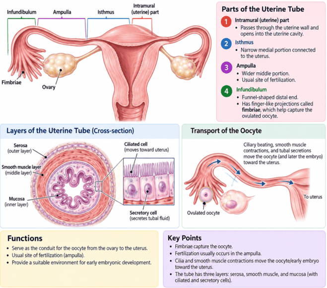
Fallopian (Uterine) Tubes: Anatomy, Structure and Function
3. Uterus
Hollow, muscular organ that supports implantation, pregnancy, menstruation, and childbirth.
Layers of the Uterine Wall
| Layer | Structural Composition | Function & Characteristics |
|---|---|---|
| 1. Perimetrium | Outermost serous covering; consists mainly of mesothelium and connective tissue | Forms external peritoneal coat |
| 2. Myometrium | Thick middle layer made of smooth muscle | Responsible for uterine contractions during: menstruation, orgasm and labour and delivery |
| 3. Endometrium | Innermost mucosal lining; composed of two distinct functional zones | Site of blastocyst implantation and menstrual shedding |
Endometrial Sub-Layers
- Stratum basalis: remains after menstruation and regenerates the endometrium.
- Stratum functionalis: thickens under the influence of estrogen and progesterone and is shed during menstruation if pregnancy does not occur.
Physical Dimensions and Gravid Expansion
- Uterus narrows to form the cervix (neck) which opens to the tubular - the vagina.
- Uterus is the only physical barrier between the vagina and uterus is a plug of cervical mucus.
- Non-pregnant state of uterus: is 7 cm long, 4 cm wide & 2.5 cm thick and weighs between 30–50 g.
- The uterus expands during pregnancy to about 1 to 1.2 kg.
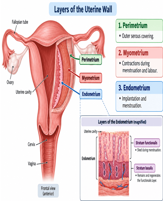
Uterus: Layers of the Uterine Wall and magnified Endometrium
Vestibule, Bartholin Glands and Clitoris
- Vaginal opening: lies posterior to the external urethral opening within the vestibule.
- Vestibule: is enclosed by the labia minora, while the labia majora form the outer folds of the vulva.
- Greater vestibular (Bartholin) glands: are paired glands located posterolateral to the vaginal opening.
- They secrete a small amount of mucus during sexual arousal, which helps lubricate the vestibule and vaginal opening.
- Clitoris: is a highly sensitive erectile organ located at the anterior junction of the labia minora.
- It contains erectile tissue and becomes engorged with blood during sexual arousal.
That is: Bartholin glands → Lubrication → Clitoris → Erectile and sensory organ important in sexual arousal
Vulva: Refers to the external female genital structures.
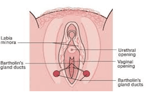
Vestibule, Bartholin Glands and Clitoris
Female Hormonal System; Ovarian Cycle, The Production, Metabolism and Actions of The Female Sex Hormones
Female Hormonal System
Female reproductive hormonal system is controlled by the hypothalamic–pituitary–ovarian (HPO) axis and involves three main levels:
- Hypothalamus: Secretes gonadotropin-releasing hormone (GnRH).
- Anterior pituitary gland: GnRH stimulates secretion of:
- Follicle-stimulating hormone (FSH)
- Luteinizing hormone (LH)
- Ovaries:
- FSH and LH stimulate the ovaries to produce: estrogens, progesterone, inhibin and other regulatory factors.
- These hormones vary cyclically throughout the menstrual cycle and regulate follicular development, ovulation, endometrial changes, and reproductive function.
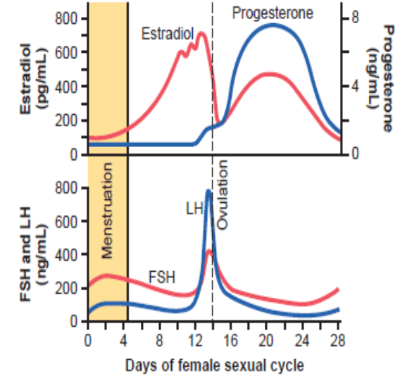
Plasma concentrations of the gonadotropins and ovarian hormones during the normal female sexual cycle. FSH, follicle-stimulating hormone; LH, luteinizing hormone.
Ovarian Cycle
Ovarian Cycle
Series of cyclic changes that occur in the ovaries during the reproductive years. It involves:
- Oogenesis: process of formation and maturation of the female germ cell (oocyte).
- Folliculogenesis: growth and development of ovarian follicles.
Ovarian cycle occurs approximately every 28 days on average, although normal cycle length varies among women.
It has three main phases:
- Follicular phase – follicular growth and maturation
- Ovulation – release of the secondary oocyte
- Luteal phase – formation and function of the corpus luteum
Oogenesis and Follicular Development in the Ovary
Oogenesis is the process by which female germ cells develop into mature ova.
1. Formation and migration of primordial germ cells
- Primordial germ cells arise early in embryonic life, temporarily localize near the yolk sac, then migrate along the hindgut mesentery to the developing gonadal ridges.
- During migration, the primordial germ cells undergo repeated mitotic divisions.
2. Formation of oogonia
- After reaching the developing ovaries, primordial germ cells differentiate into oogonia.
- Oogonia multiply rapidly by mitosis during fetal life.
3. Formation of Primary Oocytes
- Before birth, many oogonia enlarge and begin meiosis I, becoming primary oocytes.
- Meiosis I stops in prophase I and remains arrested until puberty.
4. Formation of Primordial Follicles
- Each primary oocyte becomes surrounded by a single layer of flattened follicular cells.
- This structure is called a primordial follicle.
Key Sequence: Primordial germ cell → oogonium → primary oocyte → primordial follicle.
At birth, the ovaries therefore contain mainly primary oocytes arrested in prophase I, rather than mature ova.
5. Meiotic Divisions (I & II) and Maturation of the Oocyte
- From puberty onward, selected primary oocytes complete meiosis I, forming:
- One large secondary oocyte
- One small first polar body
- Each contains 23 chromosomes, with each chromosome still composed of two sister chromatids.
- Secondary oocyte begins meiosis II but becomes arrested at metaphase II.
- Ovulation releases the secondary oocyte, not a fully mature ovum.
- Meiosis II is completed only if fertilization occurs.
- Fertilization triggers separation of the sister chromatids, producing:
- Mature ovum
- A second polar body which later degenerates
Key sequence: Secondary oocyte → metaphase II arrest → fertilization → ovum + second polar body → zygote.
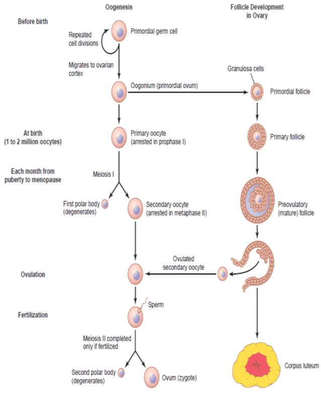
Oogenesis and Follicular Development in Fetal and Pubertal Life: Unequal cell division of oogenesis produces one to three polar bodies that later degrade, as well as a single haploid ovum, which is produced only if there is penetration of the secondary oocyte by a sperm cell.
Note: Ovarian Follicle Reserve and Atresia
- At birth, oocyte number declines from about 1–2 million at birth to 300,000–400,000 primary oocytes remaining in the ovaries at puberty.
- During the reproductive years, only about 400–500 oocytes are ovulated.
- Most follicles do not reach ovulation; instead, they undergo atresia, a process of degeneration.
- With increasing age, the ovarian follicle reserve progressively declines.
- By menopause, very few functional follicles remain, resulting in cessation of ovulation and reproductive capacity.
Key point: Most ovarian follicles undergo atresia; only a small fraction is ever ovulated.
Oocyte Arrest Points ("Pro-Meta" Rule)
- Arrest 1 (Birth to Puberty): Arrested in PROphase I of Meiosis I (PROlonged arrest).
- Arrest 2 (Ovulation to Fertilization): Arrested in METAphase II of Meiosis II (waiting until an egg MET a sperm).
Note: Comparison of Male and Female Gametogenesis
| Feature | Female (Oogenesis) | Male (Spermatogenesis) |
|---|---|---|
| Initiation | Begins during intra-uterine life (before birth) | Arrests at spermatogonia stage; commences at puberty |
| Postnatal Stem Cells | Cannot form oogonia after birth; fixed finite pool | Produces spermatogonia and primary spermatocytes continuously throughout life |
| Peak Germ Cell Count | At 5th month of gestation, ovaries contain 6 to 7 million oogonia | Spermatogonial stem cell pool constantly renews into senescence |
| Meiotic Continuity | Characterized by two prolonged arrest phases (Prophase I & Metaphase II) | Uninterrupted progression once initiated |
Ovulation
Ovulation
- Definition: Release of a mature secondary oocyte from the ovary.
- It is a key event that separates the follicular phase from the luteal phase of the ovarian cycle.
1. Timing of Ovulation
- In a typical 28-day cycle, ovulation occurs around day 14, where day 1 is the first day of menstruation.
- More generally, ovulation occurs about 14 days before the next menstrual period.
- This timing is mainly determined by the hormonal changes of the ovarian cycle.
- During the follicular phase of ovarian cycle, as the dominant follicle grows, it secretes increasing amounts of estrogen.
- When estrogen remains high for a sufficient period, it produces positive feedback on the hypothalamus and anterior pituitary.
- This leads to a sudden increase in luteinizing hormone (LH) called the LH surge.
- High estrogen → positive feedback → LH surge → ovulation
- Ovulation usually occurs about 24–36 hours after the beginning of the LH surge.
2. Development of the Mature Follicle
- During the first half of the cycle, FSH stimulates several ovarian follicles to grow.
- Usually, one becomes the dominant follicle and develops into a mature Graafian follicle.
- As it enlarges:
- Its fluid-filled cavity, the antrum, expands.
- The follicle moves closer to the surface of the ovary.
- The ovarian surface begins to bulge outward.
3. Formation of the Stigma
- Shortly before ovulation, a small area over the mature follicle becomes thin and weak. This area is called the stigma.
- The stigma forms because the LH surge stimulates:
- Proteolytic enzymes, such as collagenases, which weaken connective tissue in the follicular wall.
- Prostaglandins, which increase blood flow, vascular permeability, and local inflammatory-like changes.
- Increased follicular fluid and pressure.
- Thus: LH surge → enzymes + prostaglandins → weakening of follicular wall → stigma formation.
4. Follicular Rupture
- As the follicular wall becomes weaker, pressure within the follicle increases.
- Eventually, the stigma ruptures and follicular fluid escapes. This carries the oocyte out of the follicle.
- The released cell is not yet a completely mature ovum. It is a secondary oocyte arrested in metaphase II of meiosis.
5. What Is Released?
Ovulation releases: The secondary oocyte surrounded by:
- Its surrounding zona pellucida
- A layer of granulosa cells called the corona radiata
- The corona radiata helps protect and nourish the oocyte.
So the ovulated structure is: Secondary oocyte + zona pellucida + corona radiata.
Ovulation is fundamentally an LH-triggered rupture of the mature follicle, releasing a secondary oocyte surrounded by the zona pellucida and corona radiata.
6. Entry into the Uterine Tube
- The oocyte is initially released into the peritoneal cavity near the ovary.
- The fimbriae of the infundibulum of the uterine tube help sweep the oocyte into the tube.
- Ciliary movement and smooth-muscle contractions then transport it toward the ampulla, which is the usual site of fertilization.
7. What Happens After Ovulation?
- After the oocyte is released, the remaining follicular cells transform into the corpus luteum under the influence of LH.
- Corpus luteum secretes mainly:
- Progesterone
- Estrogen
- These hormones prepare and maintain the endometrium for possible implantation.
- If fertilization does not occur, the corpus luteum later degenerates and menstruation follows.
Key Sequence: FSH → follicular growth → ↑ estrogen → LH surge → stigma formation → follicular rupture → release of secondary oocyte → entry into uterine tube → corpus luteum formation.
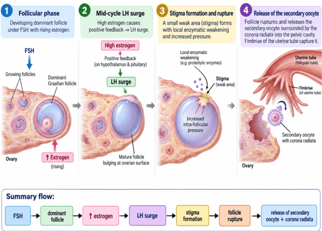
The Postulated Mechanism of Ovulation: From LH surge to release of the secondary oocyte
Features of Ovulation
| Feature | Physiological Mechanism | Clinical Characteristic |
|---|---|---|
| Timing | Follows LH peak by 24–36 hours | Day 14 in 28-day cycle; consistently 14 days prior to next menses |
| Basal Body Temperature | Thermogenic action of progesterone on hypothalamic preoptic area | Rises by 0.3–0.5°C after ovulation; persists during luteal phase |
| Cervical Mucus | High preovulatory estrogen levels | Clear, thin, watery, slippery, resembles raw egg white; facilitates sperm entry |
| Spinnbarkeit | Estrogen-induced alteration of mucin glycoproteins | Ability of cervical mucus to stretch into long strands without breaking |
| Postovulatory Mucus | Progesterone domination from corpus luteum | Becomes thick, viscous, hostile, and impermeable to sperm |
| Lower Abdominal Pain | Follicular rupture causing peritoneal irritation by fluid/blood | Mild, unilateral, mid-cycle pain (Mittelschmerz) |
Summary of Ovulatory Markers
LH surge → ovulation → corpus luteum formation → ↑ progesterone → rise in basal body temperature.
Estrogen makes cervical mucus thin and stretchy before ovulation, while progesterone makes it thick after ovulation.
Involution of the Corpus Luteum
- If pregnancy does not occur, the corpus luteum degenerates about 12–14 days after ovulation and becomes the corpus albicans (fibrous scar).
- This causes a sharp fall in progesterone, estrogen, and inhibin.
- Loss of negative feedback allows FSH and LH to rise, stimulating growth of new ovarian follicles and initiating the next ovarian cycle.
Key sequence: Corpus luteum regression → ↓ progesterone/estrogen/inhibin → ↑ FSH and LH → new follicular growth.
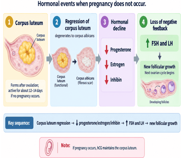
Involution of the Corpus Luteum
Disorders of Ovarian Function
Ovarian disorders may affect ovulation, hormone production, fertility, and menstrual regularity.
Ovarian Cysts
Fluid-filled sacs that develop in or on the ovary. Many are benign and asymptomatic, but some may cause pelvic pain, menstrual disturbances, or complications (e.g., rupture, torsion).
Polycystic Ovary Syndrome (PCOS)
A common endocrine disorder characterized by ovulatory dysfunction and excess androgen activity.
May cause: Irregular or absent menstruation, Infertility, Acne, Hirsutism, Polycystic ovarian morphology.
Primary Ovarian Insufficiency (POI)
Reduced or loss of normal ovarian function before age 40.
Causes / Manifestations:
- Irregular or absent menstruation
- Reduced estrogen
- Infertility
- Menopausal-type symptoms
Surgical & Infectious Ovarian Conditions
- Ovarian Cancer: Malignant growth arising from ovarian tissue. May impair ovarian and reproductive function and often requires surgery and/or systemic treatment.
- Ovarian Torsion: A painful emergency condition where an ovary twists on its supporting structures, cutting off blood supply.
- Endometriosis: When tissue similar to the lining of the uterus grows outside the uterus, including on the ovaries (forming endometriomas / "chocolate cysts").
- Pelvic Inflammatory Disease (PID): An infection and inflammation of the reproductive organs, which can include the ovaries (tubo-ovarian abscess).
Symptoms of Ovarian Disorders
Symptoms vary depending on the specific disorder but can include:
- Irregular, absent, or heavy periods
- Pelvic pain or pressure
- Bloating
- Difficulty getting pregnant
- Symptoms of hormonal imbalance like acne or excess hair growth
Ovarian Hormones
Estrogens
Major female sex hormones responsible mainly for growth, proliferation, and development of the female reproductive system and secondary sexual characteristics.
Major Estrogens
| Estrogen Type | Potency | Primary Biological Source & Predominance |
|---|---|---|
| 1. Estradiol (E2) | Most potent | Secreted mainly by the ovaries; predominant estrogen of the reproductive years |
| 2. Estrone (E1) | Intermediate (less potent than E2) | Produced by ovaries and peripheral conversion of androgens in adipose tissue; predominant after menopause |
| 3. Estriol (E3) | Weakest | Produced in large amounts by the placenta; predominant during pregnancy |
Estrogen Predominance Mnemonics
E1 = Estr-ONE: 1 letter 'O' = Old age (postmenopausal / adipose conversion).
E2 = Estr-TWO: 2 ovaries working = Reproductive years (most potent).
E3 = Estr-THREE: 3 trimesters = Pregnancy (synthesized by placenta/fetal unit).
Sources of Estrogen
- a. Ovarian follicles: principal source in non-pregnant reproductive-age women.
- b. Corpus luteum: contributes after ovulation.
- c. Adrenal glands and peripheral tissues: provide smaller amounts or androgen precursors.
- d. Placenta: major source during pregnancy.
Synthesis: Estrogens are steroid hormones synthesized from cholesterol.
Main Functions of Estrogens:
- Promote growth and maturation of the female reproductive organs.
- Produce and maintain female secondary sexual characteristics.
- Stimulate proliferation of the endometrium during the follicular phase.
- Support breast development, bone growth, and reproductive function.
Transport of Estrogen
- Circulate in the plasma mainly bound to albumin and sex hormone-binding globulin (SHBG).
- Only a small fraction circulates freely and is biologically active.
- Protein binding is reversible, allowing estrogen to be released readily to target tissues.
Plasma Estrogen Levels
Estrogen secretion varies throughout the menstrual cycle and typically shows two peaks:
- Major peak just before ovulation: Due mainly to secretion from the dominant Graafian follicle.
- Smaller peak during the mid-luteal phase: Due to secretion by the corpus luteum.
Metabolism of Estrogen
Estrogens are metabolized mainly in the liver. The liver:
- Converts estradiol and estrone into less potent metabolites, including estriol.
- Conjugates estrogens with glucuronide and sulfate.
- Conjugated metabolites are excreted mainly in the urine, with some entering the bile.
Physiological Actions of Estrogens
- 1. Growth and Development of Female Reproductive Organs:
- Estrogens promote cellular proliferation, growth, and maturation of the female reproductive system.
- At puberty, they cause enlargement and maturation of the: Ovaries, Uterine tubes (fallopian tube), Uterus, Vagina, External genitalia.
- They also contribute to development of the mons pubis, labia majora, and labia minora.
- 2. Development of Secondary Sexual Characteristics:
- Estrogens promote and maintain typical female secondary sexual characteristics, including: Breast development, Female pattern of fat distribution, Pelvic widening, Maturation of reproductive tissues.
- 3. Effects on the Vagina:
- Estrogens transform the vaginal epithelium into a thicker stratified squamous epithelium. This improves resistance to mechanical trauma and contributes to protection against infection.
- 4. Role in Ovulation:
- Rising estrogen from the dominant follicle produces positive feedback on the hypothalamus and anterior pituitary.
- This triggers the mid-cycle LH surge, which leads to ovulation and subsequent corpus luteum formation.
- 5. Effect of Estrogens on the Uterine (Fallopian) Tubes:
- Estrogens promote growth and proliferation of the tubal mucosa.
- They increase the number and activity of ciliated epithelial cells.
- Ciliary beating and tubal smooth-muscle activity help move the oocyte or early embryo toward the uterus.
- 6. Effect of Estrogens on the Breasts:
- Estrogens stimulate: Growth of the ductal system, Development of breast stromal tissue, Fat deposition in the breasts.
- Estrogens alone produce only limited development of the lobules and alveoli.
- Progesterone promotes lobuloalveolar development, while prolactin is essential for milk production.
- 7. Effect of Estrogens on the Skeleton:
- Estrogens help maintain bone mass by reducing osteoclast-mediated bone resorption.
- During puberty, estrogens act together with growth hormone and IGF-1 to produce the adolescent growth spurt.
- Estrogens also accelerate epiphyseal plate maturation and closure, eventually stopping growth in length of the long bones.
- Because girls generally enter puberty earlier than boys, linear growth usually stops earlier in females.
- Severe estrogen deficiency before epiphyseal closure can delay fusion of the growth plates and may result in continued linear growth and increased final height.
- In males, much of the effect of testosterone on epiphyseal closure is mediated after its conversion to estrogen by aromatase.
Postmenopausal Osteoporosis Due to Estrogen Deficiency
After menopause, ovarian estrogen production falls markedly. Estrogen normally helps maintain bone by limiting osteoclast-mediated bone resorption.
Estrogen deficiency causes:
- ↑ Osteoclast activity and bone resorption
- ↓ Bone mass and bone mineral density
- Progressive weakening of bone structure
This can lead to osteoporosis, increasing the risk of fractures, particularly of the: Vertebrae, Hip, Wrist. Bone loss is especially rapid during the early years after menopause.
↓ Estrogen → ↑ bone resorption → ↓ bone mineral density → osteoporosis → increased fracture risk.
The phrase "osteoporosis of old age" is better replaced with "postmenopausal osteoporosis due to estrogen deficiency."
- 8. Effect of Estrogens on Hair Distribution:
- Estrogens have little direct effect on pubic and axillary hair growth.
- After puberty, these hairs develop mainly under the influence of androgens, particularly adrenal androgens such as DHEA and DHEA-S. Estrogens may influence scalp and skin physiology, but they are not the principal regulators of androgen-dependent body hair.
- 9. Effect of Estrogens on the Skin:
- Estrogens help maintain skin thickness, collagen content, hydration, and elasticity.
- They also support cutaneous blood flow and vascularity, contributing to a soft and well-perfused skin appearance. Estrogen deficiency, especially after menopause, is associated with thinner, drier, less elastic skin.
- 10. Effect of Estrogens on Fluid and Electrolyte Balance:
- Estrogens can promote mild sodium and water retention, partly through effects on the renin–angiotensin–aldosterone system and renal sodium handling. This effect is usually modest in non-pregnant women.
- During pregnancy, high estrogen levels may contribute to increase in ECF fluid volume, although other hormones such as progesterone, aldosterone, and placental factors are also involved.
Progestins
Progesterone is the most important naturally occurring progestin in the female body.
Progestins are steroid hormones whose major functions are to:
- Prepare the endometrium for implantation and pregnancy.
- Help maintain the uterus in a state suitable for pregnancy.
- Promote development of the mammary glands in preparation for lactation.
Sources of Progesterone
- Corpus luteum of the ovary: the major source during the luteal phase of the menstrual cycle.
- Placenta: becomes the major source during pregnancy.
- Ovarian follicles: secrete small amounts, particularly around the time of ovulation.
- Small amounts are also produced by the adrenal cortex.
Synthesis: Progesterone is synthesized from cholesterol via the basic pathway: Cholesterol → Pregnenolone → Progesterone.
Transport in Blood
Progesterone is poorly soluble in water and therefore circulates mainly bound to plasma proteins:
- Approximately 98% is protein-bound:
- About 80% is bound to albumin.
- About 18% is bound to corticosteroid-binding globulin (CBG/transcortin).
- Approximately 2% circulates in the free, biologically active form.
Major Key Point: During the menstrual cycle, the corpus luteum is the principal source of progesterone, whereas during pregnancy, the placenta progressively becomes the major source.
Plasma Concentration
- During the follicular (pre-ovulatory) phase, plasma progesterone concentration is low, approximately 0.5–1 ng/mL.
- After ovulation, during the luteal phase, progesterone levels increase markedly, often by about 10–20 fold, due to secretion from the corpus luteum.
Metabolism and Fate of Progesterone
- Progesterone is rapidly metabolized, mainly in the liver.
- It is converted to inactive metabolites and conjugated mainly with glucuronic acid before excretion in the urine.
- The major urinary metabolite is pregnanediol, mainly excreted as pregnanediol glucuronide.
- Because urinary pregnanediol reflects progesterone metabolism, its excretion can be used as an indirect indicator of progesterone production in the body.
Physiological Actions of Progesterone
Progesterone plays a major role in the establishment and maintenance of pregnancy; hence, it is often referred to as the "hormone of pregnancy."
- 1. Effects on the Fallopian Tubes:
- Increases secretory activity of the mucosal lining of the fallopian tubes.
- These secretions provide nutrients for the fertilized ovum and early embryo as it moves through the fallopian tube toward the uterus before implantation.
- 2. Effects on the Breasts:
- Promotes development of the lobules and alveoli of the mammary glands.
- Stimulates proliferation and enlargement of alveolar cells and prepares them for secretory activity.
- However, progesterone alone does not initiate milk secretion. Lactation occurs mainly under the influence of prolactin after delivery.
- Progesterone may also contribute to breast enlargement and swelling through growth of glandular tissue and increased tissue fluid.
- 3. Effects on the Uterus:
- Converts the estrogen-primed endometrium into a secretory endometrium, making it suitable for implantation of the fertilized ovum.
- Increases the secretion of nutrient-rich substances by endometrial glands to support the early embryo before placental circulation is established.
- Reduces excitability and contractility of uterine smooth muscle, helping to prevent premature expulsion of the embryo or fetus.
- 4. Effects on the Cervix:
- Makes cervical mucus thick, viscous, and less permeable to sperm (contrasting with estrogen's thin, watery mucus).
- Forms a protective mucus barrier against microorganisms during pregnancy.
- 5. Effects on the Hypothalamus and Anterior Pituitary:
- Exerts negative-feedback effects on the hypothalamus and anterior pituitary gland.
- Suppresses GnRH secretion and reduces release of LH and FSH, inhibiting further follicular development and ovulation during the luteal phase and pregnancy.
- 6. Effect on Body Temperature:
- Has a thermogenic effect; increases basal body temperature by approximately 0.3–0.5°C after ovulation.
- Persists throughout luteal phase and falls when progesterone declines before menses; serves as an indirect ovulation indicator.
- 7. Role in Maintenance of Pregnancy:
- Maintaining a secretory and well-developed endometrium suitable for implantation and fetal support.
- Reducing uterine contractions and maintaining uterine quiescence.
- Supporting development of the mammary glands in preparation for lactation.
- Modulating maternal immune responses, thereby promoting tolerance of the developing fetus.
- Supporting the uterine environment until the placenta becomes the major source of progesterone.
Progesterone Key Target Summary
Progesterone primarily prepares:
- Reproductive tract for pregnancy and helps maintain pregnancy by supporting the endometrium,
- Reducing uterine contractility,
- Modifying cervical mucus,
- Suppressing further ovulation, and
- Preparing the breasts for lactation.
Prolactin
Protein hormone secreted mainly by lactotrophs (mammotrophs) (acidophilic cells of the anterior pituitary gland).
Major Actions of Prolactin
- Stimulates milk production and secretion by the mammary glands after childbirth.
- Promotes growth and development of the mammary glands, particularly during pregnancy, in cooperation with estrogen and progesterone.
- Helps maintain milk production during breastfeeding (galactopoiesis).
- High prolactin levels inhibit GnRH secretion, thereby reducing FSH and LH secretion and suppressing ovulation during breastfeeding (lactational amenorrhea).
Regulation of Prolactin Secretion
- Prolactin secretion is mainly under inhibitory control of dopamine (prolactin-inhibiting hormone, PIH) from the hypothalamus.
- Suckling reduces dopamine inhibition and markedly increases prolactin secretion.
- Estrogen also increases prolactin synthesis during pregnancy.
Prolactin vs. Oxytocin Distinction
Prolactin is primarily responsible for milk production, whereas oxytocin is responsible for milk ejection (let-down).
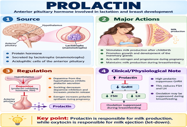
Prolactin: Anterior pituitary hormone involved in lactation and breast development
Oxytocin
Peptide hormone synthesized mainly in the paraventricular and supraoptic nuclei of the hypothalamus. It is transported along hypothalamic axons to the posterior pituitary, where it is stored and released into the bloodstream.
Physiological Actions of Oxytocin
1. Uterine Contraction
- Stimulates powerful contractions of the pregnant uterus, especially during labour.
- Near the end of pregnancy, increased uterine sensitivity to oxytocin helps initiate and strengthen labour contractions.
- During labour, cervical stretching promotes further oxytocin release through a positive-feedback mechanism (Ferguson reflex).
- After delivery, oxytocin promotes uterine contraction and involution.
- These postpartum contractions help compress uterine blood vessels and therefore reduce the risk of postpartum haemorrhage.
- During sexual activity, oxytocin may also increase uterine contractions and may assist sperm transport through the female reproductive tract.
2. Milk Ejection (Let-Down Reflex)
- Suckling stimulates sensory receptors in the nipple and areola.
- Nerve impulses travel to the hypothalamus, stimulating oxytocin release from the posterior pituitary.
- Oxytocin causes contraction of myoepithelial cells surrounding the mammary alveoli and ducts.
- This contraction forces milk from the alveoli into the ducts and toward the nipple, producing the milk ejection or let-down reflex.
- The reflex may also be triggered by stimuli such as the sight, sound, or thought of the baby.
Key Point: Prolactin produces milk, whereas oxytocin ejects milk.
Control of Oxytocin Secretion
Oxytocin secretion is mainly controlled by neuroendocrine reflexes triggered by sensory stimulation:
1. Unconditioned (Neuroendocrine) Reflexes
- a. Suckling Reflex: Suckling stimulates mechanoreceptors in nipple/areola → impulses to paraventricular/supraoptic nuclei → posterior pituitary oxytocin release → myoepithelial cell contraction → milk ejection.
- b. Labour and Coitus: Stretching of cervix/genital tract sends sensory impulses to hypothalamus → oxytocin release → uterine contraction (Ferguson positive feedback reflex).
2. Conditioned Reflexes
- In lactating women, oxytocin release and milk ejection may occur in response to the sight, sound, smell, or even thought of the baby.
- Emotional stress, anxiety, or pain may inhibit the milk-ejection reflex (via sympathetic activation).
Key Point: Suckling → Hypothalamus → Posterior pituitary → Oxytocin release → Myoepithelial contraction → Milk ejection.
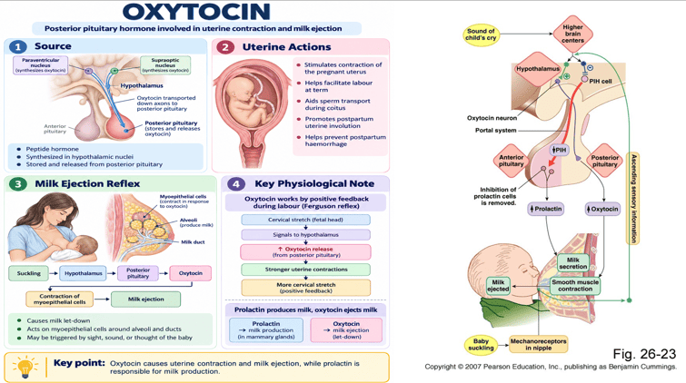
Oxytocin: Posterior pituitary hormone involved in uterine contraction and milk ejection
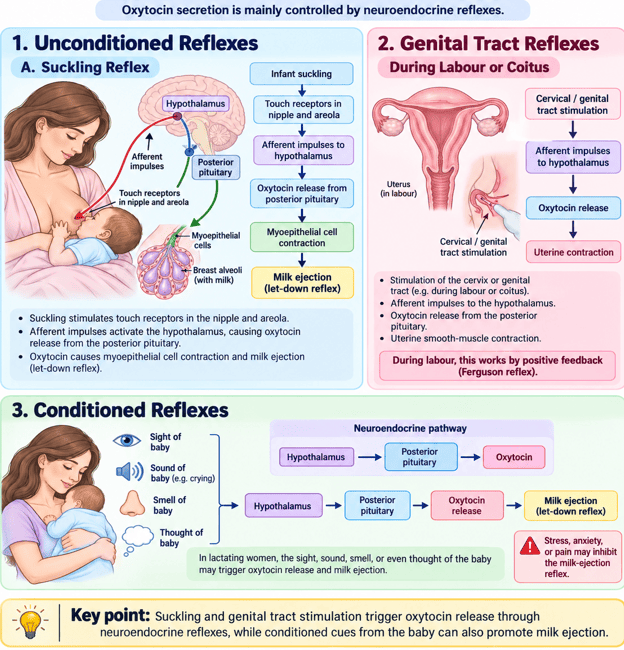
Control of Oxytocin Secretion: Unconditioned and Conditioned Pathways
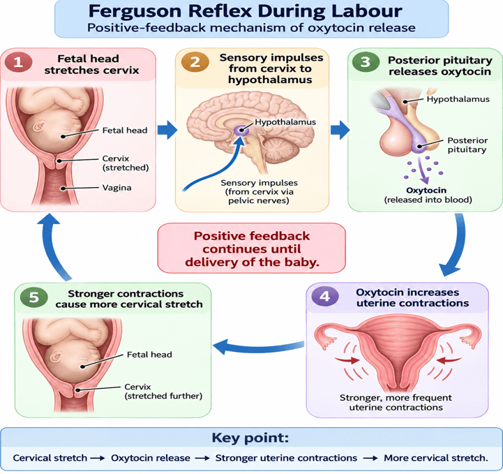
Ferguson Reflex During Labour: Positive-feedback mechanism of oxytocin release
Relaxin
Peptide hormone produced mainly by the corpus luteum during pregnancy, with additional production from the decidua and placenta.
Physiological Actions
- Relaxes and increases the flexibility of the pelvic ligaments.
- Helps soften and widen the pubic symphysis, facilitating childbirth.
- Promotes cervical softening and remodeling in preparation for labour.
- Reduces uterine smooth-muscle activity, particularly during early pregnancy, helping to maintain uterine relaxation.
- Contributes to maternal cardiovascular adaptations, including vasodilation and increased renal blood flow.
Secretion of Relaxin During Pregnancy
- Relaxin levels rise markedly in early pregnancy, with the highest concentrations generally occurring during the first trimester.
- Its effects on the cervix and pelvic tissues become particularly important as pregnancy progresses toward delivery.
Relaxin Function in Labour
Relaxin prepares the birth canal for delivery by relaxing pelvic ligaments, softening the cervix, and reducing uterine contractility.
The Menstrual Cycle
Monthly Endometrial Cycle and Menstruation
Endometrial (uterine) cycle refers to the cyclical changes that occur in the lining of the uterus during the female reproductive cycle.
These changes are regulated mainly by the ovarian hormones estrogen and progesterone. Endometrial cycle consists of three main phases:
| Phase of Endometrial Cycle | Corresponding Ovarian Phase & Days | Dominant Hormone & Morphological Changes |
|---|---|---|
| 1. Proliferative Phase | Follicular phase (Days 5–14) | Driven by Estrogen from growing follicles; stimulates regeneration of stratum functionalis; glands and vessels grow straight and lengthen |
| 2. Secretory Phase | Luteal phase (Days 15–28) | Driven by Progesterone from corpus luteum; endometrial glands become tortuous, coiled, and secrete glycogen-rich mucus; highly vascular and receptive to implantation |
| 3. Menstrual Phase | End of luteal phase (Days 1–4/5) | Driven by Hormone withdrawal (fall in estrogen and progesterone); constriction of spiral arteries → ischemia → shedding of functionalis layer with bleeding |
Hormonal Control of the Endometrium
Estrogen promotes endometrial proliferation, progesterone converts the endometrium into a secretory state, and withdrawal of both hormones leads to menstruation.
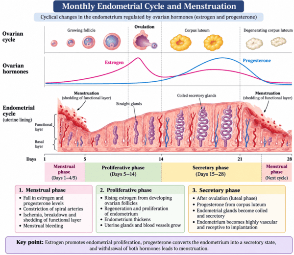
Monthly Endometrial Cycle and Menstruation: Cyclical changes in the endometrium regulated by ovarian hormones
Components of Menstrual Fluid
Menstrual fluid is a mixture of blood, endometrial tissue, cervical and vaginal secretions, cells, and biochemical substances. It is therefore not composed entirely of blood.
- Blood:
- Blood constitutes a substantial proportion of menstrual fluid, but menstrual discharge is not pure blood.
- It contains plasma and blood cells derived mainly from disrupted endometrial blood vessels.
- Endometrial Tissue:
- Menstruation involves shedding of the functional layer of the endometrium.
- Fragments of endometrial tissue therefore form an important component of menstrual discharge.
- Cervical and Vaginal Secretions:
- Menstrual fluid contains cervical mucus and vaginal secretions.
- These contribute water, electrolytes, mucus, and other substances to the menstrual flow.
- Cells: Menstrual fluid contains various cell types, including:
- Endometrial epithelial cells
- Stromal cells
- Red blood cells
- Leukocytes and other immune cells
- Some stem/progenitor cells
- Biochemical Substances:
- Contains proteolytic and fibrinolytic enzymes (e.g., plasmin), which assist in tissue breakdown and help prevent extensive clot formation under normal physiological conditions.
- Contains prostaglandins, cytokines, and other signaling molecules involved in inflammation, vascular changes, and endometrial shedding.
Common Menstrual Disorders
1. Dysmenorrhea
Painful menstruation characterized by lower abdominal or pelvic cramps occurring before or during menstruation.
- Primary: without underlying pelvic disease (mediated by excess uterine prostaglandin PGF2α).
- Secondary: due to underlying pelvic pathology such as endometriosis or uterine fibroids.
2. Menorrhagia
Excessive or prolonged menstrual bleeding occurring at regular menstrual intervals. The modern clinical term is often heavy menstrual bleeding (HMB).
3. Amenorrhea
Absence of menstruation.
- Primary amenorrhea: menstruation has not begun by approximately 15 years of age in an individual with otherwise normal development.
- Secondary amenorrhea: absence of menstruation for 3 months or more in someone who previously had regular cycles.
Clinical Nomenclature of Menstrual Variations
| Disorder Term | Clinical Definition & Diagnostic Criteria |
|---|---|
| 4. Oligomenorrhea | Infrequent menstruation, usually with menstrual cycles occurring more than 35 days apart |
| 5. Polymenorrhea | Frequent menstruation, traditionally defined as menstrual cycles occurring at intervals of less than 21 days |
| 6. Hypomenorrhea | Menstrual bleeding that is unusually light or of short duration |
| 7. Metrorrhagia | Irregular uterine bleeding occurring between expected menstrual periods (intermenstrual bleeding) |
| 8. Menometrorrhagia | Heavy, prolonged, and irregular uterine bleeding occurring at unpredictable intervals |
9. Premenstrual Syndrome (PMS)
A group of physical, emotional, and behavioral symptoms that occur during the luteal phase, usually several days before menstruation, and improve shortly after menstruation begins.
Common symptoms include: breast tenderness, bloating, headache, irritability, mood changes, and fatigue.
Menstrual Irregularity Diagnostic Quick-Check
- Poly- vs. Oligo-: Polymenorrhea = too frequent (< 21 days); Oligomenorrhea = too infrequent (> 35 days).
- -rrhagia vs. -rhea: Menorrhagia = heavy at normal times; Metrorrhagia = bleeding at wrong/irregular times; Menometrorrhagia = heavy AND irregular.
