Fertilization and Stages of Fertilization
Fertilization
- Fusion of the male and female gametes to form a zygote.
- Following ejaculation, about 200–300 million spermatozoa may be deposited in the female reproductive tract, but normally only one sperm fertilizes the secondary oocyte.
- Fertilization usually occurs in the ampulla of the uterine (fallopian) tube.
- To reach the oocyte, the sperm must penetrate:
- Corona radiata, formed by surrounding granulosa cells.
- Zona pellucida, the glycoprotein layer surrounding the oocyte.
- Penetration is facilitated by the acrosome reaction, during which enzymes such as acrosin and hyaluronidase are released from the sperm acrosome.
- After one sperm fuses with the oocyte membrane, a cortical reaction occurs.
- Cortical granules release substances that modify and harden the zona pellucida, producing a block to polyspermy and preventing additional sperm from entering.
- The male and female pronuclei subsequently fuse, restoring the diploid chromosome number and forming the zygote.
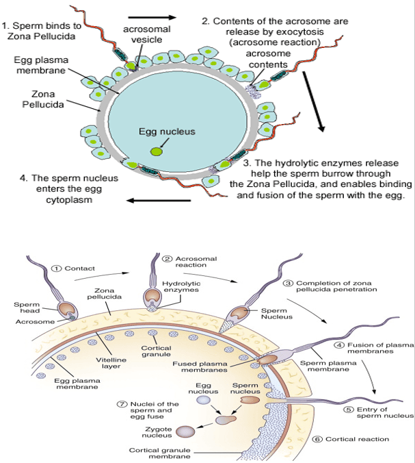
Fertilization and Acrosomal Penetration
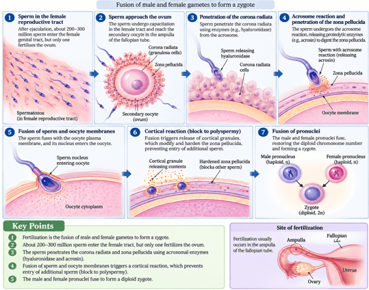
Fertilization Process and Site of Fertilization
Sperm Transit Through the Female Reproductive Tract
- Sperm transit refers to the movement of spermatozoa through the female reproductive tract toward the site of fertilization, usually the ampulla of the fallopian (uterine) tube.
- Fertilization may be described as a “numbers game” because millions of sperm are deposited during ejaculation, but only a very small proportion reach the fallopian (uterine) tube, and normally only one sperm fertilizes the oocyte.
- During ejaculation, hundreds of millions of spermatozoa are deposited in the vagina.
- Many are rapidly lost because of the acidic vaginal environment, while others fail to pass through the cervical mucus.
- Sperm that enter the uterus may also be removed by phagocytic leukocytes. As a result, only a small fraction of the original sperm population reaches the uterine (fallopian) tubes, where fertilization usually occurs.
- Sperm transport is aided by their own motility as well as uterine and tubal contractions.
- Sperm may reach the uterine (fallopian) tubes within approximately 30 minutes to a few hours after ejaculation.
Capacitation of Sperm
During their passage through the female reproductive tract, sperm undergo a series of functional changes known as capacitation, which prepares them for fertilization.
Major Changes During Capacitation
| Functional Change | Physiological Mechanism | Biological Purpose |
|---|---|---|
| Increased motility | Hyperactivation; develops enhanced, vigorous whiplash motility | Improves ability to swim through tubal fluid and penetrate the oocyte vestments |
| Changes in the sperm membrane | Cholesterol and surface glycoproteins removed from the acrosomal cap | Increases membrane fluidity; prepares the membrane for acrosome reaction |
| Preparation for enzyme release | Destabilization of acrosomal membrane allows subsequent exocytosis | Enables release of acrosin and hyaluronidase to digest outer coverings |
| Acquisition of fertilizing ability | Completion of functional maturation within female tract | Allows binding to ZP3 receptors; uncapacitated sperm cannot penetrate zona pellucida |
- Acquisition of fertilizing ability:
- Sperm must complete capacitation before they can successfully fertilize the oocyte.
- If sperm reach the oocyte before capacitation is complete, they are generally unable to undergo the acrosome reaction effectively and cannot penetrate the zona pellucida.
Capacitation Triad ("M-M-E")
M — Motility Hyperactivation: Whiplash tail movement driven by calcium influx.
M — Membrane Destabilization: Cholesterol efflux increases membrane fluidity.
E — Enzyme Competence: Prepares acrosomal cap for release of hyaluronidase and acrosin.
Other Key Points To Note
- Large numbers of sperm are required:
- Millions of sperm are ejaculated into the female reproductive tract.
- Most are lost or eliminated as they pass through the vagina, cervix, uterus, and uterine tubes.
- Very low individual success rate:
- Only a small fraction of ejaculated sperm reach the vicinity of the oocyte.
- Of these, usually only one sperm successfully penetrates and fertilizes the oocyte.
- Importance of sperm quality:
- Successful sperm transit depends on adequate sperm count, motility, morphology, and functional competence.
- Poor sperm quality can reduce the likelihood of successful fertilization.
- Female reproductive tract assists sperm transport:
- Cervical mucus, uterine contractions, and tubal movements help facilitate sperm movement toward the site of fertilization.
- Sperm also undergo capacitation within the female reproductive tract, which prepares them for fertilization.
Survival of Sperm and Oocyte
- If sperm do not encounter an oocyte immediately, they may remain viable in the female reproductive tract for approximately 3–5 days.
- Therefore, fertilization can still occur when intercourse takes place several days before ovulation.
- After ovulation, the secondary oocyte remains viable for about 12–24 hours.
- Consequently, intercourse occurring more than about 24 hours after ovulation is much less likely to result in fertilization.
Contact Between Sperm and Oocyte
- Following ovulation, the secondary oocyte is released from the ovary and captured by the fimbriae of the uterine tube.
- It is then transported along the uterine tube by ciliary action and smooth-muscle contractions.
- Fertilization usually occurs in the ampulla of the uterine tube, where sperm normally encounter the oocyte.
- Because the oocyte remains viable for only about 12–24 hours after ovulation, fertilization must occur relatively soon after its release.
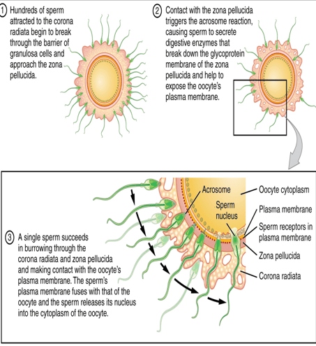
Contact Between Sperm and Oocyte and Penetration Mechanisms
Protective Layers Surrounding the Oocyte
The ovulated oocyte is surrounded by two important layers:
| Layer | Structure & Origin | Enzyme Cleaved By | Primary Physiological Role |
|---|---|---|---|
| 1. Corona radiata | Outer layer composed of granulosa cells remaining attached after ovulation | Hyaluronidase (disperses intercellular matrix) | Protects and nourishes the secondary oocyte during tubal transit |
| 2. Zona pellucida | Thick, transparent glycoprotein coat surrounding oocyte plasma membrane | Acrosin (proteolytically digests ZP matrix) | Species-specific sperm binding (ZP3), induction of acrosome reaction, and permanent block to polyspermy |
Sperm Penetration of the Corona Radiata, Zona Pellucida and Oocyte
- In the uterine tube, capacitated spermatozoa are guided toward the oocyte by chemical signals released from the oocyte–corona radiata complex.
- To reach the oocyte plasma membrane, sperm must penetrate two major protective layers:
- Corona radiata
- Zona pellucida
- Many sperm contribute to dispersing the cells of the corona radiata through vigorous motility and the action of enzymes such as hyaluronidase.
- When a capacitated sperm binds to the zona pellucida, it undergoes the acrosome reaction, releasing acrosomal enzymes (acrosin) which assist sperm penetration through the zona pellucida.
- The combined action of acrosomal enzymes and vigorous sperm motility enables the sperm to penetrate the oocyte coverings.
- Normally, only one sperm reaches and fuses with the oocyte plasma membrane.
- Sperm–oocyte fusion triggers the cortical reaction, during which cortical granules release substances that modify and harden the zona pellucida, thereby establishing the major block to polyspermy.
- The sperm nucleus enters the oocyte and forms the male pronucleus.
- Because only a small proportion of deposited sperm reach the site of fertilization, severely reduced sperm count, motility or functional competence can decrease the likelihood of successful fertilization.
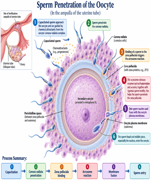
Sperm Penetration of the Oocyte
Prevention of Polyspermy
- After sperm–oocyte fusion, the oocyte undergoes a cortical reaction.
- Cortical granules modify and harden the zona pellucida, preventing entry of additional sperm.
- This block to polyspermy preserves the normal diploid chromosome number.
- Polyspermy may cause polyploidy (e.g., triploidy), leading to abnormal development and usually early pregnancy loss.
Mechanisms That Prevent Polyspermy
| Mechanism | Onset & Duration | Biochemical / Electrical Mediator | Physiological Consequence |
|---|---|---|---|
| 1. First Mechanism (Fast Block) | Sets in almost immediately; lasts for ~1 minute | Instantaneous increase in Na⁺ permeability → rapid membrane depolarization (-70 mV to +20 mV) | Prevents additional sperm from fusing with oolemma; triggers calcium wave for slow block |
| 2. Second Mechanism (Slow Block / Zona Reaction) | Initiated within minutes; provides permanent, long-lasting block | Ca²⁺ influx triggers exocytosis of cortical granules into perivitelline space | Proteases modify and harden zona pellucida; hydrolyzes ZP2 and ZP3 sperm receptors |
Mammalian Polyspermy Block
In mammals, the electrical fast block is transient and incomplete. The slow block (cortical/zona reaction) mediated by cortical granule exocytosis provides the principal, definitive barrier to polyspermy.
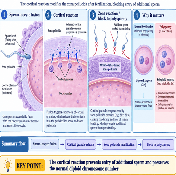
Prevention of Polyspermy and Zona Reaction
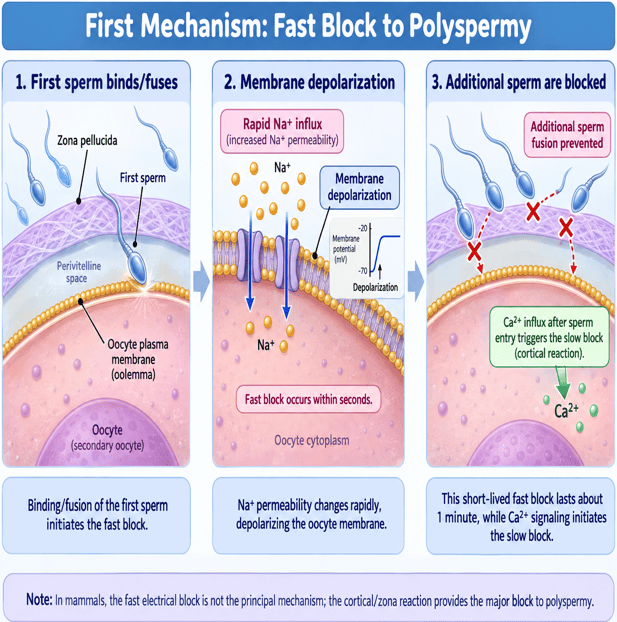
First Mechanism: Fast Block to Polyspermy
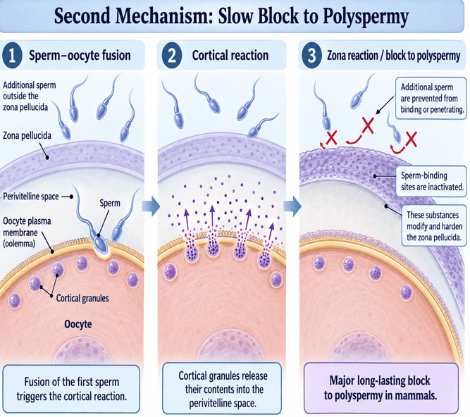
Second Mechanism: Slow Block to Polyspermy
Implantation and Placentation
Implantation
- Process by which the developing blastocyst attaches to and becomes embedded in the endometrial lining of the uterus.
- After fertilization in the fallopian tube, the zygote undergoes repeated mitotic divisions (cleavage) while moving toward the uterus.
- It usually takes about 3–5 days for the developing embryo to reach the uterine cavity.
- During its passage through the fallopian tube, the embryo receives nourishment from tubal secretions.
- The embryo develops into a morula (16–32 cells) and then a blastocyst before implantation.
- Implantation usually begins around day 6–7 after fertilization, when the blastocyst attaches to the receptive endometrium, and is generally complete by the 10th day.
- Successful implantation establishes the early physical and nutritional relationship between the embryo and the mother, allowing further development of the placenta and pregnancy.
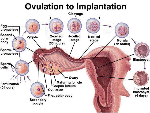
Fertilization, Cleavage & Formation of a Blastocyst Across the First Week
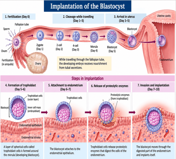
Implantation of the Blastocyst Steps
Mechanism of Implantation
- As the morula develops into a blastocyst, its outer layer of cells differentiates into the trophoblast.
- Trophoblast attaches to the endometrial epithelium and begins the process of implantation.
- Trophoblastic cells release proteolytic enzymes that digest the extracellular matrix and cells of the endometrium.
- This allows the blastocyst to invade and become embedded within the endometrial lining.
- Trophoblast subsequently differentiates into:
- Cytotrophoblast: Inner layer of individual mononucleated cells.
- Syncytiotrophoblast: Outer multinucleated invasive syncytium lacking distinct cell borders; secretes human chorionic gonadotropin (hCG).
Sequence of Implantation:
Morula → Blastocyst → Trophoblast attachment → Endometrial invasion → Implantation
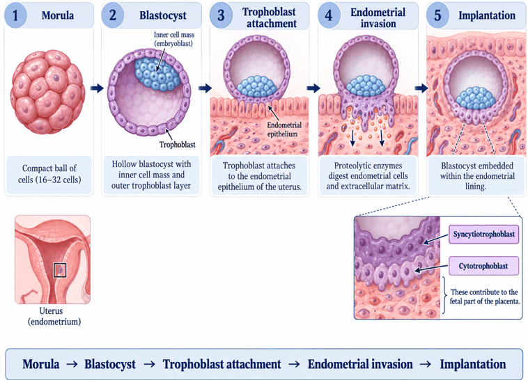
Mechanism of Implantation
Placentation and Establishment of Pregnancy
Development of the Placenta
- After implantation, the placenta develops from the fetal chorion (trophoblast) and the maternal endometrium (decidua basalis).
- Placenta is a temporary, highly vascular organ present during pregnancy and is expelled after childbirth.
- It forms the major structural and physiological connection between the mother and fetus.
- Placenta helps anchor the developing fetus to the uterus and facilitates the exchange of oxygen, nutrients, wastes, and hormones between maternal and fetal circulations.
- It is therefore essential for the maintenance and progression of pregnancy.
Sequence of Events:
1. Implantation → 2. Placental development → 3. Maternal-fetal connection → 4. Maintenance of pregnancy
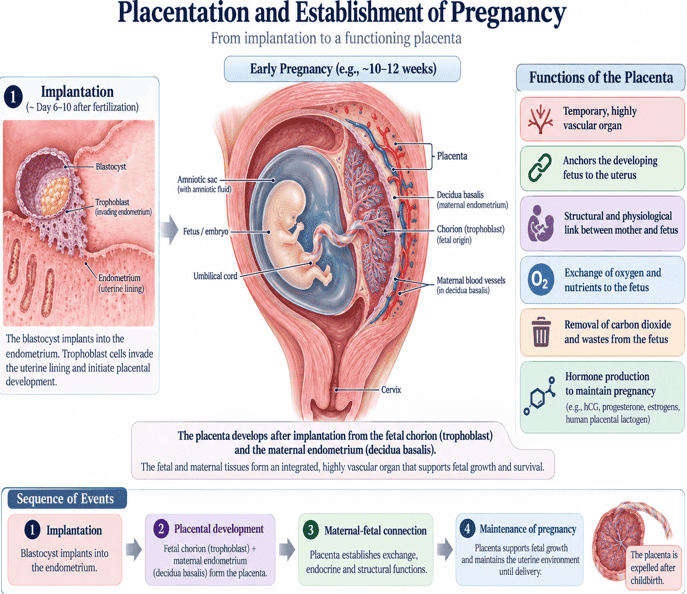
Placentation and Establishment of Pregnancy
Structure and Delivery of the Placenta
- Placenta is formed from both fetal and maternal tissues and therefore has two components:
- Fetal part: derived mainly from the chorion.
- Maternal part: derived from the uterine endometrium (decidua basalis).
- Placenta is connected to the fetus by the umbilical cord, which contains fetal blood vessels embedded in connective tissue (Wharton's jelly).
- After delivery of the baby, the placenta is expelled as the afterbirth.
- The umbilical cord is then clamped and cut. The site where the umbilical cord was attached to the fetal anterior abdominal wall becomes the umbilicus (navel).
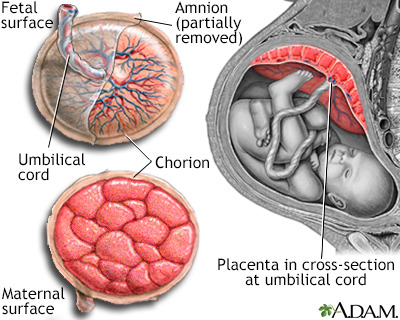
Structure and Surfaces of the Placenta
Functions of the Placenta
Placenta performs five essential functions for the developing fetus:
1. Respiratory Function
- Fetal lungs are not functional for gas exchange before birth; therefore, the placenta acts as the fetal respiratory organ.
- Oxygen (O₂) diffuses from maternal blood across the placental membrane into fetal blood.
- Carbon dioxide (CO₂) diffuses in the opposite direction, from fetal blood into maternal blood for elimination by the mother's lungs.
- Thus, the placenta enables continuous maternal–fetal gas exchange required for fetal survival and development.
2. Nutritive and Metabolic Function
- Placenta transports essential nutrients from the mother to the fetus, including:
- Carbohydrates: mainly glucose (via facilitated diffusion)
- Proteins: mainly amino acids (via active transport)
- Lipids: fatty acids and essential lipids
- Vitamins and minerals: including Na⁺, K⁺, Cl⁻, Ca²⁺ and phosphate
- The placenta also stores and metabolizes substances such as glycogen and lipids.
3. Excretory Function
- Fetal waste products diffuse across the placenta into maternal blood for elimination.
- These include urea, uric acid and creatinine.
4. Protective Function
- Placenta acts as a selective barrier, reducing the passage of some harmful substances from mother to fetus. However, it is not an absolute barrier, as some drugs, toxins and microorganisms can cross it.
- Maternal IgG antibodies cross the placenta and provide the fetus with passive immunity.
5. Endocrine Function of the Placenta
Placenta functions as an important endocrine organ during pregnancy and secretes several hormones:
| Placental Hormone | Secretory Cell Source | Key Physiological Actions |
|---|---|---|
| i. Human Chorionic Gonadotropin (hCG) | Syncytiotrophoblast | Maintains the corpus luteum in early pregnancy; supports progesterone secretion; detectable in blood shortly after implantation and in urine slightly later; basis of pregnancy tests. |
| ii. Human Placental Lactogen (hPL / hCS) | Syncytiotrophoblast | Actions similar to growth hormone and prolactin; promotes maternal insulin resistance to increase glucose availability to fetus; stimulates mammary development. |
| iii. Estrogens | Syncytiotrophoblast (fetal-placental unit) | Promote growth of the uterus, breasts, and maternal reproductive tissues during pregnancy; increase uterine blood flow. |
| iv. Progesterone | Corpus luteum (early) → Placenta (8–12 wk) | Maintains the endometrium; prevents menstruation; reduces uterine contractions (uterine quiescence); supports mammary gland development. |
| v. Relaxin | Corpus luteum, Placenta, and Decidua | Promotes relaxation of pelvic ligaments; softens the cervix in preparation for childbirth; increases pubic symphysis flexibility. |
Physiology of Pregnancy
- Results from successful fertilization followed by implantation of the developing embryo in the uterus.
- Gestational age is conventionally calculated from the first day of the last menstrual period (LMP).
- A normal pregnancy lasts approximately 280 days (40 weeks) from the LMP, equivalent to about 9 months and 7 days.
- In a regular 28-day menstrual cycle, ovulation and fertilization usually occur around day 14.
- Therefore, the actual duration from fertilization to birth is approximately 266 days (38 weeks).
- Miscarriage refers to pregnancy loss before fetal viability; WHO definitions may classify pregnancy loss before 28 completed weeks as miscarriage.
- Preterm birth is the birth of a live baby before 37 completed weeks of gestation.
Summary:
- LMP → 40 weeks (280 days) → Delivery
- Fertilization → ~38 weeks (266 days) → Delivery
- Before 37 weeks is termed preterm birth, while preterm labour specifically refers to labour beginning before 37 completed weeks.
Maternal Changes During Pregnancy
Pregnancy produces important structural, metabolic and physiological changes in the mother to support fetal growth and prepare for childbirth and lactation.
1. Structural (Anatomical) Changes
Changes occur mainly in the reproductive organs and mammary glands:
| Organ | Structural & Anatomical Adaptations | Physiological Significance |
|---|---|---|
| Ovaries | Ovulation and new follicular development cease (FSH and LH suppressed). Corpus luteum enlarges. | Secretes progesterone and estrogen to maintain the endometrium until the placenta assumes endocrine production at 8–12 weeks. |
| Vagina | Becomes more vascular, slightly enlarged; increased epithelial glycogen deposition. | Supports lactobacilli, maintaining an acidic environment to inhibit pathogenic infection. |
| Cervix | Increased vascularity and mucus secretion (cervical softening); formation of thick cervical mucus plug. | Seals the cervical canal to protect the uterine cavity from ascending bacterial infections. |
| Uterus | Enlarges from pyriform (pear-shaped) to globular, then ovoid. Weight increases from 50–70 g to 900–1,000 g at term. | Driven by myometrial hypertrophy and hyperplasia; blood flow increases markedly to supply growing placenta and fetus. |
| Mammary Glands | Ducts, lobules, and alveoli enlarge; increased fat deposition; nipples and areolae enlarge and darken. | Stimulated by estrogen, progesterone, prolactin, and hPL to prepare breasts for postpartum lactation. |
2. Metabolic Changes During Pregnancy
- i. Basal Metabolic Rate (BMR):
- BMR increases progressively, especially in the second and third trimesters, because of increased maternal and fetal metabolic demands. This provides the extra energy needed by the mother and growing fetus.
- ii. Protein Metabolism:
- Protein synthesis and retention increase to support growth of the uterus, breasts, placenta and fetus. This produces a positive nitrogen balance.
- iii. Carbohydrate Metabolism:
- During pregnancy, especially in the later months, the body becomes less sensitive to insulin (insulin resistance mediated by hPL, cortisol, and progesterone).
- Therefore, blood glucose may rise after meals, while fasting glucose may be slightly lower.
- Glucose may appear in the urine (glucosuria) because the kidneys filter more glucose and reabsorb it less efficiently.
- iv. Lipid Metabolism:
- Maternal fat stores increase during early pregnancy.
- Blood triglycerides and cholesterol increase.
- In late pregnancy, increased fat breakdown provides energy for the mother and helps preserve glucose for the fetus.
- v. Water and Mineral Metabolism:
- There is increased sodium and water retention, causing expansion of maternal blood and extracellular fluid volume.
- Requirements for minerals such as iron and calcium also increase to support maternal and fetal needs.
3. Changes in Physiological Systems
| Physiological System | Primary Adaptations | Clinical Characteristics |
|---|---|---|
| Blood | Maternal blood volume increases by 40–50%; plasma volume increases more than red cell mass. | Produces hemodilution, resulting in physiological anemia of pregnancy; iron requirements markedly increase. |
| Cardiovascular System (CVS) | Cardiac output increases by 30–50% (increased stroke volume and heart rate). | Systemic vascular resistance falls; blood pressure drops slightly in mid-pregnancy and returns to baseline near term. |
| Respiratory System | Tidal volume and minute ventilation increase; respiratory rate unchanged; O₂ consumption rises. | Mild dyspnea/breathlessness occurs commonly in late gestation due to diaphragmatic elevation by expanding uterus. |
| Renal/Excretory System | Renal blood flow and glomerular filtration rate (GFR) increase substantially. | Urinary frequency is common in early and late pregnancy due to mechanical uterine compression on the bladder. |
| Gastrointestinal System | Nausea and vomiting (morning sickness) in early weeks; progesterone relaxes smooth muscle. | Decreased gastrointestinal motility predisposing to constipation and gastroesophageal reflux (heartburn). |
| Nervous System | Fatigue, sleep disturbances, mood swings, and altered emotional responses. | Reflects hormonal and physical adaptation. Does not cause generalized excitation of the nervous system. |
Hemodynamic Summary:
Pregnancy → ↑ Blood volume → ↑ Heart rate & stroke volume → ↑ Cardiac output → Slight fall in BP during mid-pregnancy
In Summary .....
1. Physiological Basis of Pregnancy
- Pregnancy begins with fertilization of the ovum by a spermatozoon, followed by development of the embryo and successful implantation of the blastocyst in the uterine endometrium.
Fertilization and Implantation
- Fertilization usually occurs in the ampulla of the fallopian tube.
- Zygote undergoes cleavage to form the morula and subsequently the blastocyst.
- The blastocyst reaches the uterine cavity within about 3–5 days.
- Implantation begins approximately 6–7 days after fertilization.
Role of the Trophoblast
- Following implantation, trophoblastic cells differentiate into:
- Cytotrophoblast
- Syncytiotrophoblast
- Syncytiotrophoblast invades the endometrium and secretes human chorionic gonadotropin (hCG).
Role of hCG
- hCG maintains the corpus luteum of pregnancy during the early weeks.
- The corpus luteum continues to secrete progesterone and estrogen. These hormones maintain the endometrium, suppress menstruation and support early embryonic development.
- By approximately 8–12 weeks of pregnancy, the placenta becomes the major source of progesterone and estrogen.
Hormonal Maintenance of Pregnancy
- Progesterone:
- Maintains the endometrium.
- Reduces uterine contractions.
- Supports development of the mammary glands.
- Estrogens:
- Promote enlargement of the uterus.
- Increase uterine blood flow.
- Promote breast and reproductive tissue development.
- Human placental lactogen (hPL):
- Alters maternal metabolism to increase nutrient availability to the fetus.
- Contributes to mammary gland development.
- Relaxin:
- Promotes relaxation of pelvic ligaments and softening of the cervix.
2. Physiological Basis of Pregnancy Testing
- Pregnancy tests are primarily based on the detection of human chorionic gonadotropin (hCG) produced by the syncytiotrophoblast after implantation.
Human Chorionic Gonadotropin Subunits
- hCG enters the maternal circulation shortly after implantation and subsequently appears in the urine.
- hCG has two parts: alpha (α) and beta (β):
- β-hCG: commonly detected because it provides greater specificity than the common α-subunit. The beta part is unique to hCG, so pregnancy tests usually detect β-hCG to identify hCG more accurately.
- α-hCG: is similar to that of LH, FSH, and TSH, so it is not very specific.
Types of Pregnancy Tests
- A. Urine Pregnancy Test:
- Detects hCG in urine using an immunoassay and is commonly used for home and clinical pregnancy testing.
- Usually becomes reliably positive around the time of the missed menstrual period.
- First-morning urine may improve sensitivity because it contains a higher concentration of hCG.
- B. Serum β-hCG Test:
- Serum testing is more sensitive than urine testing.
- It may be:
- Qualitative: Reports whether hCG is present or absent.
- Quantitative: Measures the actual concentration of β-hCG in blood. Useful for assessing very early pregnancy and monitoring pregnancy progression.
- hCG can usually be detected in maternal blood approximately 8–10 days after ovulation/fertilization.
Changes in hCG During Pregnancy
- hCG rises rapidly during early pregnancy. In many normal early pregnancies, its concentration doubles substantially every 48–72 hours.
- Levels usually reach a peak at approximately 8–11 weeks of gestation.
- They subsequently decline and remain at a lower level for the remainder of pregnancy.
Interpretation of hCG Pregnancy Tests
| Test Outcome | Underlying Causes & Clinical Scenarios |
|---|---|
| Positive Test | Usually indicates normal pregnancy; may occur in trophoblastic disease or germ cell tumors. |
| False-Negative Result | Testing performed too early; urine excessively diluted; hCG concentration below test detection limit. |
| False-Positive Result | Recent pregnancy or pregnancy loss; administration of hCG-containing fertility medications; trophoblastic or germ-cell disorders; rare assay/heterophile antibody interference. |
Confirmation of Pregnancy
Pregnancy may be further confirmed and assessed using:
- Serial serum β-hCG measurements
- Ultrasonography
- Ultrasound is particularly important for confirming the location, development and viability of the pregnancy.
Endocrine Cascade Summary:
Fertilization → Blastocyst → Implantation → Syncytiotrophoblast → hCG secretion → Maintenance of corpus luteum → Progesterone and estrogen secretion → Maintenance of pregnancy
Pregnancy Testing Sequence:
Implantation → hCG enters blood → hCG appears in urine → Detection by serum or urine pregnancy test
Parturition
- Process of childbirth, involving the expulsion of the fetus and placenta from the uterus at the end of pregnancy.
- Near term, the uterus becomes progressively more excitable and contractile, eventually producing strong, coordinated rhythmic contractions that lead to delivery.
- The exact mechanism initiating labour is complex, but two major factors contribute:
- Hormonal changes:
- Increased uterine sensitivity to oxytocin
- Increased production of prostaglandins
- Functional reduction in progesterone influence with increasing estrogenic effects
- Mechanical factors:
- Progressive stretching of the uterus
- Pressure of the fetal head on the cervix, which further stimulates uterine contractions
- Together, these changes initiate and sustain the strong uterine contractions responsible for labour and delivery.
- Hormonal changes:
Stages of Parturition
| Stage of Labour | Onset to Endpoint | Key Mechanical & Clinical Events |
|---|---|---|
| 1. First Stage (Cervical Dilatation) | Onset of regular contractions → Full cervical dilatation (10 cm) | Contractions spread downward from fundus; fetal head pushes against cervix, causing effacement and dilatation. |
| 2. Second Stage (Delivery of Fetus) | Full cervical dilatation → Complete birth of the infant | Strong contractions assisted by maternal pushing move fetus through birth canal. Duration is variable. |
| 3. Third Stage (Delivery of Placenta) | Birth of infant → Expulsion of placenta and membranes | Uterine retraction shears placenta from uterine wall; expelled as the afterbirth within 5–30 minutes. |
True Labour vs. Braxton-Hicks Contractions
Braxton-Hicks contractions are irregular, painless "practice" contractions occurring throughout pregnancy without cervical effacement or dilatation. True labour contractions are regular, progressively increase in frequency and intensity, and cause progressive cervical dilatation.
Onset of Labour: Positive Feedback Mechanism
- During pregnancy, the uterus shows intermittent weak contractions (Braxton Hicks contractions).
- Near term, uterine contractions become stronger, more regular and coordinated, eventually developing into true labour contractions.
- As the fetal head stretches the cervix, sensory signals trigger stronger uterine contractions.
- These contractions push the fetus further downward, causing additional cervical stretch.
- Cervical stretch also stimulates release of oxytocin from the posterior pituitary, which further increases uterine contractions. This creates a positive feedback cycle that continues until the baby is delivered.
Positive Feedback Sequence (Ferguson Reflex):
Cervical stretch → Oxytocin release → Stronger uterine contractions → Greater fetal descent → More cervical stretch → Further oxytocin release → Delivery
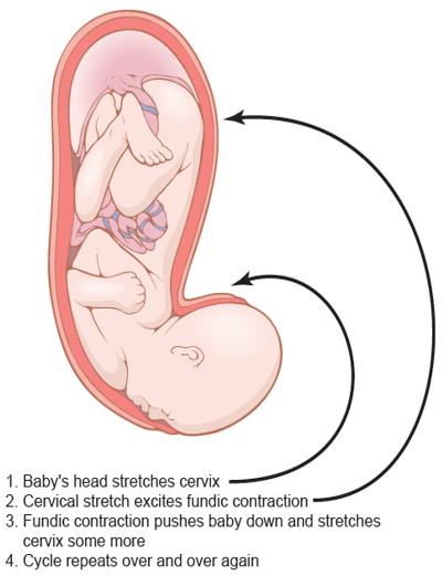
Theory for the Onset of Intensely Strong Contractions During Labor
Mechanisms of Parturition/Labour
The exact trigger for labour is not fully understood, but hormonal and mechanical factors increase uterine excitability and contractility near term.
1. Hormonal Factors
- a. Increased Estrogenic Influence Relative to Progesterone:
- Progesterone maintains uterine relaxation during pregnancy.
- Estrogen increases uterine excitability by promoting gap junction formation (connexin 43) and increasing responsiveness to oxytocin.
- Near term, the balance shifts toward greater estrogenic activity relative to progesterone (functional progesterone withdrawal), making the myometrium more contractile.
- This contributes to the onset and progression of labour.
- b. Oxytocin Increases Uterine Contractions:
- Oxytocin, released from the posterior pituitary, stimulates strong uterine contractions.
- Near term, the myometrium develops more oxytocin receptors, increasing uterine sensitivity.
- Cervical stretching stimulates further oxytocin release through the Ferguson reflex.
- Oxytocin strengthens contractions, causing more cervical stretch and further oxytocin release.
- Sequence: Cervical stretch → Oxytocin release → Stronger uterine contractions → More cervical stretch.
- c. Effect of Fetal Hormones on the Uterus:
- Fetus’s pituitary gland secretes increasing quantities of oxytocin, which might play a role in exciting the uterus.
- Also, the fetus’s adrenal glands secrete large quantities of cortisol, another possible uterine stimulant.
- In addition, the fetal membranes release prostaglandins in high concentration at the time of labor.
- These prostaglandins, too, can increase the intensity of uterine contractions.
- d. Prostaglandins:
- Prostaglandins, especially PGE₂ (and PGF₂α), help initiate and strengthen labor.
- They are produced by the uterus, fetal membranes, and placenta.
- Their levels increase in maternal blood and amniotic fluid during labor.
- They make uterine muscles contract more strongly by increasing calcium inside muscle cells.
- e. Progesterone:
- During pregnancy, progesterone keeps the uterus relaxed and reduces uterine contractions.
- Near term, its relaxing effect decreases (functional progesterone withdrawal).
- This allows increased prostaglandin activity and uterine contractions, helping labor begin.
- f. Estrogen:
- Estrogen levels and effects increase toward the end of pregnancy.
- Estrogen increases uterine excitability and contractions; increases oxytocin receptors in the uterus; promotes prostaglandin production.
- In late pregnancy, stretching of the uterus by the growing fetus also increases uterine excitability and helps promote contractions.
- g. Cortisol:
- Cortisol levels rise during labor as part of the body’s stress response.
- It helps the mother meet the increased energy and stress demands of labor.
- Fetal cortisol may also contribute to labor by promoting changes that increase prostaglandin activity.
- h. Calcium Ions (Ca²⁺):
- Calcium is essential for uterine muscle contraction.
- Oxytocin increases Ca²⁺ inside myometrial cells. The rise in intracellular Ca²⁺ causes the uterus to contract more strongly.
- i. Relaxin:
- Produced mainly by the corpus luteum and, during pregnancy, by the placenta and decidua. It helps prepare for delivery by:
- Softening the cervix
- Relaxing pelvic ligaments
- Increasing flexibility of the pubic symphysis
- These changes help the birth canal expand during labor.
- Produced mainly by the corpus luteum and, during pregnancy, by the placenta and decidua. It helps prepare for delivery by:
2. Mechanical Factors
- Stretch of the Uterine Musculature: Enlargement of the fetus stretches the uterine muscle, increasing its excitability and tendency to contract.
- Stretch or Irritation of the Cervix: Pressure of the fetal head on the cervix causes cervical stretching. This triggers the Ferguson reflex, which increases oxytocin release from the posterior pituitary gland and strengthens uterine contractions. This creates a positive-feedback cycle that continues until delivery.
- Role of Abdominal Muscles: During the second stage of labor, contraction of the abdominal muscles increases intra-abdominal pressure and helps push the fetus through the birth canal.
- Role of the Diaphragm: Contraction of the diaphragm, together with abdominal muscles, helps increase pressure during maternal pushing.
- Relaxation of the Pelvic Muscles/Tissues: Relaxation and stretching of the pelvic floor, ligaments, and soft tissues allow easier passage of the fetus.
- Weight Theory of the Baby: Refers to the idea that, near term, the increasing weight of the fetus—especially the fetal head—places greater pressure on the lower uterus and cervix.
- As the fetal head presses downward, it:
- Stretches the cervix, promoting cervical dilatation
- Stimulates the Ferguson reflex
- Increases oxytocin release from the posterior pituitary gland
- Strengthens uterine contractions
- Thus, fetal weight may help promote labor mechanically, but it is not considered the sole cause of labor.
- It has been suggested that some pregnancies last for 42 weeks because the weight of the baby is not normal.
- As the fetal head presses downward, it:
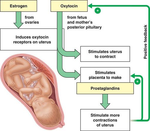
Hormonal Control and Induction of Labor Contractions
Breast Development, Lactation and Breastfeeding
Breast Development
- In childhood, the mammary glands are rudimentary, consisting mainly of a few ducts with little glandular tissue.
- At puberty, breast development begins under the influence of estrogen.
- Estrogen stimulates:
- Growth and branching of the mammary ducts
- Deposition of fat, which contributes to breast size and shape
- Progesterone promotes development of the lobules and alveoli. During pregnancy, high levels of estrogen, progesterone, prolactin, and other hormones cause marked growth of the breasts.
- Ductal system enlarges, while the lobules and alveoli become fully developed in preparation for milk production.
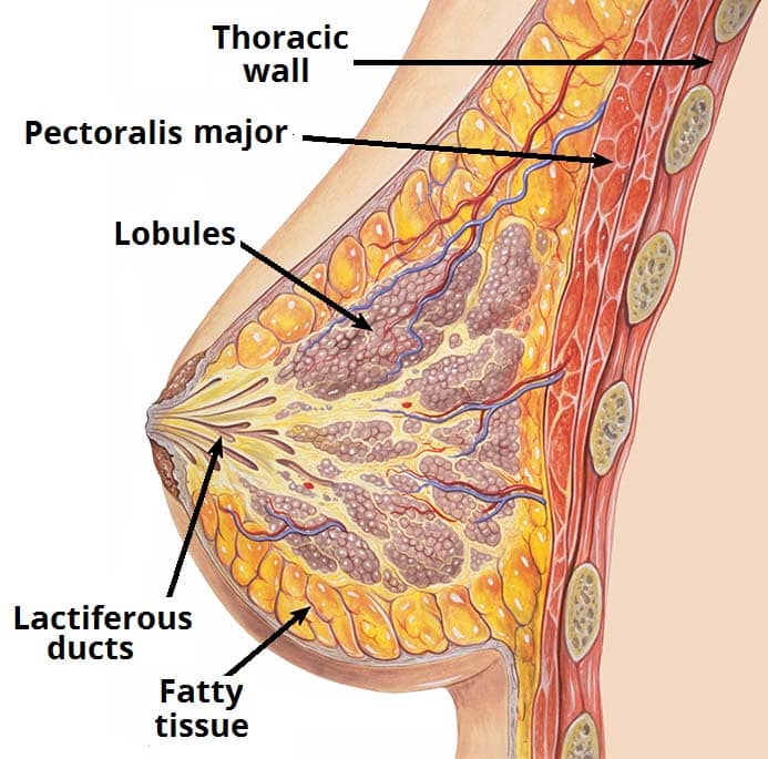
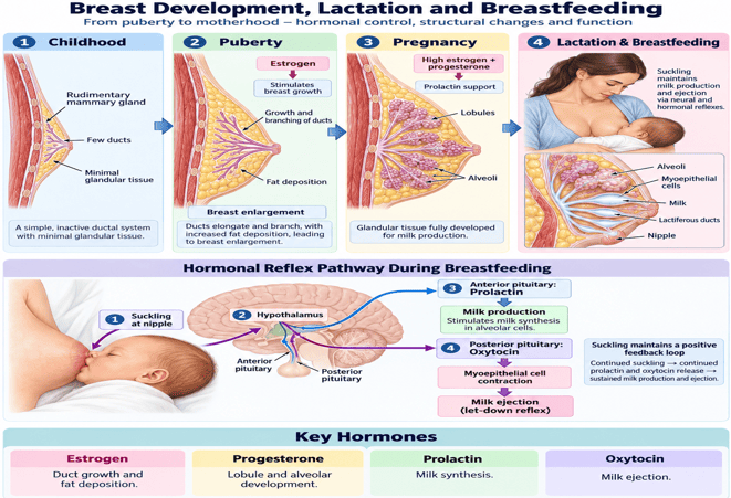
Breast Development, Lactation and Breastfeeding
Lactation
Process of milk production, maintenance, and ejection from the mammary glands. It involves three main processes:
- Lactogenesis: initiation of milk production
- Galactopoiesis: maintenance of milk production
- Galactokinesis: milk ejection or let-down
1. Lactogenesis
- Initiation of milk synthesis and secretion after delivery.
- During pregnancy, high levels of estrogen and progesterone promote breast development but inhibit full milk secretion. After delivery and expulsion of the placenta, estrogen and progesterone levels fall markedly.
- This removes their inhibitory influence on lactation, allowing prolactin to stimulate milk synthesis by the mammary alveolar cells. Prolactin is the principal hormone responsible for initiating milk production.
2. Galactopoiesis
- Maintenance and continuation of milk production after lactation has been established. Continued suckling is the major stimulus for maintaining prolactin secretion.
- Hormones that support galactopoiesis include: Prolactin, Growth hormone, Thyroid hormones, Cortisol, Insulin.
3. Galactokinesis
- Ejection of milk from the alveoli through the ducts toward the nipple.
- Suckling stimulates release of oxytocin from the posterior pituitary.
- Oxytocin causes contraction of myoepithelial cells surrounding the alveoli.
- This produces the milk ejection or let-down reflex.
Hormonal Division of Lactation
Prolactin = Production (Galactopoiesis/Lactogenesis): Secreted by anterior pituitary; acts on alveolar epithelial cells to synthesize milk.
Oxytocin = Outflow (Galactokinesis): Secreted by posterior pituitary; contracts myoepithelial cells for milk let-down.
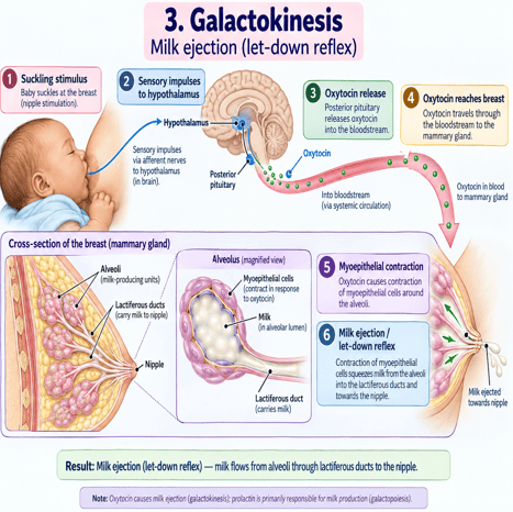
Galactokinesis: Milk Ejection (Let-Down Reflex)
Mechanisms of the Milk Ejection Reflex
Milk let-down or milk ejection reflex operates through different mechanisms:
- Unconditioned Reflex:
- Suckling stimulates sensory receptors in the nipple.
- Impulses travel to the hypothalamus, stimulating oxytocin release from the posterior pituitary.
- Oxytocin causes myoepithelial contraction → milk ejection.
- The reflex continues as long as suckling persists.
- Conditioned Reflex:
- Milk ejection may also occur without direct suckling.
- Seeing, hearing, touching, or thinking about the baby can stimulate oxytocin release and trigger the let-down reflex.
Breastfeeding Dual Reflex Loop
- Breastfeeding maintains lactation through two major hormonal reflexes:
- Suckling → Prolactin → Milk production
- Suckling → Oxytocin → Milk ejection
- Frequent breastfeeding therefore helps maintain both milk synthesis and milk release.
Breast/Human Milk
- Human milk is the natural and primary source of nutrition for infants, especially during the first months of life.
- It provides nutrients, water, immune protection, and bioactive factors needed for growth and development.
Composition
- Human milk contains approximately:
- Water: ~87–88%
- Solids: ~12–13%, including:
- Lactose: major carbohydrate
- Fat: major energy source
- Proteins: mainly whey proteins, including α-lactalbumin
- Vitamins and minerals
- Immunological and bioactive components
- Compared with cow’s milk, human milk generally contains more lactose and less protein, making it better adapted to the nutritional needs and immature kidneys of infants.
Human Milk vs. Cow's Milk Comparison
| Nutrient / Feature | Human Milk | Cow's Milk | Physiological Consequence for Infant |
|---|---|---|---|
| Carbohydrate (Lactose) | Higher (~7 g/dL) | Lower (~4.5 g/dL) | Provides optimal galactose for infant brain development and calories. |
| Protein Composition | Lower total; predominantly whey (α-lactalbumin) | Higher total; predominantly casein | Cow's casein forms tough curds, harder to digest; human whey forms soft, easily absorbed curds. |
| Renal Solute Load | Low sodium, potassium, and phosphorus | High solute and mineral load | Excess cow's milk solute strains immature neonatal renal filtration capacity. |
| Immune Defense | Rich in Secretory IgA, lactoferrin, lysozyme | Minimal maternal immune factors | Human milk directly coats GI mucosa, preventing bacterial and viral enteritis. |
| Iron Content & Bioavailability | Low absolute concentration but high bioavailability (~50%) | Low concentration and low absorption (~10%) | Cow's milk can cause occult GI blood loss and iron-deficiency anemia in infants. |
Advantages of Breast Milk
- Provides balanced nutrition appropriate for infant growth and development.
- Contains maternal antibodies, especially secretory IgA, which help protect against infections.
- Contains immune cells, including macrophages and leukocytes, that contribute to antimicrobial defense.
- Provides protective factors such as lactoferrin, lysozyme, and human milk oligosaccharides, which limit microbial growth and support healthy intestinal flora.
- Is easily digested and readily available, with appropriate temperature and composition for the infant.
- Supports development of the gastrointestinal tract and immune system.
Disadvantages of Animal Milk in Infants
- Animal milk, particularly cow’s milk, is not suitable as the main drink for young infants because its composition differs markedly from human milk.
- High protein and mineral load: Contains more protein, sodium, potassium, phosphorus, and other minerals than human milk. This may place an excessive solute load on the immature infant kidneys.
- Poorer digestibility: Cow’s milk contains a relatively high proportion of casein, which forms a firmer curd and is more difficult for infants to digest than human milk proteins.
- Risk of gastrointestinal irritation and blood loss: In some infants, cow’s milk can irritate the intestinal mucosa and cause occult gastrointestinal blood loss, contributing to anemia.
- Low iron content and bioavailability: Cow’s milk contains little iron, and prolonged use may increase the risk of iron-deficiency anemia.
- Less suitable fat and micronutrient profile: It contains lower amounts of some essential fatty acids and certain vitamins compared with appropriately fortified infant feeds.
- Not nutritionally adapted to infant needs: Its overall composition is designed for calf growth rather than human infant development.
Lactation and the Menstrual Cycle
- During pregnancy, high levels of estrogen and progesterone inhibit the full milk-producing action of prolactin.
- After delivery and expulsion of the placenta, estrogen and progesterone levels fall sharply, allowing prolactin to initiate lactation.
- During breastfeeding, elevated prolactin suppresses hypothalamic GnRH secretion.
- Reduced GnRH lowers FSH and LH secretion from the anterior pituitary.
- This suppresses ovarian follicular development and ovulation, resulting in lactational amenorrhea.
Key Pathway:
Suckling → ↑ Prolactin → ↓ GnRH → ↓ FSH/LH → ↓ Ovulation → Amenorrhea
Lactational Amenorrhea as Contraception
Lactational amenorrhea is temporary, and ovulation may resume before the first postpartum menstrual period. Consequently, barrier or progestin-only contraception is recommended to prevent unplanned pregnancy.
Abnormalities of Lactation
Chiari–Frommel Syndrome
A rare postpartum disorder characterized by persistent galactorrhea, amenorrhea, hyperprolactinemia, and infertility.
It results from persistent prolactin secretion after pregnancy.
Other causes of persistent galactorrhea, such as prolactin-secreting pituitary adenomas, hypothyroidism, and certain drugs, should be excluded.
Failure of Lactation
Refers to inadequate initiation or maintenance of milk production.
Causes include:
- Maternal or neonatal illness
- Delayed mother–infant contact or ineffective suckling
- Emotional stress
- Hormonal disorders, particularly inadequate prolactin response
- Breast or pituitary disorders
Galactocele
A milk-retention cyst of the breast caused by obstruction of a lactiferous duct during or after lactation.
It presents as a usually painless, well-defined breast swelling containing milk.
