
Temporal and infratemporal fossae: boundaries, neurovascular contents & clinical anatomy
On this page
Temporal fossa boundaries
The temporal fossa is a shallow depression on the lateral aspect of the neurocranium, situated superior to the zygomatic arch. It houses the fleshy fan of the temporalis muscle and communicates inferiorly with the infratemporal fossa via the gap medial to the zygomatic arch.
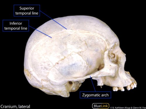
Anatomical Boundaries of the Temporal Fossa
Osteological Margins
- Anterior Margin: Formed by the posterior edge of the zygomatic process of the frontal bone and the posterior border of the frontal process of the zygomatic bone.
- Superior & Posterior Margins: Defined by the paired temporal lines (superior and inferior temporal lines) which arch across the frontal bone, cross the coronal suture onto the parietal bone, and curve downwards and forwards onto the temporal bone. The superior line anchors the investing temporal fascia; the inferior line demarcates the peripheral bony origin of the temporalis muscle.
- Lateral Boundary (Roof): Enclosed by the tough, glistening temporal fascia superiorly and the horizontal span of the zygomatic arch inferiorly.
- Inferior Boundary: Demarcated medially by the infratemporal crest on the greater wing of the sphenoid bone. Medial to the zygomatic arch, the fossa opens directly into the infratemporal fossa.
- Floor: Formed by the union of four cranial bones: the frontal bone, parietal bone, squamous part of the temporal bone, and the greater wing of the sphenoid bone. These four bones articulate at an H-shaped suture junction known as the pterion.
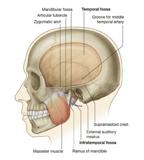
Temporal fossa contents
The temporal fossa is primarily packed by the temporalis muscle and the neurovascular structures supplying it and the overlying scalp.

Primary Contents
- 1. Temporalis Muscle: A fan-shaped muscle of mastication originating from the bony floor of the temporal fossa and the deep surface of the overlying temporal fascia. Its fibers converge inferiorly into a tendon that passes medial to the zygomatic arch to insert into the coronoid process and anterior border of the ramus of the mandible.
- 2. Deep Temporal Nerves: Typically two in number (anterior and posterior deep temporal nerves). Arise from the anterior division of the mandibular nerve (V3) within the infratemporal fossa, pass superiorly across the infratemporal crest, and run deep to the temporalis muscle to provide motor innervation.
- 3. Deep Temporal Arteries: Arise from the second (pterygoid) part of the maxillary artery. Accompany the deep temporal nerves on the bony floor of the fossa, anastomosing with the middle temporal artery to supply the muscle.
- 4. Auriculotemporal Nerve: A sensory branch of the posterior division of V3 that ascends posterior to the temporomandibular joint, passes over the root of the zygoma with the superficial temporal vessels, and distributes sensory fibers to the skin of the temple and auricle.
- 5. Superficial Temporal Vessels: The superficial temporal artery represents the smaller terminal branch of the external carotid artery. It ascends anterior to the tragus over the zygomatic arch alongside the superficial temporal vein, giving off the middle temporal artery (which pierces the temporal fascia to supply the temporalis muscle) before bifurcating into frontal and parietal branches to supply the scalp.
- 6. Zygomaticotemporal Nerve: A small sensory branch of the zygomatic nerve (from the maxillary division, V2). Pierces the temporal surface of the zygomatic bone to supply a small cutaneous area of the anterior temple.
Infratemporal fossa boundaries
The infratemporal fossa is an irregular, wedge-shaped deep facial space located inferior to the zygomatic arch, posterior to the maxilla, and deep to the ramus of the mandible. It represents a vital crossroads communicating with the middle cranial fossa, the orbit, and the pterygopalatine fossa.
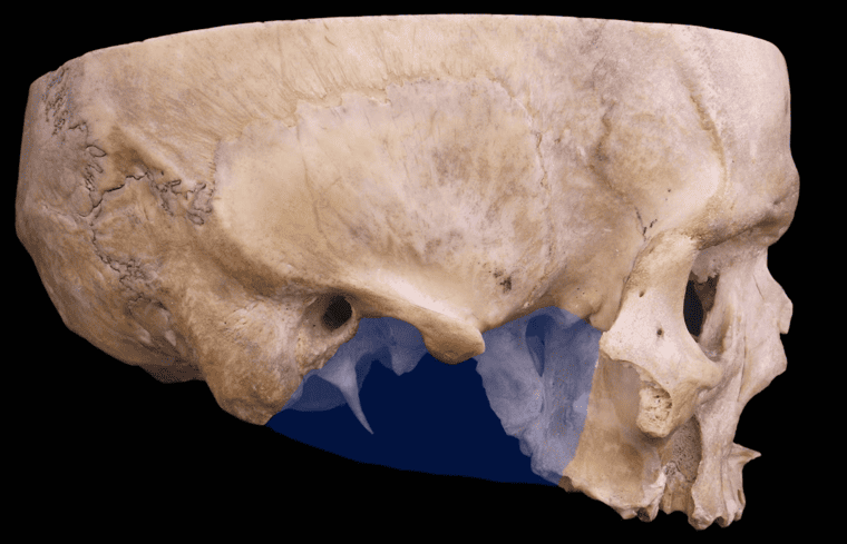
Anatomical Boundaries
Scroll the table sideways on a smaller screen.
| Boundary | Anatomical Structures Formed By |
|---|---|
| Anterior Wall | Posterior (infratemporal) surface of the body of the maxilla (pierced by posterior superior alveolar canals) and the inferior part of the zygomatic bone |
| Posterior Wall | Styloid process of the temporal bone, mastoid process, and the carotid sheath |
| Medial Wall | Lateral pterygoid plate of the sphenoid bone; more anteriorly, the pterygomaxillary fissure leading into the pterygopalatine fossa |
| Lateral Wall | Medial surface of the ramus and coronoid process of the mandible |
| Roof (Superior Wall) | Infratemporal surface of the greater wing of the sphenoid bone and the infratemporal crest; pierced by the foramen ovale and foramen spinosum |
| Floor (Inferior) | Open; continuous with the tissue spaces of the neck and the submandibular region at the angle of the mandible |
Infratemporal Fossa Contents Overview
The space is densely packed with muscles, arteries, veins, and nerves:
- Muscular Structures: The lower portion and tendon of the temporalis muscle; the lateral pterygoid muscle; and the medial pterygoid muscle.
- Arterial Tree: The maxillary artery (its first/mandibular and second/pterygoid parts) and its extensive branches.
- Venous Network: The extensive pterygoid venous plexus and the maxillary vein.
- Neural Structures: The mandibular nerve (CN V3) and its branches (auriculotemporal, inferior alveolar, lingual, buccal, and muscular nerves); the chorda tympani (branch of CN VII); and the parasympathetic otic ganglion.
Muscles of mastication
The four classic muscles of mastication develop embryologically from the first pharyngeal (mandibular) arch and are consequently innervated by the motor root of the mandibular division of the trigeminal nerve (CN V3).
Scroll the table sideways on a smaller screen.
| Muscle | Anatomical Origin | Insertion Site | Nerve Supply | Mandibular Actions |
|---|---|---|---|---|
| Temporalis | Floor of the temporal fossa and deep surface of temporal fascia | Coronoid process and anterior border of the ramus of the mandible | Deep temporal nerves (V3) | Elevates mandible (closes jaw); posterior horizontal fibers retract the protracted mandible |
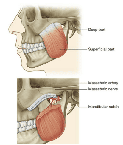
| Masseter | Lower border and medial surface of the zygomatic arch (superficial and deep parts) | Lateral surface of the coronoid process, ramus, and angle of the mandible | Masseteric nerve (V3) via mandibular notch | Powerful elevator of the mandible (closes jaw); superficial part assists in protraction; deep part retracts |
| Lateral Pterygoid | • Superior Head: Infratemporal surface of greater wing of sphenoid • Inferior Head: Lateral surface of lateral pterygoid plate |
Pterygoid fovea on the neck of the mandible; capsule and articular disc of the temporomandibular joint (TMJ) | Nerve to lateral pterygoid (V3) | Depresses mandible (opens mouth); bilateral contraction causes protraction; unilateral contraction causes contralateral chewing/grinding movements |
| Medial Pterygoid | • Superficial Head: Tuberosity of the maxilla and pyramidal process of palatine bone • Deep Head: Medial surface of the lateral pterygoid plate |
Medial surface of the angle and ramus of the mandible (mirroring the masseter laterally) | Nerve to medial pterygoid (V3 trunk) | Elevates mandible (closes jaw); assists in protraction; unilateral contraction aids in lateral grinding movements |
Mandibular nerve (V3) branches
The mandibular division of the trigeminal nerve (CN V3) is a mixed nerve containing both general somatic afferent (sensory) and special visceral efferent (branchial motor) fibers. It exits the middle cranial fossa through the foramen ovale to enter the infratemporal fossa, where it immediately gives off two initial branches from its short main trunk before dividing into anterior and posterior divisions.
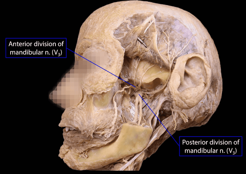
1. Branches from the Main Trunk
- Meningeal Branch (Nervus Spinosus): Re-enters the cranium through the foramen spinosum alongside the middle meningeal artery to supply the sensory innervation of the dura mater in the middle cranial fossa and mastoid air cells.
- Nerve to Medial Pterygoid: A motor branch entering the deep surface of the medial pterygoid muscle. It passes through or close to the otic ganglion without synapsing, giving off small motor twigs:
- Nerve to Tensor Veli Palatini: Tenses the soft palate during swallowing.
- Nerve to Tensor Tympani: Tenses the tympanic membrane to damp loud sounds.
2. Branches of the Anterior Division (Predominantly Motor)
- Masseteric Nerve: Passes laterally above the lateral pterygoid muscle, traverses the mandibular notch, and enters the deep surface of the masseter. Also sends an articular twig to the temporomandibular joint.
- Deep Temporal Nerves (Anterior & Posterior): Pass superior to the lateral pterygoid to enter the deep surface of the temporalis muscle.
- Nerve to Lateral Pterygoid: Enters the deep surface of both the superior and inferior heads.
- Buccal Nerve: The only sensory branch of the anterior division. It descends between the two heads of the lateral pterygoid muscle, crosses the anterior border of the ramus of the mandible, and courses over the buccinator muscle. It innervates the skin of the cheek, the fascia over the buccinator, and pierces the muscle without innervating it to supply general sensation to the mucous membrane of the cheek and the buccal gingiva of the mandibular molars. (Motor supply to the buccinator is provided exclusively by the buccal branch of the facial nerve, CN VII).

3. Branches of the Posterior Division (Predominantly Sensory)
The posterior division is predominantly sensory, giving origin to three major nerves and one minor motor branch:
- Auriculotemporal Nerve: Arises characteristically by two roots that encircle the middle meningeal artery. The roots unite into a single trunk that runs posteriorly medial to the neck of the mandible, turns superiorly posterior to the temporomandibular joint, and ascends over the zygomatic arch behind the superficial temporal vessels.
- Sensory (GSA): Innervates the skin of the auricle (helix, tragus), the external acoustic meatus, the tympanic membrane, the TMJ, and the scalp of the temple.
- Autonomic Hitchhikers (GVE): Transports postganglionic parasympathetic secretomotor fibers (derived from the otic ganglion) and postganglionic sympathetic vasoconstrictor fibers directly into the parotid gland.
- Lingual Nerve: Detailed below in Section 06.
- Inferior Alveolar Nerve: Detailed below in Section 07.
Lingual nerve & chorda tympani
The lingual nerve is a major sensory branch of the posterior division of V3. It mediates general somatic sensation (touch, pain, temperature) from the anterior two-thirds of the tongue, the floor of the mouth, and the mandibular lingual gingiva.
Anatomical Course & Relationships
- Descends in the infratemporal fossa deep to the lateral pterygoid muscle, lying anterior and slightly medial to the inferior alveolar nerve.
- On the lateral surface of the medial pterygoid muscle, it is joined at an acute angle from behind by the chorda tympani (a branch of the facial nerve, CN VII, which exits the middle ear via the petrotympanic fissure).
- Passes beneath the lower border of the superior pharyngeal constrictor, lying directly against the medial surface of the mandible adjacent to the roots of the third lower molar tooth (vulnerable to iatrogenic transection during wisdom tooth extraction).
- Enters the oral cavity, crossing lateral to the styloglossus and hyoglossus muscles, lying deep to the mylohyoid muscle.
- Crucially, the lingual nerve descends lateral to, loops underneath, and then ascends medial to the submandibular duct (“the lingual nerve winds around Wharton’s duct”) before breaking into terminal sensory branches across the dorsum of the tongue.

Functional Fiber Components Carried by the Lingual Nerve
Composite Modalities
- 1. General Somatic Afferent (GSA): Derived directly from V3 (trigeminal ganglion). Conveys discriminative touch, pain, and temperature from the anterior two-thirds of the tongue and floor of the mouth.
- 2. Special Visceral Afferent (SVA - Taste): Contributed by the chorda tympani (CN VII). Conveys taste sensation from the taste buds of the anterior two-thirds of the tongue (cell bodies in the geniculate ganglion, synapsing in the rostral solitary nucleus).
- 3. General Visceral Efferent (GVE - Parasympathetic): Contributed by the chorda tympani (superior salivatory nucleus). Conveys preganglionic parasympathetic secretomotor fibers to the submandibular ganglion, which synaptically relays postganglionic secretomotor fibers to the submandibular and sublingual salivary glands.
Inferior alveolar nerve
The inferior alveolar nerve is the largest branch of the posterior division of V3. It is a mixed nerve that conveys sensory innervation from the mandibular dentition and chin, and provides motor innervation to two suprahyoid muscles.
Course & Branches
- Trajectory: Descends deep to the lateral pterygoid muscle, and then passes between the sphenomandibular ligament and the medial surface of the ramus of the mandible. It enters the mandibular canal through the mandibular foramen alongside the inferior alveolar artery and vein.
- 1. Mylohyoid Nerve (Motor): Arises immediately before the inferior alveolar nerve enters the mandibular foramen. It pierces the sphenomandibular ligament, descends along the mylohyoid groove on the medial mandibular ramus, and enters the submandibular triangle to innervate the mylohyoid muscle and the anterior belly of the digastric muscle.
- 2. Inferior Dental Plexus: Inside the mandibular canal, branches form a plexus that innervates the lower molar and premolar teeth and their corresponding adjacent gingiva.
- 3. Mental Nerve (Terminal Sensory): Exits the mandible through the mental foramen (below the second premolar tooth) to supply the skin of the chin, the skin and mucous membrane of the lower lip, and the labial gingiva.
- 4. Incisive Branch (Terminal Sensory): Remains within the bony incisive canal to supply the mandibular canine and incisor teeth and surrounding periodontal ligaments.
Otic ganglion
The otic ganglion is a small, flattened parasympathetic peripheral ganglion located in the infratemporal fossa immediately inferior to the foramen ovale. Topographically, it is intimately associated with the medial side of the main trunk of the mandibular nerve (V3), lying lateral to the tensor veli palatini muscle and anterior to the middle meningeal artery.
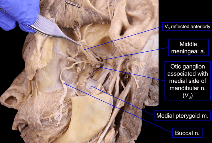
Parasympathetic Pathway to the Parotid Gland
Although anatomically plastered to CN V3, the otic ganglion is functionally a glossopharyngeal (CN IX) parasympathetic relay station:
- Central Origin: Preganglionic parasympathetic neurons originate in the inferior salivatory nucleus of the rostral medulla.
- Tympanic Route: Fibers exit via the glossopharyngeal nerve (CN IX), branch into the tympanic nerve (of Jacobson), enter the middle ear cavity, and participate in forming the tympanic plexus on the promontory.
- Lesser Petrosal Nerve: Fibers reform as the lesser petrosal nerve, pass through the middle cranial fossa, and traverse the foramen ovale (or canaliculus innominatus) to enter the infratemporal fossa.
- Synaptic Relay: Preganglionic fibers synapse upon postganglionic parasympathetic cell bodies inside the otic ganglion.
- Distribution to Parotid: Unmyelinated postganglionic secretomotor axons exit the ganglion, join the auriculotemporal nerve, and travel with it to supply secretomotor drive to the parotid gland.
Maxillary artery: first (mandibular) part
The maxillary artery is the larger terminal branch of the external carotid artery, arising within the parotid gland behind the neck of the mandible. It traverses the infratemporal fossa and ends within the pterygopalatine fossa. For descriptive purposes, it is divided into three parts relative to the lateral pterygoid muscle.
The First (Mandibular) Part
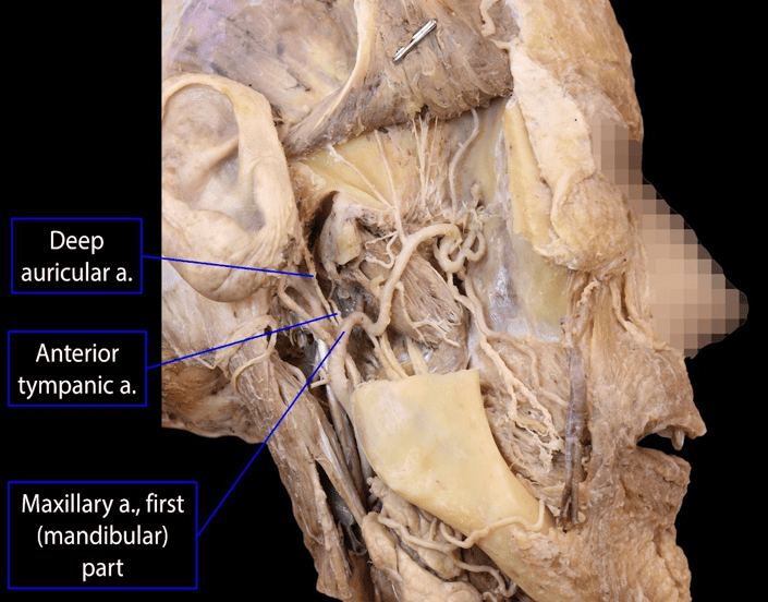
Runs horizontally forward between the neck of the mandible laterally and the sphenomandibular ligament medially, running parallel to the auriculotemporal nerve. All its branches pass into bony canals or foramina:
- 1. Deep Auricular Artery: Ascends to pierce the cartilaginous or bony wall of the external acoustic meatus, supplying its lining and the outer surface of the tympanic membrane.
- 2. Anterior Tympanic Artery: Ascends through the petrotympanic fissure into the tympanic cavity to supply the inner surface of the tympanic membrane.
- 3. Middle Meningeal Artery: The most clinically vital branch. Ascends vertically, embraced by the two roots of the auriculotemporal nerve, and enters the middle cranial fossa via the foramen spinosum. Runs between the endosteal dura mater and periosteum, dividing into anterior (frontal) and posterior (parietal) terminal branches across the pterion.
- 4. Accessory Meningeal Artery: Ascends through the foramen ovale into the middle cranial fossa to supply the trigeminal ganglion and adjacent dura.
- 5. Inferior Alveolar Artery: Descends alongside the inferior alveolar nerve, gives off the mylohyoid artery (which runs in the mylohyoid groove to supply the mylohyoid muscle), and enters the mandibular foramen. It traverses the mandibular canal to vascularize the lower teeth, bone, and periodontal structures, terminating as the mental artery on the chin.

Maxillary artery: second (pterygoid) part
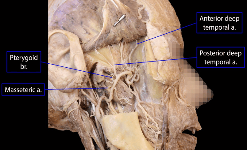
The second (pterygoid) part of the maxillary artery runs obliquely anteriorly and superiorly, positioned either superficial (two-thirds of cases) or deep (one-third of cases) to the lower head of the lateral pterygoid muscle, running deep to the temporalis muscle.
Branches Supplying Muscles of Mastication
Unlike the first and third parts, the branches of the second part are primarily muscular branches that supply masticatory and facial muscles:
- 1. Anterior Deep Temporal Artery: Ascends between the temporalis muscle and the pericranium to supply the anterior temporalis.
- 2. Posterior Deep Temporal Artery: Ascends to supply the posterior fibers of the temporalis muscle; anastomoses with the middle temporal artery.
- 3. Pterygoid Branches: Irregular small arterial twigs supplying both the lateral and medial pterygoid muscles.
- 4. Masseteric Artery: Passes laterally through the mandibular notch with the masseteric nerve to enter the deep surface of the masseter muscle.
- 5. Buccal Artery: Runs obliquely forward with the buccal nerve onto the buccinator muscle, supplying the buccinator and buccal fat pad.
Maxillary artery: third (pterygopalatine) part
The third (pterygopalatine) part of the maxillary artery passes between the two heads of the lateral pterygoid muscle and enters the pterygopalatine fossa through the pterygomaxillary fissure. Here, it branches extensively to supply the midface, palate, nasal cavity, and paranasal sinuses.

Branches of the Pterygopalatine Part
- 1. Posterior Superior Alveolar (PSA) Artery: Arises just before the artery enters the fissure. Descends on the tuberosity of the maxilla, penetrates alveolar canals, and supplies the maxillary molar and premolar teeth, gingiva, and mucosal lining of the maxillary sinus.
- 2. Infraorbital Artery: Enters the orbit through the inferior orbital fissure, traverses the infraorbital groove and canal in the orbital floor, and emerges onto the face via the infraorbital foramen. Supplies the lower eyelid, lacrimal sac, upper lip, and cheek, and gives off anterior and middle superior alveolar arteries to the maxillary incisors, canines, and maxillary sinus.
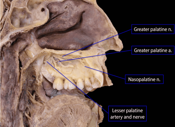
- 3. Descending Palatine Artery: Descends through the greater palatine canal, dividing into:
- Greater Palatine Artery: Emerges via the greater palatine foramen to run forward along the hard palate, supplying gingiva and palatal mucosa, and anastomosing through the incisive canal with the sphenopalatine artery.
- Lesser Palatine Arteries: Emerge through lesser palatine foramina to vascularize the soft palate and palatine tonsil.
- 4. Artery of the Pterygoid Canal (Vidian Artery): Passes backward through the pterygoid canal to supply the upper pharynx, auditory (Eustachian) tube, and tympanic cavity.
- 5. Pharyngeal Artery: Traverses the palatovaginal canal to supply the roof of the nasopharynx, sphenoid sinus, and auditory tube.
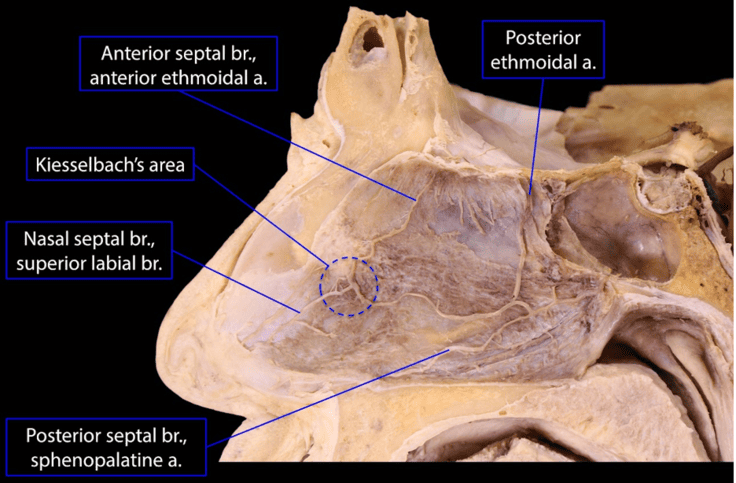
- 6. Sphenopalatine Artery: The terminal branch of the maxillary artery. Traverses the sphenopalatine foramen into the nasal cavity alongside the nasopalatine nerve. It is the principal arterial supply of the nasal cavity, supplying nasal conchae, meatuses, and dividing into posterior septal branches across the nasal septum.
Pterygoid plexus & venous drainage
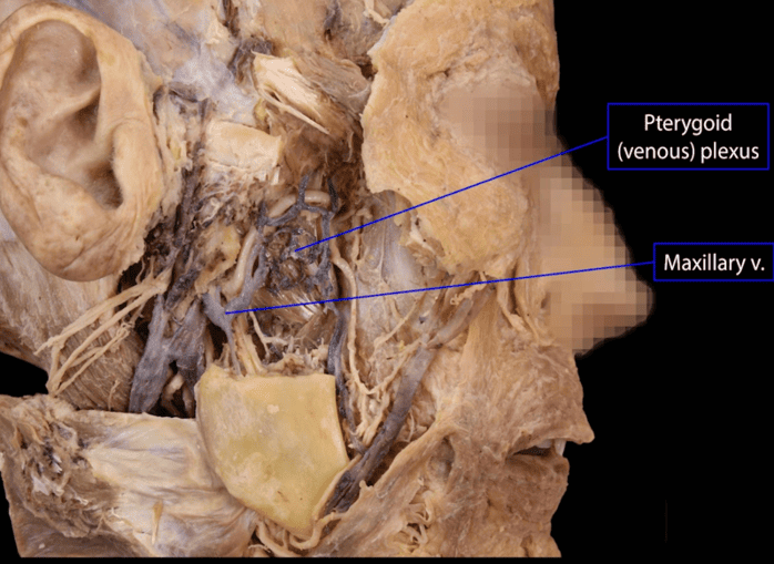
The venous drainage of the infratemporal fossa centers on the pterygoid venous plexus, an extensive network of valve-free veins wrapped around and within the lateral pterygoid muscle and resting on the lateral surface of the medial pterygoid muscle.
Tributaries & Anastomotic Connections
- Inflow & Drainage: Receives veins corresponding to the branches of the maxillary artery (deep temporal, sphenopalatine, alveolar, and middle meningeal veins). The plexus coalesces posteriorly into a short, wide trunk: the maxillary vein.
- Critical Anastomoses: The pterygoid plexus lacks valves, permitting bidirectional blood flow based on pressure gradients:
- Cavernous Sinus: Communicates directly via emissary veins passing through the foramen ovale, foramen lacerum, and the sphenoidal emissary foramen (of Vesalius).
- Inferior Ophthalmic Vein: Communicates via connections coursing through the inferior orbital fissure.
- Facial Vein: Communicates directly via the large deep facial vein, which passes backward across the buccinator muscle.

The Retromandibular Vein
Formed within the substance of the parotid gland by the confluence of the superficial temporal vein and the maxillary vein. It descends through the gland, emerging at its inferior pole to divide into two terminal branches:
- 1. Anterior Branch: Unites with the facial vein to form the common facial vein, which crosses the hypoglossal nerve to drain directly into the internal jugular vein.
- 2. Posterior Branch: Unites with the posterior auricular vein to form the external jugular vein, which descends superficially across the sternocleidomastoid muscle to drain into the subclavian vein.
Clinical correlations
Temporomandibular Joint (TMJ) Dislocation
The TMJ is a modified hinge (ginglymoarthrodial) synovial joint between the condylar process of the mandible, the mandibular fossa, and the articular tubercle of the temporal bone. An internal fibrocartilaginous articular disc divides the cavity into upper (gliding/translation) and lower (hinge/rotation) compartments.
- Dislocation Mechanism: During maximal mouth opening (e.g., yawning, dental extractions, trauma), excessive contraction of the lateral pterygoid muscle pulls the mandibular condyle forward over the summit of the articular tubercle into the infratemporal fossa. The condyle slips anteriorly into the infratemporal space.
- Spasm & Fixation: The masseter, temporalis, and medial pterygoid muscles immediately enter protective reflex spasm, locking the jaw wide open (“open lock”) and preventing condylar return.
- Reduction Technique: The clinician places gloved thumbs intraorally onto the lower molar teeth and wraps fingers around the inferior border of the mandible. Downward pressure is applied to overcome the spasm of the elevators, followed by backward pressure to seat the condyles into the mandibular fossae.
Trigeminal Neuralgia (Tic Douloureux)
A debilitating neurological condition characterized by paroxysms of severe, sharp, lancinating pain along the dermatomal distribution of CN V (most commonly V2 and V3). Triggered by light tactile stimulation (shaving, brushing teeth, chewing). Often caused by vascular compression of the trigeminal nerve root entry zone by an aberrant loop of the superior cerebellar artery. In refractory cases, surgical access to V3 at the foramen ovale or radiofrequency rhizotomy can be performed through the infratemporal approach.
Quick recap
References
The following authoritative anatomical reference texts support this curriculum note:
- Gray’s Anatomy: Standring, S. (2021) Gray’s Anatomy: The Anatomical Basis of Clinical Practice. 42nd edn. London: Elsevier. [Section 4: Head and Neck; Chapter 31: Infratemporal and Pterygopalatine Fossae and Temporomandibular Joint].
- Moore’s Clinically Oriented Anatomy: Dalley, A.F. and Agur, A.M.R. (2023) Moore’s Clinically Oriented Anatomy. 9th edn. Philadelphia: Wolters Kluwer. [Chapter 8: Head; Temporal and Infratemporal Regions].
- Snell’s Clinical Anatomy by Regions: Waugh, A. and Grant, A. (2019) Snell’s Clinical Anatomy by Regions. 10th edn. Philadelphia: Wolters Kluwer. [Chapter 11: The Head and Neck; The Pterygopalatine and Infratemporal Fossae].
- Last’s Anatomy: Sinnatamby, C.S. (2011) Last’s Anatomy: Regional and Applied. 12th edn. Edinburgh: Churchill Livingstone. [Chapter 6: Head and Neck and Spine; Infratemporal Fossa].