
Triangles of the neck: anterior, lateral & neurovascular contents
On this page
Quadrilateral neck space
The anterolateral aspect of the human neck is topographically organized as a large quadrilateral space. This structural framework allows clinicians, anesthetists, and surgeons to map vital neurovascular pathways, identify surgical landmarks, and isolate pathological cervical masses.
Boundaries of the Anterolateral Quadrilateral Space
- Superior Boundary: Formed by the inferior margin of the mandible anteriorly, and an imaginary horizontal line extending from the angle of the mandible to the mastoid process of the temporal bone posteriorly.
- Inferior Boundary: Formed by the superior margin of the manubrium sterni in the midline anteriorly, and the superior border of the middle third of the clavicle laterally.
- Posterior Boundary: Defined by the anterior border and attachment of the trapezius muscle.
- Anterior Boundary: Marked by the anterior median midline of the neck, projecting vertically from the symphysis menti of the mandible down to the jugular (suprasternal) notch of the manubrium sterni.
- Diagonal Bisection: The oblique trajectory of the sternocleidomastoid (SCM) muscle courses from the mastoid process and superior nuchal line down to the sternum and clavicle. This divides the quadrilateral space into two primary anatomical regions: the anterior cervical triangle and the posterior cervical triangle (lateral cervical region).

Anterior triangle boundaries
The anterior cervical triangle houses the visceral compartment of the neck (larynx, trachea, pharynx, esophagus, thyroid and parathyroid glands) along with major neurovascular conduits supplying the head, face, and brain.
Anatomical Boundaries
- Lateral Border: Formed by the anterior border of the sternocleidomastoid muscle.
- Superior Border (Base): Formed by the inferior border of the mandible and its continuation to the mastoid process.
- Medial Border: Formed by the anterior midline of the neck.
- Apex: Directed inferiorly toward the jugular notch of the manubrium sterni.
- Roof: Formed sequentially by the skin, superficial fascia containing the platysma muscle, cutaneous cervical nerves, and the investing layer of deep cervical fascia.
Subdivisions of the anterior triangle
The digastric muscle and the superior belly of the omohyoid muscle further subdivide the anterior triangle into four smaller, surgically distinct secondary triangles: the submandibular (digastric), submental, carotid, and muscular triangles.

1. Submandibular (Digastric) Triangle
- Superior Boundary: Inferior border of the mandible and a line projecting to the mastoid process.
- Inferoanterior Boundary: Anterior belly of the digastric muscle.
- Inferoposterior Boundary: Posterior belly of the digastric muscle and the stylohyoid muscle.
- Floor: Formed predominantly by the mylohyoid and hyoglossus muscles, with a contribution from the middle pharyngeal constrictor posteriorly.
- Key Contents: Submandibular gland (superficial and deep lobes), facial artery and vein, submandibular lymph nodes, hypoglossal nerve (CN XII), and the nerve to the mylohyoid.
2. Submental Triangle (Unpaired Midline)
- Inferior Boundary (Base): Body of the hyoid bone.
- Lateral Boundaries: Anterior bellies of the right and left digastric muscles.
- Apex: Directed superiorly toward the mandibular symphysis menti.
- Floor: Formed by the two mylohyoid muscles meeting at the midline median fibrous raphe.
- Key Contents: Submental lymph nodes and small tributaries that unite to form the anterior jugular veins.
3. Carotid Triangle
- Anteroinferior Boundary: Superior belly of the omohyoid muscle.
- Superior Boundary: Posterior belly of the digastric muscle and the stylohyoid muscle.
- Posterior Boundary: Anterior border of the sternocleidomastoid muscle.
- Floor: Formed by the thyrohyoid membrane, hyoglossus muscle, and inferior and middle pharyngeal constrictors.
- Key Contents: Carotid sheath contents (common carotid artery bifurcating into internal and external carotids, internal jugular vein, vagus nerve), ansa cervicalis, hypoglossal nerve, superior laryngeal nerve, and deep cervical lymph nodes.
4. Muscular (Omotracheal) Triangle
- Superior Boundary: Hyoid bone.
- Superolateral Boundary: Superior belly of the omohyoid muscle.
- Inferolateral Boundary: Anterior border of the sternocleidomastoid muscle.
- Medial Boundary: Anterior midline of the neck extending from hyoid to jugular notch.
- Key Contents: Infrahyoid strap muscles (sternohyoid, sternothyroid), thyroid gland, parathyroid glands, larynx, trachea, and esophagus.
Suprahyoid & infrahyoid muscles
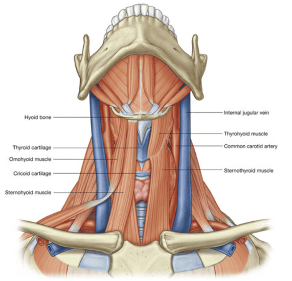
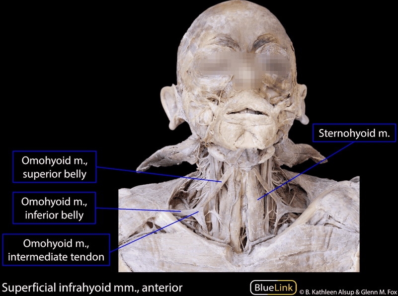
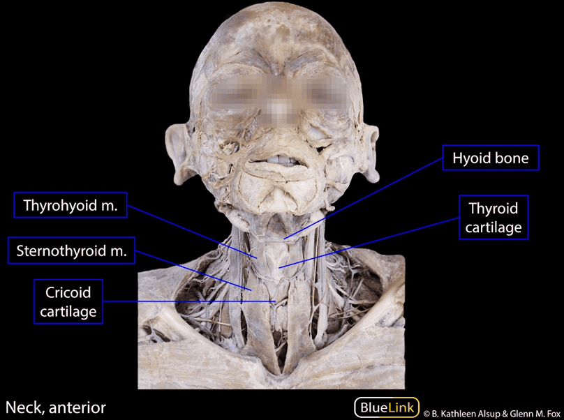
The muscles of the anterior triangle stabilize, elevate, and depress the hyoid bone and laryngeal skeleton during mastication, deglutition (swallowing), and phonation (speech). They are divided relative to the hyoid bone into suprahyoid and infrahyoid groups.

Scroll the table sideways on a smaller screen.
| Muscle | Origin | Insertion | Innervation | Primary Action(s) |
|---|---|---|---|---|
| Mylohyoid | Mylohyoid line on internal surface of mandible | Mylohyoid raphe and body of hyoid bone | Nerve to mylohyoid (branch of inferior alveolar nerve from CN V3) | Elevates hyoid bone, floor of mouth, and tongue during swallowing and phonation |
| Geniohyoid | Inferior mental spine (genial tubercle) of mandible | Anterior surface of body of hyoid bone | C1 spinal nerve traveling via hypoglossal nerve (CN XII) | Pulls hyoid anterosuperiorly; shortens floor of mouth; widens pharynx during deglutition |
| Stylohyoid | Posterior aspect of styloid process of temporal bone | Body of hyoid bone at junction with greater horn | Stylohyoid branch of facial nerve (CN VII) | Elevates and retracts hyoid bone, elongating the floor of the mouth |
| Digastric (Anterior Belly) | Digastric fossa on inner lower border of mandibular symphysis | Intermediate tendon bound to hyoid body/greater horn by fibrous fascial sling | Nerve to mylohyoid (CN V3) | Depresses mandible against resistance; elevates and steadies hyoid bone during swallowing |
| Digastric (Posterior Belly) | Mastoid notch (digastric groove) on medial mastoid process | Intermediate tendon attached to hyoid bone | Digastric branch of facial nerve (CN VII) | Pulls hyoid bone posterosuperiorly; acts synergistically with anterior belly |
| Sternohyoid | Posterior surface of manubrium sterni & medial clavicle | Medial lower border of body of hyoid bone | Ansa cervicalis (C1–C3 ventral rami) | Depresses hyoid bone after its elevation during swallowing |
| Omohyoid | Superior border of scapula near suprascapular notch | Inferior border of body of hyoid bone lateral to sternohyoid | Ansa cervicalis (Superior root to superior belly; inferior root to inferior belly) | Depresses, retracts, and steadies hyoid bone; tenses deep cervical fascia |
| Sternothyroid | Posterior surface of manubrium sterni inferior to sternohyoid | Oblique line on lamina of thyroid cartilage | Ansa cervicalis (C2–C3 branches) | Depresses hyoid bone and laryngeal thyroid cartilage |
| Thyrohyoid | Oblique line of lamina of thyroid cartilage | Inferior border of body and greater horn of hyoid bone | C1 nerve fibers via hypoglossal nerve (CN XII) | Depresses hyoid bone; elevates larynx when hyoid is fixed |
Common & internal carotid arteries
The arterial supply to the head and neck travels primarily through the carotid arterial system. Both common carotid arteries ascend within the deep neck enclosed inside the carotid sheath, lying immediately lateral to the cervical trachea, larynx, and esophagus.
Origins and Course
- Right Common Carotid Artery (RCCA): Originates from the brachiocephalic trunk (artery) immediately posterior to the right sternoclavicular joint. It is entirely cervical throughout its anatomical course.
- Left Common Carotid Artery (LCCA): Arises directly from the arch of the aorta within the superior mediastinum of the thorax. It ascends through the superior thoracic aperture to enter the neck near the left sternoclavicular joint.
- Absence of Cervical Branches: Neither common carotid artery gives off any collateral branches in the neck prior to its terminal bifurcation.
The Carotid Bifurcation: Level C3/C4
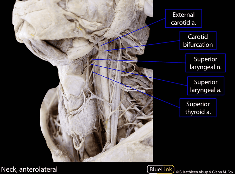
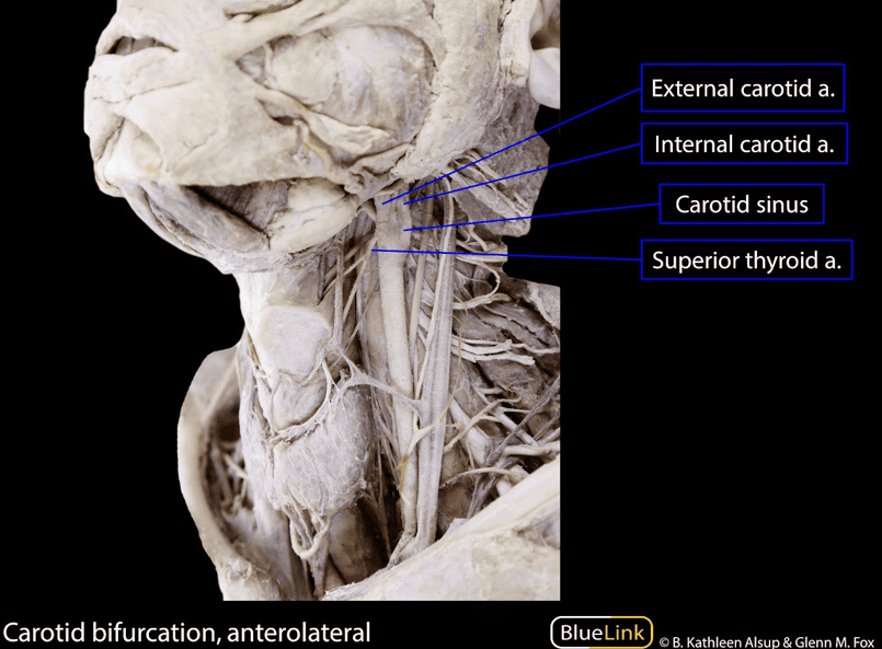
Near the superior border of the thyroid cartilage (vertebral level C3/C4), within the carotid triangle, each common carotid artery divides into two terminal branches: the internal carotid artery (ICA) and the external carotid artery (ECA).
Specialized Sensory Receptors at the Bifurcation
- 1. Carotid Sinus (Baroreceptor): A localized dilation situated within the terminal segment of the common carotid artery and the proximal bulb of the internal carotid artery.
Function: Acts as a specialized baroreceptor that monitors systemic arterial blood pressure. Increased stretch due to elevated arterial pressure stimulates stretch-sensitive nerve endings.
Innervation: Primarily innervated by the sinus nerve of Hering, a branch of the glossopharyngeal nerve (CN IX), with secondary contributions from the vagus nerve (CN X). Afferents project to the nucleus tractus solitarii (NTS) in the medulla to modulate autonomic vasomotor and cardiac tone. - 2. Carotid Body (Chemoreceptor): A small, reddish-brown, highly vascularized ovoid mass of neuroendocrine glomus cells situated in the bifurcation cleft behind the carotid vessels.
Function: Acts as a peripheral chemoreceptor detecting changes in blood chemistry, primarily monitoring decreased arterial oxygen tension (hypoxemia, low PaO2), increased carbon dioxide (hypercapnia, high PaCO2), and decreased pH (acidosis).
Innervation: Dual sensory innervation provided by branches of both the glossopharyngeal nerve (CN IX) and the vagus nerve (CN X). Stimulation initiates hyperventilation, systemic vasoconstriction, and increased sympathetic drive.
Internal Carotid Artery (ICA)
- Cervical Course: Ascends vertically from the bifurcation within the carotid sheath toward the skull base. The ICA gives off no collateral branches in the neck.
- Cranial Entry: Enters the cranial cavity by traversing the tortuous carotid canal in the petrous part of the temporal bone.
- Supply Territory: Forms the anterior circulation of the brain, supplying the cerebral hemispheres, eyes and orbital contents (via the ophthalmic artery), and the skin of the forehead.
External carotid artery & branches
The external carotid artery (ECA) supplies structures external to the cranium, including the face, scalp, tongue, maxilla, and neck viscera. In contrast to the internal carotid, the ECA begins giving off branches immediately upon its origin within the carotid triangle.
Eight Branches of the External Carotid Artery
The ECA yields three anterior branches, one medial branch, two posterior branches, and two terminal branches:
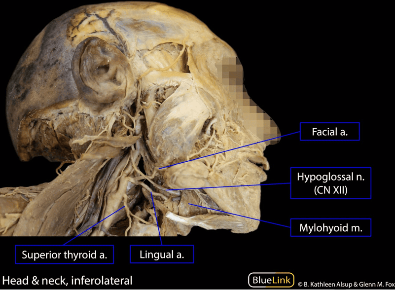
- 1. Superior Thyroid Artery (Anterior): The first branch of the ECA. Arises from the anterior aspect at or near the bifurcation, descending forward deep to the infrahyoid muscles to reach the superior pole of the thyroid gland. Gives origin to the superior laryngeal artery, which pierces the thyrohyoid membrane with the internal laryngeal nerve to supply the internal larynx.
- 2. Ascending Pharyngeal Artery (Medial): The second branch and the smallest branch of the ECA. Arises from its deep medial surface, ascending vertically between the internal carotid artery and the pharynx to supply the pharyngeal wall, tonsils, and middle ear.
- 3. Lingual Artery (Anterior): Arises from the anterior surface at the level of the tip of the hyoid greater horn, just superior to the superior thyroid artery. Courses deep to the hypoglossal nerve (CN XII), passes between the middle pharyngeal constrictor and the hyoglossus muscle, and turns upward at the anterior border of the hyoglossus to divide into the deep lingual and sublingual arteries to supply the tongue and floor of the mouth.
- 4. Facial Artery (Anterior): Arises anteriorly just superior to the lingual artery. Passes deep to the stylohyoid and posterior belly of the digastric, loops through a deep groove in the submandibular gland, hooks over the inferior border of the mandible immediately anterior to the masseter muscle, and ascends tortuously across the face. Cervical branches include the ascending palatine artery, tonsillar artery, and submental artery.
- 5. Occipital Artery (Posterior): Arises from the posterior surface opposite the origin of the facial artery. Courses posterosuperiorly parallel and medial to the posterior belly of the digastric in the occipital groove of the temporal bone, crossing superficial to the internal carotid artery and cranial nerves IX, X, and XI. Supplies the posterior scalp and musculature.
- 6. Posterior Auricular Artery (Posterior): A small branch arising above the digastric muscle, ascending between the external acoustic meatus and the mastoid process. Supplies the auricle, scalp, parotid gland, and facial nerve (via the stylomastoid artery).
- 7 & 8. Terminal Branches: Within the substance of the parotid gland behind the mandibular neck, the ECA divides into the maxillary artery (supplying deep infratemporal structures, meninges, and teeth) and the superficial temporal artery (supplying the frontotemporal scalp).

Internal jugular vein & tributaries
The internal jugular vein (IJV) is the largest vein in the neck. It collects venous blood from the brain, anterior face, cervical viscera, and deep cervical muscles.
Origin, Course, and Relations in the Carotid Sheath
- Origin: Commences in the posterior cranial fossa at the jugular foramen as the direct anatomical continuation of the sigmoid sinus.
- Superior Bulb: Marked at its origin by a localized dilation, the superior bulb of the IJV, which occupies the jugular fossa of the temporal bone.
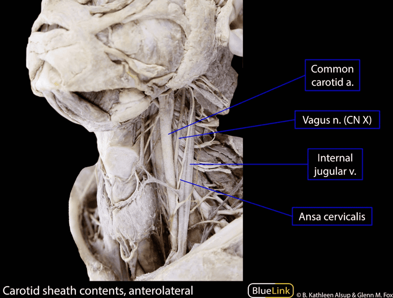
- Carotid Sheath Architecture: The IJV descends vertically within the tubular fibrous carotid sheath:
- Superiorly, the vein lies lateral to the internal carotid artery.
- Inferiorly, the vein lies lateral to the common carotid artery.
- The vagus nerve (CN X) courses posteriorly between the artery and vein throughout its cervical descent.
- Termination & Inferior Bulb: The IJV passes deep to the sternocleidomastoid muscle, traversing the gap between its sternal and clavicular heads. Immediately posterior to the sternal end of the clavicle, it dilates to form the inferior bulb of the IJV before uniting with the subclavian vein to form the brachiocephalic vein.
- Bicuspid Venous Valve: The inferior bulb contains a functional bicuspid valve that prevents retrograde regurgitation of venous blood toward the head during episodes of elevated intrathoracic pressure (e.g., coughing, Valsalva maneuver, inversion).
Tributaries of the Internal Jugular Vein
- Inferior Petrosal Sinus: Leaves the skull through the anterior part of the jugular foramen to enter the superior bulb.
- Facial Vein: Enters near the hyoid bone, often uniting with the anterior branch of the retromandibular vein.
- Lingual Vein: Drains the tongue; frequently unites with the facial vein to form a common trunk (common facial vein).
- Pharyngeal Veins: Arise from the venous plexus on the external wall of the pharynx.
- Superior Thyroid Vein: Accompanies the superior thyroid artery.
- Middle Thyroid Vein: Courses horizontally from the thyroid lateral lobe directly into the IJV, crossing anterior to the common carotid artery (a critical landmark in thyroid surgery).
- Occipital Vein: Usually drains into the suboccipital venous plexus (drained by the deep cervical and vertebral veins), but occasionally enters the IJV directly.
Nerves of the anterior cervical region
The anterior cervical region contains cranial nerves IX, X, and XII, components of the cervical spinal nerves, and the autonomic motor loops of the ansa cervicalis.
1. Hypoglossal Nerve (CN XII)
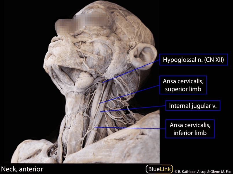
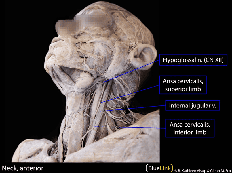
- Course: Leaves the cranial cavity via the hypoglossal canal, descends between the internal carotid artery and internal jugular vein, and enters the submandibular triangle by hooking under the occipital artery and passing deep to the posterior belly of the digastric muscle.
- Motor Innervation: Supplies all intrinsic and four of the five extrinsic striated muscles of the tongue (genioglossus, hyoglossus, styloglossus). The palatoglossus muscle is the sole exception, innervated by the vagus nerve (CN X) via the pharyngeal plexus.
- C1 Nerve Hitchhiking: Fibers from the C1 ventral ramus join the hypoglossal nerve near the skull base. As CN XII courses through the anterior triangle, these C1 fibers leave it as two distinct branches:
- The superior root of the ansa cervicalis (descendens hypoglossi).
- The nerve to the geniohyoid muscle (and nerve to thyrohyoid).
2. Glossopharyngeal (CN IX) and Vagus (CN X) Nerves
- Glossopharyngeal Nerve (CN IX): Emerges through the jugular foramen and descends between the internal carotid artery and internal jugular vein. Passes between the superior and middle pharyngeal constrictors to supply sensory innervation to the posterior third of the tongue, palatine tonsil, and pharyngeal mucosa, motor innervation to the stylopharyngeus muscle, and parasympathetic secretomotor fibers to the parotid gland (via the otic ganglion).
- Vagus Nerve (CN X): Descends within the posterior part of the carotid sheath between the carotid artery and internal jugular vein. In the neck, it yields:
- Pharyngeal branches: Form the pharyngeal plexus (motor to pharyngeal constrictors and soft palate, except tensor veli palatini [CN V3]).
- Superior laryngeal nerve: Divides into the internal laryngeal nerve (sensory to laryngeal mucosa above vocal folds) and the external laryngeal nerve (motor to cricothyroid muscle).
- Superior and inferior cervical cardiac branches: Convey parasympathetic preganglionic fibers to the cardiac plexus.
Lateral cervical region (posterior triangle)
The lateral cervical region (clinically designated as the posterior triangle of the neck) is a major anatomical space situated on the posterolateral aspect of the neck, transmitting nerves and vessels to the upper limb and shoulder girdle.

Boundaries, Apex, Roof, and Floor
- Anterior Boundary: Formed by the posterior border of the sternocleidomastoid muscle.
- Posterior Boundary: Formed by the anterior border of the trapezius muscle.
- Inferior Boundary (Base): Formed by the middle third of the clavicle between the attachments of the SCM and trapezius.
- Apex: Directed superiorly where the SCM and trapezius muscles meet on the superior nuchal line of the occipital bone.
- Roof: Formed by the investing layer of deep cervical fascia, covered superficially by the platysma, superficial fascia, and skin.
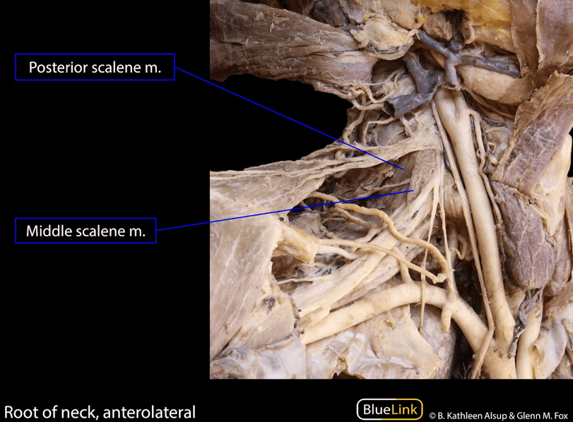
- Muscular Floor: Formed by deep cervical muscles enveloped by the prevertebral layer of deep cervical fascia. From superior to inferior, the floor consists of:
- Splenius capitis
- Levator scapulae
- Middle scalene (scalenus medius)
- Posterior scalene (scalenus posterior)

Occipital & omoclavicular triangles
The inferior belly of the omohyoid muscle crosses the posterior triangle obliquely, dividing it into an upper occipital triangle and a lower omoclavicular (subclavian) triangle.
1. Occipital Triangle
- Boundaries: Anterior border by SCM, posterior border by trapezius, inferior border by the inferior belly of omohyoid.
- Nomenclature: So named because the occipital artery crosses through its apex.
- Key Content: The most important nerve crossing the occipital triangle is the spinal accessory nerve (CN XI), which courses embedded within or immediately deep to the investing layer of deep cervical fascia.
2. Omoclavicular (Subclavian) Triangle
- Boundaries: Superior border by inferior belly of omohyoid, inferior border by middle third of clavicle, anterior border by SCM.
- Surface Landmark: Manifests externally as the supraclavicular fossa.
- Fascial Separation of Vessels: The inferior segment of the external jugular vein (EJV) crosses superficially, whereas the third part of the subclavian artery lies deep within the triangle. These vessels are separated by the investing layer of deep cervical fascia.
- Clinical Relevance: Because the third part of the subclavian artery traverses this space, it is frequently referred to as the subclavian triangle. Pulsations of the subclavian artery can be palpated here, and compressing it against the first rib controls distal upper limb hemorrhage.
Subclavian & thyrocervical branches
Arteries traversing the lateral cervical region include the third part of the subclavian artery, branches of the thyrocervical trunk, and the occipital artery.
1. Thyrocervical Trunk Branches
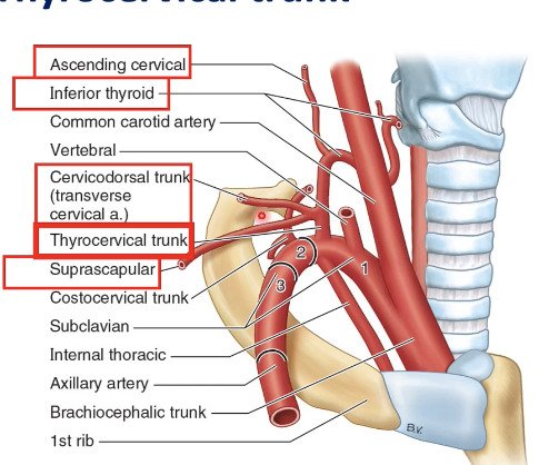
Arising from the first part of the subclavian artery medial to the anterior scalene, the thyrocervical trunk gives off two lateral branches that cross the posterior triangle:
- Suprascapular Artery: Passes inferolaterally across the anterior scalene muscle and phrenic nerve, crosses the third part of the subclavian artery and cords of the brachial plexus, and runs posterior to the clavicle to reach the suprascapular notch, contributing to the scapular collateral anastomosis. (May occasionally arise directly from the third part of the subclavian).
- Cervicodorsal Trunk (Transverse Cervical Artery): Arises laterally, coursing superficially across the phrenic nerve and anterior scalene muscle 2 to 3 cm superior to the clavicle. It crosses or passes through the trunks of the brachial plexus, supplying branches to their vasa nervorum. It bifurcates into:
- Superficial cervical artery: Passes deep (anterior) to the trapezius accompanying the spinal accessory nerve (CN XI).
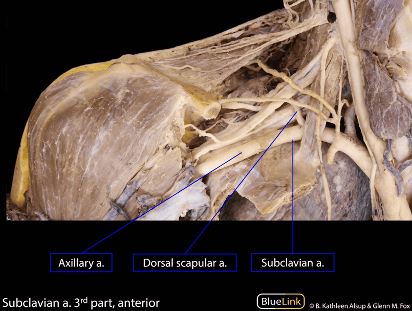
- Dorsal scapular artery: Runs deep to the levator scapulae and rhomboid muscles, supplying both and participating in the scapular anastomosis. (May alternatively arise independently from the second or third part of the subclavian artery).
- Terminal Branches: The thyrocervical trunk terminates by dividing into the inferior thyroid artery and the ascending cervical artery.

2. Third Part of the Subclavian Artery
- Course: Begins approximately one finger’s breadth superior to the clavicle at the lateral border of the anterior scalene muscle. It is the longest and most superficial part of the artery.
- Skeletal Contact & Hemostasis: Rests directly on the superior surface of the first rib posterosuperior to the subclavian vein. Compression of the artery against the first rib in the omoclavicular triangle effectively halts arterial bleeding throughout the upper extremity.
- Brachial Plexus Relation: The inferior trunk of the brachial plexus (C8–T1) lies directly posterior to the third part of the artery on the first rib.
External jugular & subclavian veins
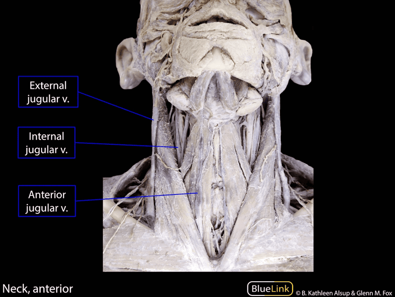
1. External Jugular Vein (EJV)
- Formation: Formed near the angle of the mandible (inferior to the ear auricle) by the union of the posterior division of the retromandibular vein with the posterior auricular vein.
- Course: Descends obliquely across the superficial surface of the SCM deep to the platysma. At the posterior border of the SCM, it pierces the investing layer of deep cervical fascia (the roof of the lateral cervical region).
- Termination: Descends into the omoclavicular triangle to terminate in the subclavian vein (or occasionally the IJV). Drains the scalp and lateral face.
- Tributaries: Just superior to the clavicle, the EJV receives the cervicodorsal (transverse cervical), suprascapular, and anterior jugular veins.
2. Subclavian Vein
- Course: The direct continuation of the axillary vein at the outer border of the first rib. Curves through the inferior part of the lateral cervical region.
- Scalene Relationships: Passes anterior to the anterior scalene muscle and phrenic nerve (separated by the muscle from the subclavian artery, which lies posterior to the anterior scalene).
- Termination: Unites at the medial border of the anterior scalene with the internal jugular vein posterior to the sternal end of the clavicle to form the brachiocephalic vein.
Nerves of the lateral cervical region
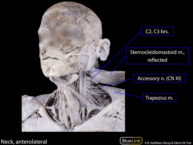
1. Spinal Accessory Nerve (CN XI)
- Entry: Emerges through the jugular foramen, passes deep to the SCM (supplying it), and enters the lateral cervical region at or just inferior to the junction of the superior and middle thirds of the posterior border of the SCM.
- Trajectory: Descends obliquely posteroinferiorly within or immediately deep to the investing layer of deep cervical fascia. It courses across the surface of the levator scapulae muscle, separated from it by the prevertebral layer of fascia.
- Exit & Innervation: Disappears deep to the anterior border of the trapezius muscle at the junction of its superior two-thirds and inferior one-third, supplying motor fibers to it.
- Surgical Vulnerability: Due to its superficial subcutaneous position within the investing fascia of the posterior triangle, CN XI is the most commonly injured nerve during posterior cervical lymph node biopsies.
2. Brachial Plexus Roots and Trunks
- Roots: Formed by the anterior rami of C5, C6, C7, C8, and T1. They emerge into the lateral cervical region through the interscalene triangle (between the anterior scalene and middle scalene muscles), lying superior and posterior to the subclavian artery.
- Trunks: The five rami unite within the inferior part of the lateral cervical region to form three trunks:
- Superior Trunk: Union of C5 and C6 rami.
- Middle Trunk: Direct continuation of C7 ramus.
- Inferior Trunk: Union of C8 and T1 rami (rests on the first rib behind the subclavian artery).
- Cervico-Axillary Canal: The trunks descend inferolaterally, passing through the cervico-axillary canal (between the first rib, clavicle, and superior border of the scapula) to enter the axilla.
- Suprascapular Nerve: Arises from the superior trunk (C5, C6) in the lateral cervical region. Runs laterally across the triangle to pass through the suprascapular foramen, innervating the supraspinatus and infraspinatus muscles and providing articular branches to the glenohumeral joint.
Cervical plexus & cutaneous branches
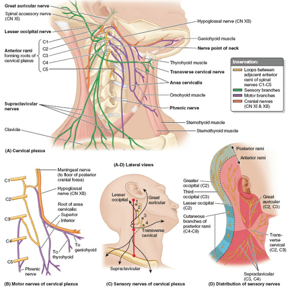
The cervical plexus is formed by the anterior rami of C1 through C4 spinal nerves, situated deep to the SCM and anteromedial to the levator scapulae and middle scalene muscles. Each participating ramus (except C1) divides into ascending and descending branches that anastomose to form primary loops.
1. Cutaneous (Sensory) Branches & Erb’s Point
The superficial sensory branches emerge around the midpoint of the posterior border of the SCM, a critical clinical landmark termed the nerve point of the neck (Erb’s point):

- 1. Lesser Occipital Nerve (C2): Arises from the C2 loop, ascends along the posterior border of the SCM to supply the skin of the neck and scalp posterosuperior to the auricle.
- 2. Great Auricular Nerve (C2, C3): Ascends vertically across the oblique surface of the SCM toward the inferior pole of the parotid gland. Divides to supply the skin overlying the parotid gland, parotid fascia, mastoid process, both surfaces of the auricle, and the angle of the mandible.
- 3. Transverse Cervical Nerve (C2, C3): Curves around the midpoint of the posterior border of the SCM, passing horizontally across the muscle deep to the EJV and platysma. Divides into superior and inferior branches supplying the skin of the anterior cervical region.
- 4. Supraclavicular Nerves (C3, C4): Emerge as a common trunk, descending under cover of the SCM to divide into medial, intermediate, and lateral supraclavicular branches. They cross the clavicle to supply the skin of the base of the neck, upper thorax, and shoulder overlying the deltoid and pectoralis major muscles.
2. Deep (Motor) Branches
- The Ansa Cervicalis: A nerve loop embedded in the anterior wall of the carotid sheath:
- Superior Root: Conveys C1 (and C2) fibers that briefly hitchhike with CN XII before descending across the carotid vessels.
- Inferior Root: Arises from a loop formed by C2 and C3 ventral rami, descending lateral to the IJV before looping forward to join the superior root.
- Innervation: Branches from the loop innervate the sternohyoid, sternothyroid, and omohyoid (both bellies) muscles. The fourth strap muscle, the thyrohyoid, receives C1 fibers that descend independently from CN XII distal to the ansa origin.
- The Phrenic Nerve: Originates primarily from the C4 ventral ramus, with secondary contributions from C3 and C5 (“C3, 4, 5 keeps the diaphragm alive”).
Course: Formed at the superolateral border of the anterior scalene muscle, descending vertically across its anterior surface under cover of the prevertebral fascia.
Composition: Contains motor, sensory, and postganglionic sympathetic fibers. Provides the sole motor innervation to the diaphragm, and sensory innervation to the central diaphragmatic tendon, mediastinal pleura, and pericardium.
Cervical lymph nodes
Lymphatic drainage from the head and neck is organized into superficial and deep tiers that correspond to the external and internal jugular venous axes.
1. Superficial Cervical Lymph Nodes
- Location: Lie in the subcutaneous tissue along the path of the external jugular vein, superficial to the sternocleidomastoid muscle.
- Drainage: Receive lymphatic fluid from superficial cutaneous tissues of the lateral neck, postauricular region, and parotid area.
- Efferent Flow: Efferent vessels pass around the borders of the SCM to empty directly into the deep cervical lymph nodes.
2. Deep Cervical Lymph Nodes
- Location: Form an extensive vertical chain embedded along the course of the internal jugular vein within the fibrous carotid sheath, extending from the base of the skull to the root of the neck.
- Subdivisions: Divided by the intermediate tendon of the omohyoid muscle into:
- Superior Deep Cervical Nodes: Clustered around the upper IJV; includes the prominent jugulodigastric node (tonsillar node), which drains the palatine tonsil.
- Inferior Deep Cervical Nodes: Located along the lower IJV; includes the jugulo-omohyoid node (related to lingual drainage) and supraclavicular nodes.
- Terminal Drainage: Efferent vessels from the deep cervical chain unite to form the jugular lymphatic trunk. On the left side, this trunk joins the thoracic duct; on the right side, it enters the right lymphatic duct or empties directly into the junction of the IJV and subclavian vein (the venous angle).
Surgical pearls & clinical correlations
Internal Jugular Vein Cannulation (Central Venous Line)
The right IJV is preferred for central venous catheterization because it provides a direct, straight pathway through the right brachiocephalic vein into the superior vena cava. The landmark for puncture is the apex of the minor supraclavicular triangle, formed by the sternal and clavicular heads of the SCM and the clavicle. Palpating the common carotid artery medially ensures needle insertion is directed laterally at a 30–45 degree angle toward the ipsilateral nipple, avoiding common carotid artery puncture.
Carotid Sinus Hypersensitivity & Endarterectomy
Excessive manual pressure over the carotid triangle (e.g., tight collars, aggressive carotid massage) can provoke exaggerated baroreceptor discharge via CN IX in patients with carotid sinus hypersensitivity. This triggers profound vagal slowing of the heart rate (severe bradycardia) and systemic vasodilation, precipitating acute cerebral hypoperfusion and carotid sinus syncope. During carotid endarterectomy, local anesthetic infiltration into the carotid sinus nerve prevents intraoperative hemodynamic instability.
Cervical Plexus Block at Erb’s Point
Superficial cervical plexus anesthesia is achieved by injecting local anesthetic along the posterior border of the SCM at its midpoint (Erb’s point). This provides cutaneous anesthesia across the anterolateral neck, auricle, and shoulder, facilitating carotid endarterectomy, lymph node excision, and superficial neck procedures in awake patients. Care must be taken to avoid deep injection into the prevertebral fascia, which could paralyze the phrenic nerve.
Quick recap
References
Verified anatomical reference texts supporting this curriculum note:
- Moore’s Clinically Oriented Anatomy: Dalley, A.F. and Agur, A.M.R. (2022) Moore’s Clinically Oriented Anatomy. 9th edn. Philadelphia: Wolters Kluwer. [Chapter 9: Neck – Cervical Fascia, Anterior Triangle, and Lateral Cervical Region].
- Gray’s Anatomy: Standring, S. (2021) Gray’s Anatomy: The Anatomical Basis of Clinical Practice. 42nd edn. London: Elsevier. [Section 4: Head and Neck – Neck Triangles, Carotid Arteries, and Cervical Plexus].
- Snell’s Clinical Anatomy by Regions: Splittgerber, R. (2019) Snell’s Clinical Anatomy by Regions. 10th edn. Philadelphia: Wolters Kluwer. [Chapter 11: The Neck].
- Netter Atlas of Human Anatomy: Netter, F.H. (2023) Atlas of Human Anatomy. 8th edn. Philadelphia: Elsevier. [Section 1: Head and Neck – Carotid Triangle and Posterior Triangle Dissections].