
Development of salivary glands, nasal cavity & palate: embryogenesis & congenital anomalies
On this page
Overview & facial primordia
Craniofacial morphogenesis begins during the fourth week of intrauterine life around the primitive mouth cavity, known as the stomodeum. The stomodeum is bounded rostrally by the frontonasal prominence, laterally by paired maxillary prominences, and caudally by paired mandibular prominences (derivatives of the first pharyngeal arch).
The Five Primary Facial Processes
Normal development of the human face and nasal apparatus depends upon the coordinated growth, migration, and mesenchymal merging of five prominent facial swellings:
- 1. Frontonasal Process: An unpaired midline ectomesenchymal elevation overlying the developing prosencephalon (forebrain). It forms the forehead, the bridge of the nose, and the primary nasal septum.
- 2 & 3. Paired Maxillary Processes: Derived from the dorsal part of the first pharyngeal arch (mandibular arch). They grow ventromedially to form the upper cheeks, lateral aspects of the upper lip, maxillae, zygomatic bones, and secondary palate.
- 4 & 5. Paired Mandibular Processes: Derived from the ventral part of the first pharyngeal arch. They merge across the midline early in the fourth week to form the lower jaw (mandible), lower lip, chin, and lower cheeks.
Development of the external nose
During the late fourth week, bilateral surface ectodermal thickenings called nasal placodes (olfactory placodes) develop on the inferolateral aspects of the frontonasal process. During the fifth week, mesenchymal proliferation around the margins of each placode produces horseshoe-shaped elevations, converting the placodes into deepened nasal pits:
- Medial Nasal Processes: The inner elevations bordering the nasal pits.
- Lateral Nasal Processes: The outer elevations bordering the nasal pits.
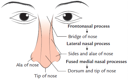
Anatomical Contributions of the Facial Processes
The external nose develops through the integration of five distinct processes:
- Bridge of the Nose: Derives directly from the central, inferior part of the unmodified frontonasal process.
- Dorsum and Tip (Apex) of the Nose: Formed by the downward migration and midline fusion of the two medial nasal processes.
- Sides and Alae of the Nose: Formed by the two lateral nasal processes, which surround the lateral boundaries of the nasal apertures.
Nasal cavities & choanae
The development of the nasal cavities proceeds from external ectodermal pits to deep internal air passages lined by specialized respiratory and sensory epithelia.

1. Deepening of Nasal Pits into Nasal Sacs
- During the sixth week of intrauterine life, the nasal pits deepen dorsally and posteriorly through the surrounding facial mesenchyme to form tubular nasal sacs.
- Each nasal sac grows dorsoventrally toward the floor of the developing forebrain, establishing the primitive nasal cavity.
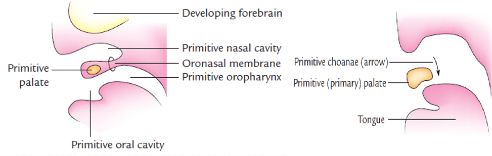
- At this stage, the primitive nasal cavity ends blindly; its dorsal floor is separated from the primitive oral cavity (stomodeum) by a thin bi-layered cellular partition called the oronasal membrane.
2. The Oronasal Membrane & Primitive Choanae
- Membrane Rupture: At the end of the sixth week, the oronasal membrane thins and ruptures, creating an open communication between the primitive nasal cavity and the primitive oral cavity.
- Primitive Choanae: These newly created internal apertures are the primitive choanae. They lie at the junction of the primitive nasal cavity and oral cavity, immediately posterior to the primary palate.
3. Transition to Definitive Choanae
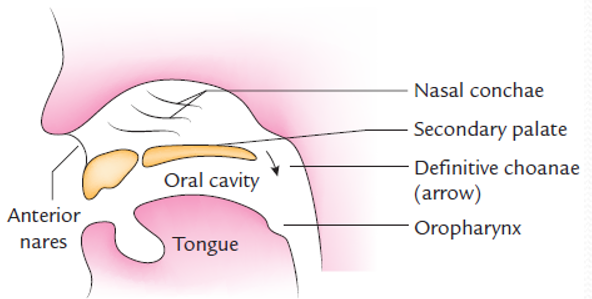
As development advances into the seventh to ninth weeks, the primitive nasal cavity expands longitudinally. With the development and midline horizontal fusion of the secondary palate, the floor of the nasal cavity is extended substantially backward. Consequently, the internal openings are displaced far posteriorly. The definitive choanae now lie at the junction of the permanent nasal cavities and the nasopharynx.

4. Lateral Wall Differentiation & Olfactory Specialization
- External Nares: The original external apertures of the nasal pits persist throughout life as the anterior nares (nostrils).
- Development of Nasal Conchae: On the lateral wall of each nasal cavity, three horizontal, curved mucosal elevations develop from the ethmoid and inferior turbinate mesenchyme to form the superior, middle, and inferior nasal conchae (turbinates). These projections disrupt laminar airflow, humidifying and warming inspired air.
- Olfactory Neuroepithelium: The ectodermal cells lining the roof of each nasal cavity specialize into pseudostratified olfactory neuroepithelium. Bipolar sensory neurons differentiate here, sending their central axonal processes through the cribriform plate of the ethmoid bone to form the olfactory nerve (CN I) fibers terminating in the olfactory bulb.
Scroll the table sideways on a smaller screen.
| Adult Nasal Component | Embryological Source |
|---|---|
| Dorsum and tip (apex) of nose | Frontonasal process (via fused medial nasal processes) |
| Nasal cavities (mucosal lining) | Nasal sacs formed by dorsal elongation of ectodermal nasal pits |
| Nasal septum | Frontonasal process (median downgrowth) |
| Nostrils (anterior nares) | External openings of the ectodermal nasal pits |
| Primitive choanae (posterior nares) | Rupture of the oronasal membrane (end of 6th week / second month) |
| Definitive choanae | Posterior elongation following secondary palatal fusion (nasopharyngeal junction) |
Paranasal air sinuses
The paranasal air sinuses are bilateral, air-filled expansions of the nasal cavity that pneumaticize the surrounding cranial and facial bones. They develop as tubular diverticulae (outgrowths) arising from the lateral and superior walls of the nasal cavity.
Mechanism of Development & Anatomy
- Bone Invasion (Pneumatization): The mucosal diverticulae extend into the adjacent skeletal elements, slowly eroding and hollowing out the maxilla, ethmoid, frontal, and sphenoid bones.
- Nomenclature: The sinuses are named according to the specific bone they invade:
- Maxillary air sinuses (antrum of Highmore) in the maxillae.
- Frontal air sinuses in the frontal bone.
- Ethmoid air cells in the ethmoidal labyrinth.
- Sphenoid air sinuses in the body of the sphenoid bone.
- Epithelial Lining: Because they arise as direct evaginations of the nasal wall, the mucosal lining of all paranasal sinuses is ectodermal, consisting of ciliated pseudostratified columnar respiratory epithelium rich in goblet cells.
- Sinus Orifices (Ostia): The original site of evagination from the nasal cavity wall persists throughout life as the permanent drainage orifice (ostium) connecting each adult sinus to its corresponding nasal meatus (e.g., middle meatus for frontal, maxillary, and anterior ethmoidal sinuses).
Chronological Development of the Sinuses
The timing of sinus development follows a predictable prenatal and postnatal schedule:
- Prenatal Development: Most paranasal sinuses begin developing before birth.
- The maxillary air sinuses are the first to develop. They appear during the third month of intrauterine life as shallow grooves on the medial nasal surfaces of each maxilla.
- Ethmoid and sphenoid sinuses develop during late fetal life as smaller evaginations.
- Postnatal Development (The Frontal Sinus Exception): All sinuses begin developing before birth except the frontal air sinuses, which develop entirely after birth. Frontal sinuses begin invading the frontal bone around the fifth or sixth postnatal year, expanding through childhood.
- Pubertal Expansion: All paranasal sinuses undergo a second rapid growth spurt during puberty, coinciding with secondary dentition eruption and definitive facial elongation, reaching their maximum anatomical size.
Physiological Functions of Paranasal Sinuses
- Facial Contouring: Contribute to the definitive shape, vertical dimension, and aesthetic profile of the adult face.
- Weight Reduction: Significantly reduce the physical weight of the heavy anterior facial skeleton and skull, balancing the head on the cervical spine.
- Vocal Resonance: Act as acoustic resonating chambers that modulate the tonal quality and resonance of the human voice.
- Climatic Conditioning: Provide supplemental humidification and thermal buffering for inhaled air before it enters the lower respiratory tract.
Development of the primary palate
The human palate separates the nasal cavities from the oral cavity, allowing mastication and respiration to occur simultaneously. Palatogenesis begins in the sixth week and is completed by the twelfth week of gestation. Embryologically, it develops in two stages: the primary palate and the secondary palate.
Mesenchymal Fusion of the Medial Nasal Processes
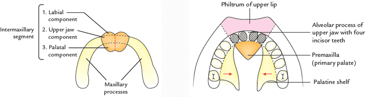
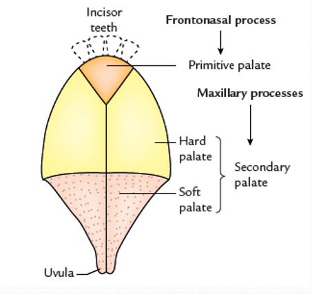
- The primary palate (primitive palate / median palatine process) is formed by the midline fusion of the two medial nasal processes (derivatives of the frontonasal process) at a deep anatomical level.
- This deep union forms a cohesive, wedge-shaped mass of mesenchyme known as the intermaxillary segment, situated in the midline of the upper jaw opposite the stomodeum.
Three Components of the Intermaxillary Segment
- 1. Labial Component: Forms the central grooved portion of the upper lip, known as the philtrum.
- 2. Upper Jaw Component: Forms the primary premaxillary part of the maxilla, which carries the four upper incisor teeth (two central and two lateral incisors) and their associated labial gingiva.
- 3. Palatal Component: Forms the triangular, wedge-shaped primary palate, which ossifies via intramembranous ossification to form the adult premaxilla. It lies anterior to the future incisive fossa.
Development of the secondary palate
The secondary palate is the primordium of the vast majority of the hard palate and the entirety of the soft palate. It develops from the bilateral maxillary processes of the first pharyngeal arches.
Stepwise Elevation and Fusion of Palatine Shelves
- 1. Shelf Outgrowth (6th Week): During the sixth week, two shelf-like medial projections—the palatine shelves (lateral palatine processes)—grow from the inner, medial aspects of the maxillary processes.
- 2. Vertical Descent Beside the Tongue: Initially, because the developing tongue is disproportionately large and positioned high within the stomodeum, the palatine shelves grow downward vertically on either side of the tongue.
- 3. Shelf Elevation (7th to 8th Week): During the seventh and eighth weeks, the embryonic mandible enlarges, allowing the tongue to descend to the floor of the mouth. Relieved of mechanical obstruction, the palatine shelves rotate upward and swing into a horizontal position above the dorsum of the tongue.
- 4. Midline Epithelial Fusion: The medial edges of the two horizontal shelves meet in the midline. The covering medial edge epithelial seams degenerate via apoptosis and epithelial-to-mesenchymal transition (EMT), fusing to form the cohesive secondary palate.
Ossification: Hard Palate vs. Soft Palate
- Formation of the Hard Palate: Intramembranous ossification extends medially from the developing maxillae and palatine bones into the anterior three-fourths of the fused shelves, forming the palatine processes of the maxillae and the horizontal plates of the palatine bones (hard palate).
- Formation of the Soft Palate & Uvula: The posterior one-fourth of the fused shelves extends posteriorly beyond the caudal border of the nasal septum. This posterior portion fails to ossify. Mesenchymal cells within this unossified shelf condense and differentiate into the striated muscles of the soft palate (tensor veli palatini, levator veli palatini, palatoglossus, palatopharyngeus, and musculus uvulae). The extreme posterior midline margin forms a free, conical projection: the uvula.
Definitive palate integration
The definitive adult palate is formed by the fusion of three distinct embryonic masses: the anterior primary palate and the two lateral secondary palatine shelves.
The Y-Shaped Suture & Incisive Fossa
- Y-Shaped Suture Line: Anteriorly, the horizontal secondary palatine shelves fuse with the posterior edge of the wedge-shaped primary palate along a distinct Y-shaped suture.
- The stem of the “Y” runs along the intermaxillary median palatine suture.
- Each oblique limb of the “Y” passes anterolaterally between the lateral incisor tooth and the canine tooth on each side.
- Adult Incisive Fossa: The anatomical junction where the primary palate meets the secondary palatine processes is marked in the adult skeleton by the incisive fossa (incisive foramen), through which the nasopalatine nerve and sphenopalatine vessels pass between the nasal and oral cavities.
Integration of the Nasal Septum
- While palatine fusion is underway, the nasal septum grows vertically downward as a median plate of mesenchyme from the internal aspect of the frontonasal process and medial nasal processes.
- The inferior margin of the growing septum joins and fuses with the superior midline surface of the anterior three-fourths of the hard palate.
- Timeline of Fusion: Septal-palatal fusion begins anteriorly during the ninth week and advances progressively backward, completing posteriorly at the posterior margin of the hard palate by the twelfth week.
- The posterior one-fourth of the palate (the soft palate) remains completely free and unattached to the nasal septum, hanging as a dynamic muscular curtain between the nasopharynx and oropharynx.
Scroll the table sideways on a smaller screen.
| Adult Palatal Component | Specific Anatomical Boundary | Embryological Source of Development |
|---|---|---|
| Hard Palate: Premaxilla | Anterior to incisive fossa; carries four incisor teeth | Fused medial nasal processes of the frontonasal process (intermaxillary segment) |
| Hard Palate: Palatine Processes | Posterior to incisive fossa; carries canines, premolars, molars | Fused horizontal palatine shelves of bilateral maxillary processes (intramembranous ossification) |
| Soft Palate & Uvula | Posterior one-fourth of palate; posterior to nasal septum | Unossified posterior extensions of the fused palatine shelves of bilateral maxillary processes |
Cleft lip & facial clefts
Failure of facial processes to merge or fuse results in facial clefts. Cleft lip (cheiloschisis) is one of the most common congenital craniofacial malformations, occurring in approximately 1 in 1000 live births. There is a marked sex predilection: 60% to 80% of affected infants are males.
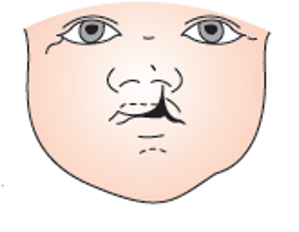
1. Unilateral Cleft Lip
- Embryological Defect: Occurs when the maxillary process fails to fuse with the medial nasal process on one side.
- Morphology: Characterized by a vertical fissure through the vermilion border and upper lip lateral to the philtrum. The alar cartilage on the affected side is flattened and displaced laterally. It is more common on the left side and may involve the alveolar ridge.
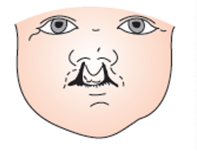
2. Bilateral Cleft Lip
- Embryological Defect: Occurs when both maxillary processes fail to fuse with the medial nasal processes of the frontonasal process.
- Morphology: Features bilateral gaps through the upper lip, completely isolating the central prolabium and underlying premaxilla. This central segment projects anteriorly due to unrestrained premaxillary growth.
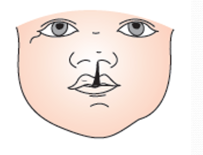
3. Central Cleft Lip (True Median Cleft / “Hare Lip”)
- Embryological Defect: Caused by the complete failure of development or merging of the two medial nasal processes of the frontonasal process in the midline.
- Clinical Features: Results in a deep midline notch or complete absence of the philtrum and premaxilla. Often associated with holoprosencephaly and severe underlying prosencephalic forebrain anomalies.
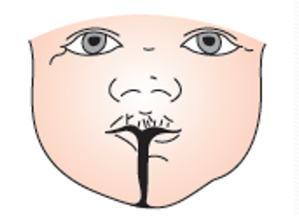
4. Cleft Lower Lip (Mandibular Cleft)
- Embryological Defect: Caused by the failure of the two mandibular processes to fuse in the midline.
- Incidence: Extremely rare anomaly presenting with a complete or partial vertical fissure splitting the lower lip, mandible, and occasionally the tongue in the midline.
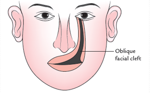
5. Oblique Facial Cleft
- Embryological Defect: Occurs when the maxillary process fails to fuse with the lateral nasal process along the line of the nasolacrimal groove (naso-optic furrow).
- Clinical Features: A severe, disfiguring fissure extending diagonally from the medial angle of the eye (inner canthus) down to the upper lip. The nasolacrimal duct is exposed to the exterior as an open mucosal gutter; this malformation is frequently bilateral.
Cleft palate varieties
Cleft palate (palatoschisis) results from defective fusion between the various embryonic palatal processes. It occurs in roughly 1 in 2500 live births, is more common in females, and presents in a spectrum ranging from minor uvular notches to complete bilateral fissures.
1. Complete Cleft Palate
A cleft that extends through the entire length of both the secondary and primary palates, directly reaching the incisive foramen and alveolar arch.
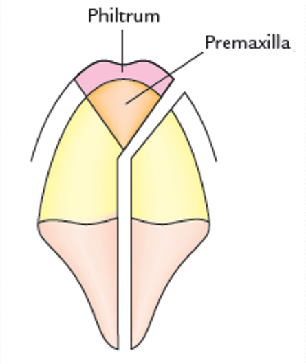
- Unilateral Complete Cleft Palate: Occurs when the maxillary process on one side fails to fuse with the premaxilla. The palatal shelf on the opposite side fuses normally with the premaxilla and the nasal septum. It is almost always associated with a unilateral cleft lip on the same side. One nasal cavity communicates directly with the oral cavity.
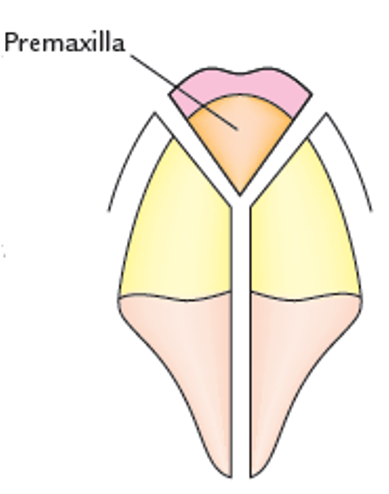
- Bilateral Complete Cleft Palate: Occurs when both maxillary processes fail to fuse with the premaxilla. The secondary palate is split into two equal lateral halves by a wide median cleft. Anteriorly, bilateral clefts diverge in an inverted V-shape between the lateral incisors and canines, isolating the central premaxillary bone, which hangs unsupported from the nasal septum.
2. Incomplete (Partial) Cleft Palate
Clefts that involve only the secondary palate, lying entirely posterior to the incisive foramen (due to failed or incomplete midline fusion of the bilateral palatine shelves):
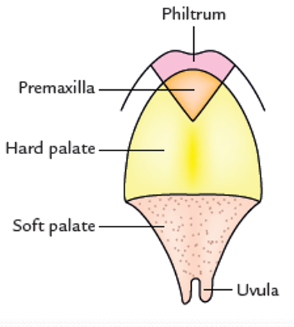
- Bifid Uvula: The mildest manifestation; a cleft restricted entirely to the uvula. It results from incomplete fusion of the caudalmost tips of the palatine shelves and has no functional clinical significance.
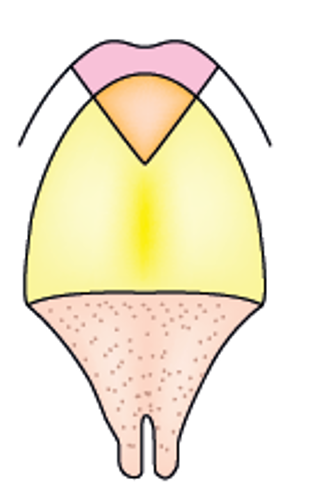
- Cleft of the Soft Palate: The cleft involves the uvula and extends through the muscular substance of the soft palate, impairing velopharyngeal sphincter competence during swallowing and speech.
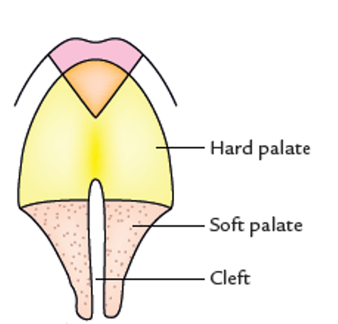
- Cleft of the Soft and Hard Palate: The cleft involves the uvula, the entirety of the soft palate, and extends forward through the bony hard palate, terminating immediately posterior to the incisive fossa.
Major salivary glands
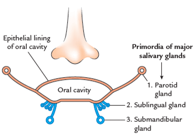
The human body possesses three pairs of major salivary glands: the parotid, submandibular, and sublingual glands. They develop as solid epithelial cords that bud from the primitive oral cavity into the underlying mesenchyme, followed by branching morphogenesis and canalization.
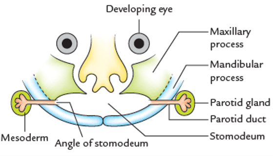
1. The Parotid Gland (Ectodermal Origin)
- Time of Appearance: The parotid is the first major salivary gland to appear, developing during the fifth week of intrauterine life.
- Germ Layer & Site: Arises as an ectodermal furrow (outgrowth) from the inner mucosal surface of the cheek at the lateral angle of the stomodeum.
- Duct Morphogenesis:
- The ectodermal furrow grows outward into the mesoderm between the mandibular and maxillary processes.
- The furrow invaginates and closes off to form a tube: the parotid duct (Stensen’s duct).
- The medial end of the duct maintains its opening into the oral cavity. The lateral end branches repeatedly into the surrounding mesenchyme.
- These branched solid cords subsequently canalize to form the ductules and acini of the gland. The surrounding mesenchyme condenses to form the fibrovascular stroma and the dense parotid capsule.
- Elongation of Jaws & Shifting Orifice: As the mandible and maxilla elongate, the parotid duct elongates substantially. The body of the gland remains anchored in the retromandibular space near its site of origin. Subsequent medially directed fusion of the mandibular and maxillary processes shifts the angle of the mouth medially, leaving the parotid duct opening in the adult oral vestibule opposite the crown of the second upper molar tooth.
2. The Submandibular Gland (Endodermal Origin)
- Time of Appearance: Develops late in the sixth week of intrauterine life.
- Germ Layer & Site: Develops as a solid endodermal outgrowth from the floor of the stomodeum, specifically from the floor of the alveololingual groove lateral to the developing lingual swellings.
- Duct Morphogenesis:
- The endodermal buds grow posteriorly into the submandibular triangle, lateral to the tongue musculature.
- A linear groove forms in the floor of the mouth lateral to the tongue; this groove closes from behind forward to form the tubular submandibular duct (Wharton’s duct).
- The anterior orifice opens on a prominent mucosal papilla—the sublingual papilla (caruncle)—on each side of the frenulum of the tongue.
3. The Sublingual Gland (Endodermal Origin)
- Time of Appearance: Develops in the eighth week of intrauterine life (approximately two weeks later than the submandibular and parotid glands).
- Germ Layer & Site: Arises as multiple (8 to 20) separate endodermal outgrowths from the floor of the linguogingival sulcus and the distal portion of the submandibular duct.
- Duct Morphogenesis:
- Each independent endodermal bud branches and canalizes separately into the underlying connective tissue.
- Consequently, the sublingual gland retains multiple excretory ducts: ducts of Rivinus (8 to 20 small ductules) that open independently along the summit of the sublingual fold in the floor of the mouth.
- Some anterior ducts frequently merge to form a principal greater sublingual duct (duct of Bartholin), which drains into the submandibular duct or directly onto the sublingual papilla.
Scroll the table sideways on a smaller screen.
| Salivary Gland | Germ Layer Origin | Anatomical Site of Origin | Time of Appearance |
|---|---|---|---|
| Parotid Gland | Ectoderm | Mucosal lining of cheek at the lateral angle of the stomodeum | 5th week of gestation (First to appear) |
| Submandibular Gland | Endoderm | Floor of stomodeum (alveololingual groove lateral to tongue) | 6th week of gestation |
| Sublingual Gland | Endoderm (multiple buds) | Floor of linguogingival sulcus and submandibular duct | 8th week of gestation (Two weeks after others) |
Clinical pearls & surgical management
Quick recap
References
The following authoritative medical embryology and anatomy reference texts support this curriculum note:
- Langman’s Medical Embryology: Sadler, T.W. (2019) Langman’s Medical Embryology. 14th edn. Philadelphia: Wolters Kluwer. [Chapter 17: Head and Neck – Pharyngeal Apparatus, Face, and Palate].
- The Developing Human: Moore, K.L., Persaud, T.V.N. and Torchia, M.G. (2020) The Developing Human: Clinically Oriented Embryology. 11th edn. Philadelphia: Elsevier. [Chapter 9: Development of the Pharyngeal Apparatus, Face, and Neck].
- Gray’s Anatomy: Standring, S. (2021) Gray’s Anatomy: The Anatomical Basis of Clinical Practice. 42nd edn. London: Elsevier. [Section 4: Head and Neck; Chapter 27: Face and Scalp; Chapter 32: Oral Cavity and Salivary Glands].
- Larsen’s Human Embryology: Schoenwolf, G.C., Bleyl, S.B., Brauer, P.R. and Francis-West, P.H. (2021) Larsen’s Human Embryology. 6th edn. Philadelphia: Elsevier. [Chapter 12: Development of the Head, the Neck, the Eyes, and the Ears].