
Development of the eye: organogenesis, tissue induction & congenital anomalies
On this page
Overview & germ layers
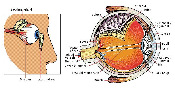
The human visual apparatus comprises two interdependent anatomical systems: the eyeball (globe) and its accessory structures (ocular adnexa), which include the eyelids, conjunctival sac, and the lacrimal apparatus. Morphogenesis of the human eye begins on approximately day 22 of embryonic life (early week 4), when the embryo possesses approximately eight somite pairs, manifested by the appearance of paired shallow neuroectodermal grooves in the forebrain neural folds.
The Four Embryonic Tissue Progenitors
The definitive eyeball is a composite organ assembled through precise, reciprocal inductive signaling cascades coordinated among four primary embryological tissues:
- 1. Neuroectoderm of the Forebrain (Prosencephalon / Diencephalon): Forms the sensory retina, the retinal pigment epithelium (RPE), the posterior pigmented epithelial layers of the ciliary body and iris, the intrinsic intraocular smooth muscles (sphincter pupillae and dilator pupillae), and the axons and glia of the optic nerve.
- 2. Surface Ectoderm of the Head: Gives rise to the crystalline lens, the anterior stratified squamous corneal epithelium, the conjunctival epithelium, the lacrimal gland, and the glandular/epidermal lining of the eyelids.
- 3. Mesoderm (Head Mesenchyme): Contributes to the corneal stroma, the endothelial lining of the anterior chamber, the primary angioblastic vascular networks (including the hyaloid artery and embryonic tunica vasculosa lentis), the temporal aspects of the sclera, and the striated extraocular muscles (derived from prechordal mesoderm paraxial head somites, innervated by cranial nerves III, IV, and VI).
- 4. Cranial Neural Crest Cells: Ectomesenchyme migrating from the neural fold margins into the circumocular space to generate the corneal stroma (substantia propria), corneal endothelium (Descemet's endothelium), trabecular meshwork, sclera, choroidal stroma, ciliary body stroma and ciliary muscle, iris stroma, and the meningeal coverings of the optic nerve.
Optic vesicle & lens placode
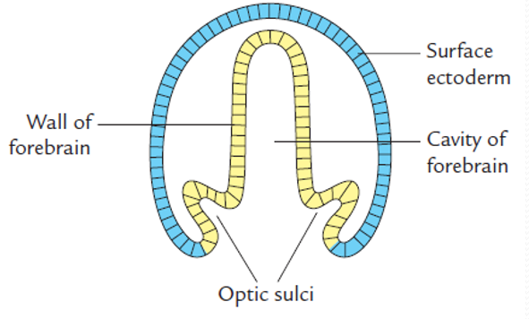
Organogenesis begins as bilateral depressions, the optic sulci (optic grooves), appear on the lateral aspects of the expanding forebrain neural folds on day 22. As the neural folds elevate and close to form the prosencephalic vesicle, these sulci evaginate outward toward the overlying surface ectoderm, expanding into hollow, bulbous diverticula termed optic vesicles.


Lens Placode Induction
As the distal portion of each optic vesicle expands, its connecting proximal stem constricts to form the narrow optic stalk. The expanding distal neuroectodermal surface comes into direct physical apposition with the overlying cephalic surface ectoderm.
- Reciprocal Molecular Induction: Secretion of Fibroblast Growth Factors (FGFs) and Bone Morphogenetic Proteins (BMPs) by the optic vesicle neuroectoderm induces the cuboidal cells of the surface ectoderm to elongate into a thickened, tall columnar epithelial disc termed the lens placode (the primordium of the crystalline lens).
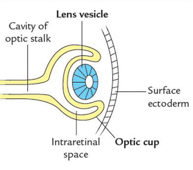
- Reciprocal Response: In turn, signals from the newly formed lens placode stimulate the underlying optic vesicle to initiate inward invagination, converting the single-walled spherical vesicle into a double-walled cup.
Optic cup & choroid fissure

Formation of the Choroid (Retinal) Fissure
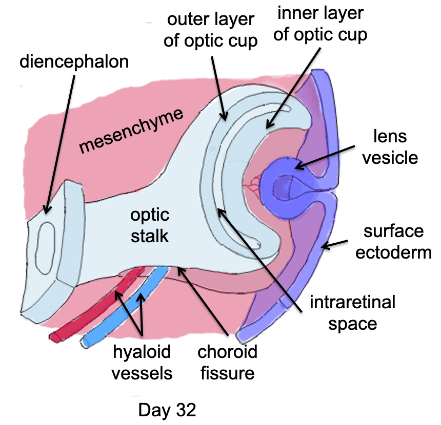
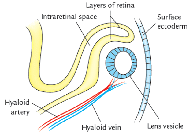
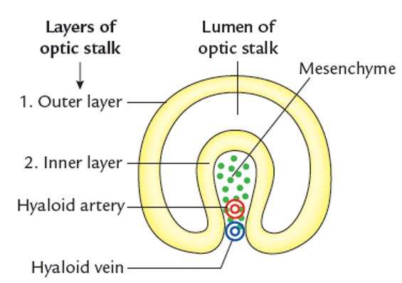
Invagination does not involve the central distal surface of the optic vesicle alone; it extends eccentrically along its ventral (inferior) aspect and continues along the ventral surface of the optic stalk. This produces a linear, longitudinal groove called the choroid fissure (retinal fissure / fetal ocular fissure).

Dynamics of Fissure Closure & Hyaloid Encasement
- Vascular Ingrowth: A core of vascular mesoderm migrates into the open choroid fissure. These vessels differentiate into the hyaloid artery and hyaloid vein, which arborize to supply the developing inner retinal layer, the vitreous mesenchyme, and the posterior surface of the lens vesicle (via the embryonic tunica vasculosa lentis).
- Fissure Closure Timeline: During gestational week 7, the opposing lips of the choroid fissure approach each other, fuse, and obliterate the seam. Fusion begins centrally in the middle of the optic stalk and proceeds simultaneously in both rostral (anterior) and caudal (posterior) directions.
- Vascular Encasement: Closure of the fissure completely encloses the hyaloid vessels within the center of the optic stalk. The distal arborizations of the hyaloid vessels subsequently regress during the third trimester, while their proximal, encased segments persist postnatally as the central artery and central vein of the retina.
Retinal morphogenesis
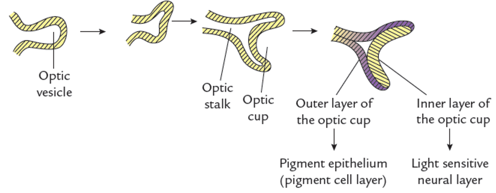
The definitive retina develops entirely from the two neuroectodermal layers of the optic cup:
- 1. Outer Thin Layer: Retains a single-cell-thick columnar profile. Its neuroepithelial cells synthesize melanin granules by week 6, differentiating into the definitive retinal pigment epithelium (RPE).
- 2. Inner Thick Layer: Undergoes active neuroblastic proliferation, mirroring the developing neural tube, to form the stratified neural retina (sensory retina). Differentiates into an outer nuclear zone (giving rise to modified neuroepithelial photoreceptor cells: rods and cones), an intermediate layer (bipolar neurons, horizontal cells, and amacrine interneurons), and an inner layer of ganglion cells and supporting Müller glia.
The Intraretinal Space & Secondary Apposition
Throughout embryonic and early fetal life, the outer pigment layer and inner neural layer are separated by an anatomical cleft termed the intraretinal space (representing the original invaginating lumen of the primary optic vesicle). Before birth, rapid cellular proliferation and elongation of the neural retina obliterates this space, bringing the outer segments of rods and cones into close physical contact with the microvilli of the retinal pigment epithelium.
Optic nerve development
The optic nerve develops as axons of retinal ganglion cells congregate within the inner nerve fiber layer, traverse the optic cup margins, and penetrate the inner wall of the tubular optic stalk. As hundreds of thousands of unmyelinated axons course through the stalk toward the diencephalon, the central lumen of the optic stalk is progressively compressed and obliterated. By week 7, with complete closure of the choroid fissure, the optic stalk and its enclosed ganglion cell axonal bundles transform into the definitive optic nerve.
Why the Optic Nerve is a CNS Tract (Not a True Peripheral Nerve)
Anatomically and developmentally, Cranial Nerve II is an evaginated central white matter tract of the diencephalon rather than a true peripheral nerve:
- Central Oligodendrocyte Myelination: The axons of the optic nerve are myelinated by oligodendrocytes (which differentiate from neuroectodermal cells of the optic stalk in the 3rd to 7th fetal month) rather than peripheral neural crest-derived Schwann cells. Myelination is incomplete at birth, completing after exposure to light postnatally.
- Absence of Peripheral Regeneration: Unlike peripheral nerves encased in endoneurial/Schwann cell sheaths, optic nerve axons lack a neurilemmal sheath and cannot functionally regenerate after surgical transection or ischemic necrosis. Injury stimulates reactive astrocyte gliosis.
- Meningeal Encasement & Subarachnoid Space: The optic stalk evaginates into head mesenchyme, carrying with it all three concentric layers of the cranial meninges: the outer dura mater (fusing anteriorly with the sclera), intermediate arachnoid mater, and inner pia mater. The intervening subarachnoid space surrounding the optic nerve is directly continuous with the intracranial subarachnoid space and filled with cerebrospinal fluid (CSF).
Lens formation & cataracts
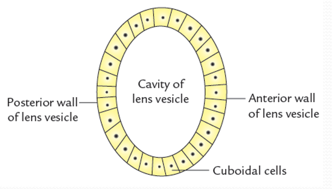
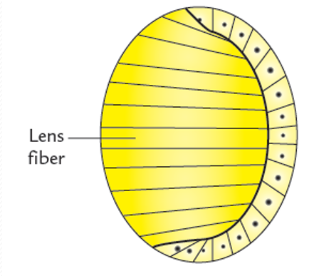
The crystalline lens originates exclusively from the surface ectoderm. Once the hollow spherical lens vesicle pinches off from the surface ectoderm during week 5, it sits suspended within the opening of the optic cup.

Primary vs. Secondary Lens Fibers
- Anterior Epithelial Layer: The cells of the anterior wall of the lens vesicle remain cuboidal throughout life, persisting as the single-layered subcapsular anterior lens epithelium.
- Primary Lens Fibers: The cells of the posterior wall elongate anteriorly, transforming into transparent, ribbon-like primary lens fibers. As they elongate, these cells synthesize optical proteins (crystallins) and lose their cell nuclei, mitochondria, and intracellular organelles. By gestational week 7, their apical ends contact the anterior epithelium, completely obliterating the central lumen of the lens vesicle to form the dense, central embryonic lens nucleus.
- Secondary Lens Fibers: Lifelong lens growth is maintained by mitotic cells located at the equatorial zone (the germinative perimeter where the anterior epithelium meets the posterior fiber mass). Epithelial cells at the equator elongate, lose their nuclei, and lay down concentric layers of secondary lens fibers over the older primary fibers, forming the fetal and adult lens cortex. As new fibers accumulate peripherally, the older, centrally positioned primary fibers become progressively compressed, dehydrated, and rigid.
Uveal tract & fibrous coats
During week 5, the developing optic cup is enveloped by a condensation of loose head mesenchyme derived predominantly from cranial neural crest cells with paraxial mesodermal contributions. Under inductive signals from the retinal pigment epithelium, this mesenchyme cleaves into two distinct morphological tunics:
- Inner Vascular Tunic (Choroid): Differentiates into a thin, pigmented vascular lamina. Anteriorly, it is continuous with the ciliary body and iris stroma; posteriorly, it is continuous with the pia-arachnoid mater enclosing the optic nerve.
- Outer Fibrous Tunic (Sclera): Differentiates into dense, collagenous connective tissue that maintains ocular shape. Anteriorly, it becomes continuous with the corneal stroma (substantia propria) at the corneoscleral limbus; posteriorly, it condenses to form the lamina cribrosa and blends directly into the dura mater sheath of the optic nerve.
1. Morphogenesis of the Ciliary Body
The ciliary body develops as an anterior wedge-shaped expansion of the choroid, containing both neuroectodermal and mesenchymal tissues:
- Dual Ciliary Epithelium (Neuroectoderm): Derived from the anterior rim of the optic cup. Differentiates into a distinctive bilayered epithelium:
- Outer Pigmented Epithelium: Derived from the outer layer of the optic cup; continuous with the retinal pigment epithelium.
- Inner Non-Pigmented Epithelium: Derived from the inner layer of the optic cup; represents an anterior, non-visual continuation of the neural retina. Its cells become highly folded into ciliary processes, developing ion transport machinery to secrete aqueous humor.
- Ciliary Muscle & Stroma (Neural Crest Mesenchyme): The smooth muscle fibers of the ciliary muscle (responsible for near lens accommodation via parasympathetic fibers of CN III) and the vascular connective tissue stroma differentiate from neural crest mesenchyme situated outside the optic cup.
2. Morphogenesis of the Iris & Pupillary Aperture
- Iris Epithelium: The anterior margins of both layers of the optic cup grow centripetally over the anterior surface of the crystalline lens, forming the double-layered pigmented posterior epithelium of the iris (the iridial retina). The failure of this rim to close completely in the center preserves a circular opening: the pupil.
- Neuroectodermal Origin of Iris Muscles: Uniquely among visceral smooth muscles in the human body (which almost universally derive from mesoderm), the sphincter pupillae (causing miosis via parasympathetic stimulation) and the dilator pupillae (causing mydriasis via sympathetic stimulation) differentiate directly from the neuroectoderm of the anterior optic cup.
- Iris Stroma: Derived from the migration of neural crest mesenchyme anterior to the expanding cup margins, forming the vascular, pigmented anterior stroma of the iris.
Cornea & aqueous chambers
Trilaminar Embryonic Origin of the Cornea
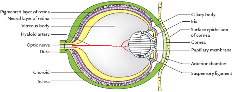
The development of the cornea is induced by the underlying detached lens vesicle. It develops from three distinct embryological sources:
- Anterior Corneal Epithelium: Stratified squamous non-keratinized epithelium derived directly from surface ectoderm.
- Substantia Propria (Corneal Stroma) & Bowman's Layer: Developed from mesenchyme (predominantly neural crest cells with mesodermal contributions) continuous with the developing sclera.
- Posterior Corneal Endothelium & Descemet's Membrane: A single layer of simple squamous/cuboidal cells derived exclusively from neural crest cells that migrate along the posterior corneal surface.
Formation of the Anterior and Posterior Chambers
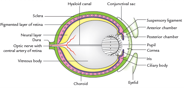
- The Anterior Chamber: Mesenchyme situated between the developing cornea and the iris undergoes progressive vacuolization and splitting, creating a fluid-filled cleft termed the anterior chamber. The mesoderm on its anterior wall forms the corneal stroma, while the posterior mesoderm condensation forms the transient pupillary membrane overlying the lens.
- The Posterior Chamber: Mesenchyme located between the developing iris and the anterior surface of the crystalline lens vacuolizes to form the posterior chamber.
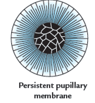
- Pupillary Membrane Resorption: Prior to the 6th gestational month, the pupil is occluded by the vascularized pupillary membrane. In late fetal life, this vascular arcade and its central connective tissue completely resorb via macrophage-mediated apoptosis, establishing a patent opening through the pupil that connects the posterior and anterior chambers.
- Aqueous Humor Drainage: Fluid is secreted into the posterior chamber by the non-pigmented ciliary epithelium of the ciliary processes, passes through the pupil into the anterior chamber, traverses the neural-crest-derived trabecular meshwork at the iridocorneal filtration angle, and drains into the sinus venosus sclerae (canal of Schlemm), a circumferential venous channel encircled by the limbic sclera.
Vitreous body & hyaloid canal
The vitreous body (vitreous humor) is an extracellular gel-like transparent matrix filling the posterior segment of the eye between the lens and the retina. Its development occurs in two sequential phases:
Biphasic Vitreous Development
- 1. Primary Vitreous: Forms during weeks 5 to 6 from mesenchymal tissue derived from neural crest cells that migrate through the choroid fissure into the optic cup. Highly vascular, containing branches of the hyaloid artery.
- 2. Secondary Vitreous: Formed during the second and third months, derived predominantly from the inner neuroectodermal layer of the optic cup with contributions from the lens vesicle. Composed of delicate, transparent collagen fibrils (predominantly type II) and hydrophilic glycosaminoglycans (hyaluronic acid). As the secondary vitreous expands, it compresses the primary vitreous into a central, narrow anteroposterior core.
- The Hyaloid Canal (Cloquet's Canal): During late fetal life, the distal portion of the hyaloid artery ceases blood flow and undergoes complete apoptotic degeneration. The collapsed avascular sheath of the regressed primary vitreous and hyaloid vessel persists through adult life as a transparent, fluid-filled central channel—the hyaloid canal (canal of Cloquet)—extending from the optic disc to the posterior pole of the crystalline lens.
Tissue origin mapping
The following structured reference table categorizes the definitive adult anatomical structures of the human eye according to their embryological tissue progenitors.
Scroll the table sideways on a smaller screen.
| Embryonic Progenitor Tissue | Embryonic Substructure | Definitive Adult Ocular Derivatives |
|---|---|---|
| Neuroectoderm (of Forebrain) | Optic Cup |
• Neural (sensory) retina (photoreceptors, bipolars, ganglion cells) • Retinal pigment epithelium (RPE) • Pigmented & non-pigmented ciliary epithelium • Posterior double-layered pigment epithelium of the iris • Sphincter pupillae and dilator pupillae muscles |
| Optic Stalk |
• Axons of the optic nerve (CN II) • Optic chiasm and optic tract neuroglia (oligodendrocytes, astrocytes) |
|
| Surface Ectoderm (of Head) | Lens Placode & Head Ectoderm |
• Crystalline lens (anterior epithelium, primary & secondary fibers) • Anterior stratified squamous corneal epithelium • Bulbar and palpebral conjunctival epithelium • Lacrimal gland acini and drainage ducts • Epidermis of the eyelids, cilia (eyelashes), and glands (Meibomian, Zeis, Moll) |
| Mesoderm (Head Somites & Mesenchyme) | Angiogenic Mesenchyme |
• Central artery and vein of the retina (proximal hyaloid remnants) • Embryonic hyaloid vascular system (transient) |
| Prechordal Paraxial Mesoderm | • Extraocular muscles (superior, inferior, medial, lateral recti; superior, inferior obliques; levator palpebrae superioris) | |
| Cranial Neural Crest Cells (Ectomesenchyme) | Circumocular Mesenchyme |
• Sclera • Corneal substantia propria (stroma) and Bowman's layer • Corneal endothelium and Descemet's membrane • Choroid vascular stroma and melanocytes • Ciliary body connective tissue stroma and ciliary muscle • Iris stroma and chromatophores • Primary vitreous body • Meningeal sheaths (dura, arachnoid, pia) of the optic nerve • Trabecular meshwork and canal of Schlemm endothelium |
Eyeball congenital anomalies
Errors during ocular morphogenesis produce structural ocular malformations. These dysgeneses stem from chromosomal aneuploidies, single-gene mutations (especially PAX6, SHH, SOX2), and intrauterine teratogenic insults.
1. Primary Malformations of Ocular Primordia
- Anophthalmia (Absence of the Eye): Complete absence of the eyeball. Results from total failure of the optic vesicle to evaginate from the lateral wall of the forebrain during week 4. May be unilateral or bilateral. Orbit and ocular adnexa (eyelids) are present but hypoplastic.
- Microphthalmia (Small Eye): A condition in which the globe is significantly reduced in volume (axial length >2 standard deviations below normal). Arises when the optic vesicle evaginates incompletely or development arrests following vesicular formation. The crystalline lens is frequently absent or severely hypoplastic. Strongly linked to the maternal TORCH infection complex (toxoplasmosis, rubella virus, cytomegalovirus, herpes simplex virus).
2. Midline Cleavage Defects: Cyclopia & Synophthalmia
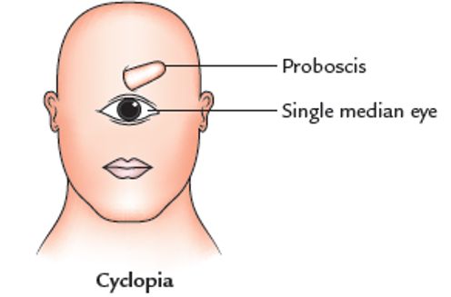
- Cyclopia: Rare congenital facial anomaly marked by the presence of a single median eye housed in a solitary central orbit. The nose is absent from its anatomical location and typically represented by a non-functional, blind tubular appendage called a proboscis positioned directly superior to the median eye.
- Synophthalmia: Represents incomplete fusion of the bilateral globes, where two partially merged eyes occupy a single shared central orbit.
- Pathophysiology: Both conditions represent the extreme ocular manifestation of holoprosencephaly (alobar holoprosencephaly), resulting from failure of the embryonic prechordal plate to cleave the single midline prosencephalic eye field into paired lateral optic vesicles during gastrulation. Associated with loss-of-function mutations in the Sonic Hedgehog (SHH) signaling pathway, maternal ingestion of teratogenic plant alkaloids (cyclopamine), and chromosomal trisomies (Trisomy 13 / Patau syndrome). Usually fatal shortly after birth due to associated severe cerebral malformations.
3. Defects of Ocular Cleavage & Fissure Closure
Coloboma (Defective Choroid Fissure Closure)
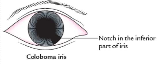
The term coloboma (Greek for “curtailed” or “mutilated”) defines a congenital notch, cleft, or sectoral tissue defect caused by failure of the embryonic choroid (retinal) fissure to fuse completely in week 7:
- Coloboma Iridis (Iris Coloboma): The most common form; manifests as a keyhole-shaped pupillary notch located strictly in the inferior-nasal quadrant of the iris (the anatomical location of the embryonic fissure).
- Complete Coloboma: If failure of closure extends along the entire length of the fissure, the segmental defect involves the ciliary body, choroid, retina, and the ventral margin of the optic nerve head, causing permanent visual field deficits or scotomas.
- Syndromic Association: Classic component of the CHARGE association (Coloboma, Heart defects, Atresia choanae, Retardation of growth, Genital hypoplasia, Ear abnormalities), linked to CHD7 gene mutations.
Other Anterior Segment Malformations
- Persistent Pupillary Membrane: Incomplete apoptotic involution of the anterior vascular mesenchyme overlying the lens, leaving fine, pigmented connective tissue threads across the pupil. While small strands rarely impair vision, dense fibrous webs cause visual deprivation amblyopia requiring laser photolysis or surgical excision.
- Congenital Aniridia: Near-total bilateral absence of the iris. Results from developmental arrest at the rim of the optic cup during week 8. Strongly linked to heterozygous loss-of-function mutations in the PAX6 gene on chromosome 11p13. Forms a cornerstone of WAGR syndrome (Wilms tumor, Aniridia, Genitourinary anomalies, and mental Retardation).
- Congenital Glaucoma (Buphthalmos): Elevated intraocular pressure in neonates caused by genetic dysgenesis of the anterior chamber angle (trabeculodysgenesis). Neural crest cells fail to differentiate the trabecular meshwork and canal of Schlemm, obstructing aqueous outflow. Elevated IOP stretches the compliant infant sclera and cornea, causing gross enlargement of the globe (buphthalmos, or “ox-eye”) and corneal clouding.
- Congenital Aphakia: Extremely rare absence of the crystalline lens. Caused by failure of the optic vesicle to induce the overlying surface ectoderm, or early apoptotic degeneration of the lens placode.
Ocular adnexa & anomalies
The accessory structures of the eye comprise the eyelids, conjunctival sac, lacrimal glands, lacrimal sac, and nasolacrimal duct.
1. Development of the Eyelids & Conjunctival Sac
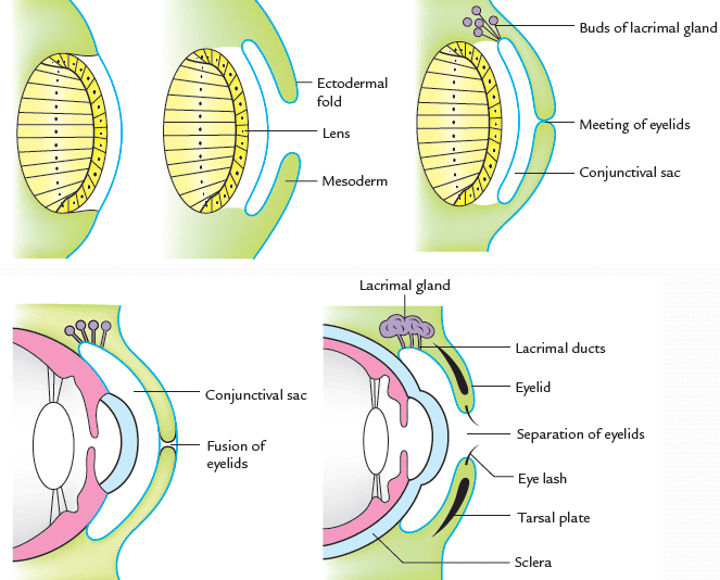
- Ectodermal Reduplication: During week 6, upper and lower eyelid primordia arise as transverse folds of surface ectoderm containing an internal core of neural crest mesenchyme, protruding immediately superior and inferior to the cornea.
- Eyelid Fusion: As these folds grow, their margins meet and fuse during week 10 of embryonic life. Fusion protects the delicate developing cornea and lens from amniotic fluid abrasions.
- The Conjunctival Sac: Fusion encloses an ectoderm-lined space between the inner surface of the eyelids and the anterior surface of the cornea: the conjunctival sac. Its anterior lining forms the palpebral conjunctiva, while its posterior reflection forms the bulbar conjunctiva.
- Eyelid Separation: The fused eyelids remain adhered until the beginning of the 28th week (7th month) of intrauterine life. Epithelial breakdown, aided by lipid secretions from differentiating Meibomian (tarsal) glands, breaks the junctional seam, separating the margins into distinct upper and lower eyelids.
2. Development of the Lacrimal Apparatus
- Lacrimal Gland: Originates as 15 to 20 solid, branching cords of surface ectoderm that bud from the superolateral fornix of the conjunctival sac into the adjacent mesenchyme during early fetal life. These cords elongate, branch, and canalize into the definitive acini and excretory ducts of the lacrimal gland, opening into the superior conjunctival fornix. Secretory function is rudimentary at birth; infants typically produce no emotional tears until 1 to 3 months of age.
- Nasolacrimal Duct & Lacrimal Sac: Develop from a solid cord of surface ectoderm that sinks into the mesenchyme along the nasolacrimal (naso-optic) groove, situated at the fusion boundary between the maxillary process and the lateral nasal process. This cord canalizes in late fetal life to form the nasolacrimal duct, connecting the lacrimal sac to the inferior nasal meatus. Failure of canalization of the mucosal membrane at the distal opening (valve of Hasner) is the most common cause of congenital epiphora (excessive tearing) and dacryocystitis in neonates.
- Lacrimal Canaliculi: Ectodermal buds sprout medially from the margins of each eyelid, canalizing to form the superior and inferior lacrimal puncta and canaliculi that drain into the lacrimal sac.
3. Clinical Congenital Anomalies of the Ocular Adnexa
Anomalies of the Eyelids
- Coloboma of the Eyelid: A full-thickness notched cleft in the eyelid margin. Usually manifests as a small defect in the upper eyelid (caused by defective fusion of the embryonic eyelid folds); coloboma of the lower eyelid is rarer and typically associated with systemic facial clefting syndromes (e.g., Treacher Collins syndrome).
- Cryptophthalmos: Complete absence of the eyelids and palpebral fissure. The skin of the forehead extends continuously over a malformed, microphthalmic eyeball. Often inherited as an autosomal recessive trait (Fraser syndrome).
- Congenital Ptosis: Drooping of the upper eyelid present from birth. Stemming from agenesis, hypoplasia, or muscular dystrophy of the levator palpebrae superioris muscle, or defective innervation by the superior division of the oculomotor nerve (CN III). Frequently transmitted as an autosomal dominant trait. Surgical levator resection is required to prevent amblyopia.
- Epicanthus (Epicanthic Fold): A crescentic cutaneous fold extending downward from the upper eyelid over the medial canthus, concealing the caruncle. A typical anatomical variation in East Asian populations; in non-Asian populations, it serves as an important clinical dysmorphic marker for Down syndrome (Trisomy 21) and Fetal Alcohol Syndrome (FAS).
- Entropion & Ectropion: Congenital malformations of lid margin orientation:
- Entropion: Inward turning of the lid margin, causing eyelashes to abrade the cornea (trichiasis).
- Ectropion: Outward eversion of the lid margin, exposing the palpebral conjunctiva to drying and chronic keratinization.
Anomalies of the Lacrimal Apparatus
- Aplasia or Hypoplasia: Total absence or functional agenesis of the lacrimal gland, producing congenital alacrima.
- Congenital Nasolacrimal Duct Obstruction: Incomplete canalization of the nasolacrimal cord (most frequently at the distal valve of Hasner), leading to chronic neonatal tearing (epiphora), purulent reflux, and recurrent dacryocystitis. Most cases resolve spontaneously by age 1; persistent cases require lacrimal duct probing.
- Supernumerary Puncta / Canaliculi: Accessory puncta or duplicated canalicular pathways arising from ectopic epithelial budding along the medial eyelid margins.
- Congenital Dacryops: Benign cystic dilations arising from ductal occlusion within the lacrimal gland or canaliculi.
Quick recap
References
The embryological and clinical descriptions in this study note have been verified against authoritative reference texts:
- Langman's Medical Embryology: Sadler, T.W. (2019) Langman's Medical Embryology. 14th edn. Philadelphia: Wolters Kluwer. [Chapter 20: Eye, pp. 385–398].
- The Developing Human: Moore, K.L., Persaud, T.V.N. and Torchia, M.G. (2020) The Developing Human: Clinically Oriented Embryology. 11th edn. Philadelphia: Elsevier. [Chapter 18: The Eye and Ear, pp. 415–430].
- Larsen's Human Embryology: Schoenwolf, G.C., Bleyl, S.B., Brauer, P.R. and Francis-West, P.H. (2021) Larsen's Human Embryology. 6th edn. Philadelphia: Elsevier. [Chapter 17: Development of the Eyes, pp. 453–470].
- Vaughan & Asbury's General Ophthalmology: Riordan-Eva, P. and Augsburger, J.J. (2018) Vaughan & Asbury's General Ophthalmology. 19th edn. New York: McGraw-Hill Education. [Chapter 1: Anatomy & Embryology of the Eye].