Structure of the Ear: External, Middle and Inner Ear Anatomy
The ear is a specialised sensory organ for hearing and balance. Its structures are arranged in three connected regions: the external ear gathers sound, the middle ear transfers vibrations, and the inner ear converts mechanical movement into nerve signals and detects head motion. Most of the middle and inner ear lie within the temporal bone.
Following the structures from the auricle to the brain helps explain how sound is conducted, where common ear disorders arise, and why injury to different parts produces different symptoms.
Learning objectives
- Describe the external, middle and inner parts of the ear and the structures in each.
- Trace the route of sound through the external auditory canal, tympanic membrane, ossicles and cochlea.
- Identify the main parts of the tympanic membrane, tympanic cavity and labyrinth.
- Relate ear anatomy to conductive hearing loss, sensorineural hearing loss, infection and vertigo.
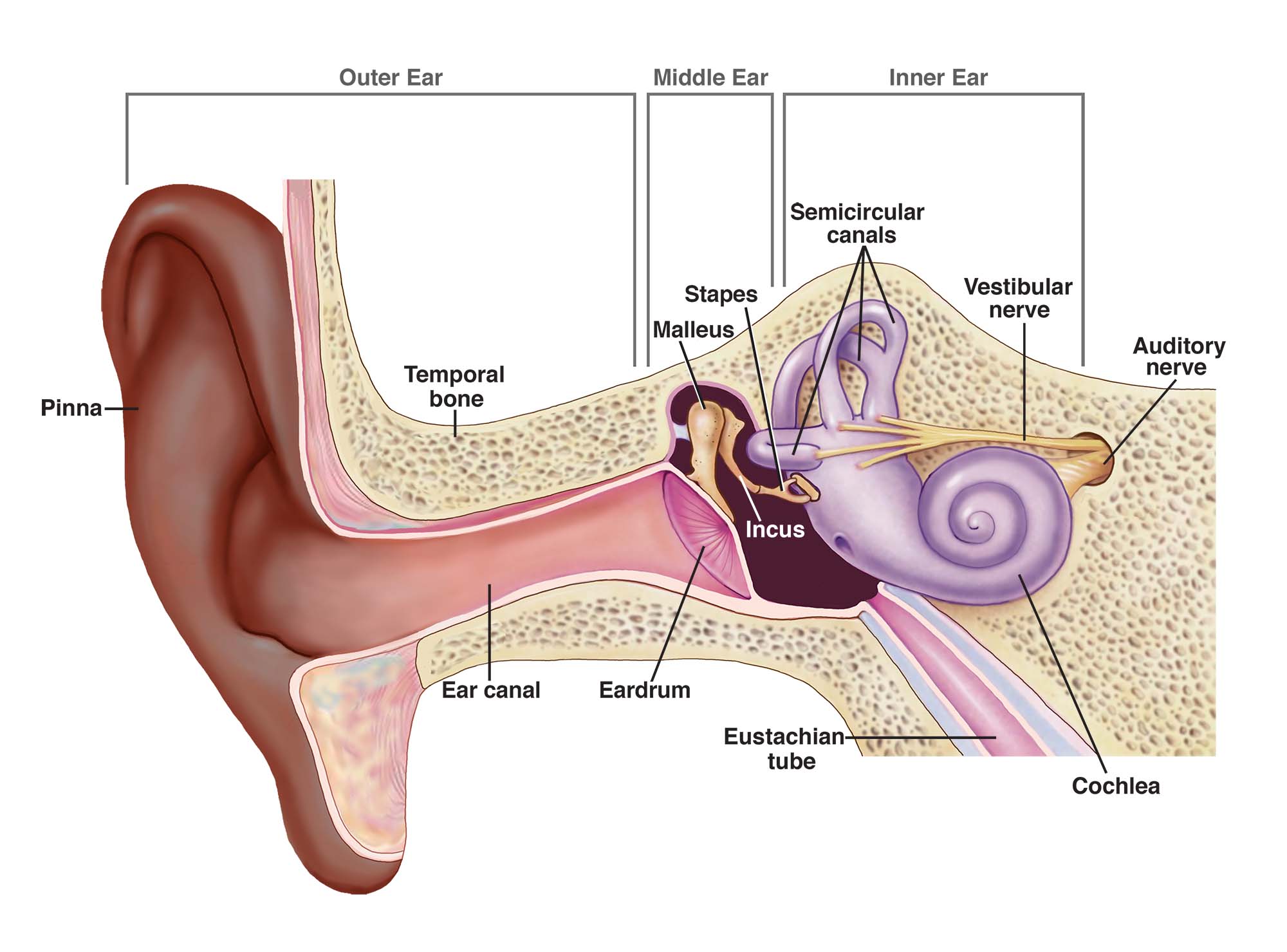
Overview: the three regions of the ear
The ear is commonly divided by the tympanic membrane and the inner-ear windows. These regions work together as one pathway, but each has different tissues and functions.
Collects and directs sound waves to the tympanic membrane. The outer surface of the tympanic membrane is conventionally included as the terminal part of the external ear.
An air-filled space that transmits tympanic membrane movement through the malleus, incus and stapes to the oval window. The auditory tube connects it with the nasopharynx.
A fluid-containing sensory system in the petrous temporal bone. The cochlea supports hearing; the vestibule and semicircular canals contribute to balance and spatial orientation.
Auricle → external auditory canal → tympanic membrane → malleus → incus → stapes → oval window → cochlear fluids and sensory cells → cochlear nerve → brainstem and auditory pathways.
Movement of endolymph and otolithic material stimulates sensory hair cells in the semicircular ducts, utricle and saccule. Signals travel in the vestibular division of cranial nerve VIII.
1. External ear
The external ear extends from the visible auricle to the tympanic membrane. It provides a protective route for sound, helps with sound localisation, and contains skin, cartilage and glands.
A. Auricle (pinna)
The auricle is the visible, irregularly shaped part on the side of the head. Its framework is mainly elastic cartilage covered by skin; the lobule (earlobe) has no cartilage and contains fibrofatty tissue. Its curved surface funnels sound into the external auditory canal.
- Helix: the curved outer rim.
- Antihelix: the raised inner curve, running roughly parallel to the helix.
- Concha: the hollow bowl leading to the external auditory canal.
- Tragus: the small projection in front of the canal opening.
- Antitragus: the prominence opposite the tragus, above the lobule.
- Lobule: the soft, inferior part of the auricle.
The shape of the auricle contributes to directional hearing by modifying incoming sound. Its exposed position makes it vulnerable to trauma, frostbite and infection. A subperichondrial haematoma can separate the cartilage from its blood supply and, if untreated, deform the auricle.
B. External auditory canal (external acoustic meatus)
The canal runs from the concha to the tympanic membrane. In adults it is approximately 2.5 cm long, though individual dimensions vary. It is slightly curved rather than a straight tube.
The outer part has a cartilaginous framework. Its skin is relatively thick and contains hair follicles, sebaceous glands and ceruminous glands. Cerumen (ear wax) helps trap debris and protects the canal skin.
The inner part is formed by the temporal bone. Its skin is thin and tightly attached to the underlying periosteum, so inflammation or pressure here may be very painful.
The cartilaginous and bony portions meet at a narrow bend. This junction is important during examination and procedures: the canal should be visualised gently, and instruments should not be advanced blindly. The canal conducts sound and provides a degree of resonance before sound reaches the tympanic membrane.
C. Tympanic membrane (eardrum)
The tympanic membrane is a thin, obliquely placed membrane separating the external auditory canal from the middle-ear cavity. Sound pressure makes it vibrate, and its medial surface is attached to the handle of the malleus.
- Pars tensa: the larger, taut portion, supported by a fibrous layer.
- Pars flaccida: the smaller, more flexible superior portion, above the lateral process of the malleus.
- Umbo: the central point where the membrane is drawn inward by the attached malleus.
- Handle of the malleus: visible through the membrane and running down to the umbo.
The pars tensa has an outer epithelial layer continuous with canal skin, a middle fibrous layer that provides strength, and an inner mucosal layer continuous with the middle-ear lining. The pars flaccida has a less substantial fibrous layer.
For otoscopy, the membrane can be described in four quadrants using an imaginary line along the malleus handle and a second line at right angles through the umbo. The cone of light is a normal reflected light seen anteroinferiorly in many healthy ears; its appearance varies with viewing angle and should not be interpreted in isolation.
External ear nerves and blood supply: a useful overview
Sensation around the auricle and external canal is shared between several nerves, including branches of the trigeminal nerve, the auricular branch of the vagus, and upper cervical nerves. This overlap helps explain why disorders of the ear can cause cough or referred pain, and why pain from nearby structures may be felt at the ear. The auricle receives blood mainly from branches of the external carotid artery, particularly the posterior auricular and superficial temporal arteries.
2. Middle ear
The middle ear, or tympanic cavity, is a small air-containing space within the temporal bone. It lies between the tympanic membrane laterally and the bony labyrinth medially. It houses the ossicles and communicates posteriorly with mastoid air spaces and anteriorly with the nasopharynx through the auditory tube.
A. Parts of the middle-ear space
- Epitympanum (attic): the upper part of the cavity, above the level of most of the tympanic membrane; it contains the head of the malleus and body of the incus.
- Mesotympanum: the central part, opposite the tympanic membrane.
- Hypotympanum: the lower recess below the level of the membrane.
B. Walls and important relations of the tympanic cavity
Learning the six walls helps locate structures that may be affected by disease spreading from the middle ear.
A thin bony plate, the tegmen tympani, separates the cavity from the middle cranial fossa and temporal lobe.
A thin bony floor lies above the jugular bulb. The relationship is clinically relevant when infection or a vascular variant is present.
Formed chiefly by the tympanic membrane, with the bony lateral wall above it in the epitympanum.
Faces the inner ear. It shows the promontory, the bulge over the basal cochlear turn, plus the oval and round windows.
Related to the internal carotid artery. The openings of the auditory tube and the canal for tensor tympani are found in this region.
Communicates with the mastoid antrum through the aditus. The pyramidal eminence, containing stapedius, and the facial canal are nearby.
C. Auditory ossicles
The three small bones form a mobile chain from the tympanic membrane to the oval window. Their joints transmit sound while helping overcome the difference between sound travelling in air and movement of fluid in the cochlea.
The handle (manubrium) is embedded in the tympanic membrane and ends at the umbo. The head articulates with the incus in the epitympanum.
The body articulates with the head of the malleus. Its long process connects to the stapes, completing the middle link of the chain.
The smallest ossicle. Its footplate fits into the oval window and moves there to deliver vibrations to the inner-ear fluids.
Sound sequence: tympanic membrane → malleus → incus → stapes footplate → oval window. The round window provides a flexible pressure-release membrane that allows cochlear fluid displacement.
D. Middle-ear muscles
Attaches to the malleus and tenses the tympanic membrane. It is supplied by the mandibular division of the trigeminal nerve (cranial nerve V3).
Attaches to the stapes and limits its movement. It is supplied by the facial nerve (cranial nerve VII). The acoustic reflex reduces transmission of some sustained sounds, but does not reliably protect against sudden or very intense noise.
E. Auditory (pharyngotympanic or Eustachian) tube
This passage connects the anterior middle ear with the nasopharynx. Its functions are to equalise pressure across the tympanic membrane, ventilate the middle ear and help clear secretions. It is usually closed at rest and opens briefly during swallowing or yawning. In children the tube is shorter and more horizontal than in adults, which contributes to the tendency for middle-ear infection in childhood.
F. Mastoid antrum and air cells
The mastoid antrum lies posterior to the middle ear and communicates with the epitympanum through the aditus. It connects to a variable system of mastoid air cells within the mastoid part of the temporal bone. Their shared lining means that middle-ear infection may extend into the mastoid air-cell system, producing mastoid inflammation.
3. Inner ear (labyrinth)
The inner ear lies within the dense petrous part of the temporal bone. Its complex spaces make up the bony labyrinth, which contains the membranous labyrinth. The space around the membranous labyrinth contains perilymph; the membranous structures contain endolymph.
Comprises the cochlea, vestibule and three semicircular canals. It forms the rigid outer enclosure within the petrous temporal bone.
Includes the cochlear duct, utricle, saccule and semicircular ducts. Sensory epithelium within these structures responds to sound-related vibration or head movement.
A. Cochlea: hearing apparatus
The cochlea is a coiled, snail-shaped structure making about two and a half turns around a central bony core called the modiolus. The cochlear duct and its membranes divide the cochlear space into three longitudinal compartments:
- Scala vestibuli: an upper perilymph-filled channel beginning at the oval window.
- Scala media (cochlear duct): the endolymph-filled middle channel, containing the organ of Corti.
- Scala tympani: a lower perilymph-filled channel that ends near the round window.
The organ of Corti rests on the basilar membrane within the cochlear duct. It contains inner and outer hair cells. Movement of the basilar membrane bends the hair-cell stereocilia against the tectorial membrane, beginning conversion of mechanical energy into electrical activity. The cochlear division of cranial nerve VIII carries auditory information toward the brain.
B. Vestibule: utricle and saccule
The vestibule is the central part of the bony labyrinth, between the cochlea and semicircular canals. The membranous utricle and saccule contain sensory areas called maculae. These detect head position relative to gravity and linear acceleration. Small calcium carbonate crystals (otoconia) add weight to the sensory gel, helping the maculae detect movement.
C. Semicircular canals and ducts
Three semicircular canals—anterior (superior), posterior and lateral—are arranged approximately at right angles. Each encloses a membranous semicircular duct. At one end, the duct expands into an ampulla containing a sensory ridge, the crista ampullaris. Movement of endolymph during head rotation deflects the sensory structure and signals angular acceleration.
The semicircular ducts chiefly detect rotational movements, while the utricle and saccule chiefly detect linear acceleration and head position relative to gravity. Vestibular input is integrated with vision and proprioception to help maintain balance and stabilise gaze during head movement.
D. Vestibulocochlear nerve and internal acoustic meatus
The vestibulocochlear nerve (cranial nerve VIII) has a cochlear division for hearing and a vestibular division for balance. The nerve, facial nerve (VII), and accompanying vessels pass through the internal acoustic meatus in the petrous temporal bone. This close relationship is important when considering lesions affecting hearing, balance or facial movement.
How anatomy produces hearing
The auricle gathers sound and directs it down the external auditory canal.
Sound pressure moves the tympanic membrane and attached malleus.
The malleus and incus transmit movement to the stapes footplate at the oval window.
Movement of cochlear fluid displaces the basilar membrane and stimulates hair cells in the organ of Corti.
Electrical signals travel in the cochlear nerve and are processed along auditory pathways in the brain.
The middle-ear ossicles are not simply a set of levers: their geometry and the area difference between the tympanic membrane and stapes footplate help transfer energy efficiently from air to cochlear fluid. The NIDCD describes the step-by-step conversion of sound vibration into electrical signals carried by the auditory nerve.
Clinical correlations: why the three-part structure matters
Canal obstruction, cerumen or inflammation may prevent sound from reaching the tympanic membrane. Otitis externa affects the canal skin; pain can be marked because the bony canal skin is tightly attached to periosteum.
Fluid, infection, perforation or ossicular fixation can disrupt vibration transfer and produce a conductive hearing deficit. A retraction pocket involving the pars flaccida may be associated with cholesteatoma.
Damage to cochlear hair cells or the cochlear nerve can cause sensorineural hearing loss. Disturbance of vestibular structures may produce vertigo, imbalance or motion-related symptoms.
Conductive: sound transmission through the external or middle ear is impaired. Sensorineural: the cochlea, auditory nerve or more central auditory pathway is affected. Mixed loss contains both components.
The facial nerve runs in a bony canal close to the middle ear and mastoid. Severe ear disease, trauma or surgery in this region can therefore threaten facial movement.
Shared sensory pathways from the ear, teeth, temporomandibular joint, pharynx and larynx mean that pain may be felt in the ear even when the ear itself is normal.
Quick revision: compare the three regions
- Main structures: auricle, canal, outer surface of tympanic membrane.
- Main role: collect, direct and shape sound.
- Common problem: canal obstruction or otitis externa.
- Main structures: tympanic cavity, ossicles, muscles, auditory tube and mastoid connection.
- Main role: transmit and efficiently couple tympanic membrane vibration to the oval window.
- Common problem: otitis media, effusion or ossicular disruption.
- Main structures: cochlea, vestibule, semicircular canals and membranous labyrinth.
- Main role: transduce sound and detect head movement and position.
- Common problem: sensorineural hearing loss or vestibular dysfunction.
Key exam points
- The tympanic membrane separates the external auditory canal from the middle-ear cavity and is attached to the handle of the malleus.
- The ossicles, in order from lateral to medial, are malleus, incus and stapes.
- The stapes footplate is seated in the oval window; the round window permits pressure displacement in the cochlea.
- The auditory tube connects the middle ear with the nasopharynx and helps equalise middle-ear pressure.
- The cochlea is primarily concerned with hearing; the vestibule and semicircular ducts provide vestibular sensory information.
- The semicircular ducts detect angular head movement; the utricle and saccule detect linear acceleration and head position relative to gravity.
- External or middle-ear transmission problems commonly cause conductive hearing loss; cochlear or auditory-nerve injury causes sensorineural hearing loss.
Self-test questions
- What are the three anatomical regions of the ear?
- Which part of the malleus is attached to the tympanic membrane?
- Name the ossicles in order from the tympanic membrane to the inner ear.
- Which middle-ear structure opens into the nasopharynx?
- Which inner-ear structure contains the organ of Corti?
- Which structures detect angular rotation, and which detect linear acceleration and gravity?
- Give one anatomical reason why disease of the middle ear may affect mastoid air cells or facial nerve function.
References and further reading
- Sutton AE, Geiger Z. External Ear Anatomy. StatPearls, NCBI Bookshelf.
- Anatomy, Head and Neck: Middle Ear. StatPearls, NCBI Bookshelf.
- Sutton AE, Peterson DC. Anatomy, Head and Neck: Inner Ear. StatPearls, NCBI Bookshelf.
- National Institute on Deafness and Other Communication Disorders (NIDCD). How Do We Hear?
- National Institute on Deafness and Other Communication Disorders (NIDCD). Parts of the Ear: Medical Illustration.
- MSD Manual Professional Edition. Introduction to Middle Ear and Tympanic Membrane Disorders.
- MSD Manual Consumer Version. Overview of the Inner Ear.
These notes are for study and revision. Clinical examination and interpretation of ear findings should be performed in the context of appropriate training and local guidance.
