
The ear: external, middle & internal ear anatomy
On this page
Overview & acoustic pathway
The human ear is a compound peripheral organ dedicated to two separate physiological modalities: hearing (audition) and equilibrium (balance). Structurally and topographically, it is divided into three distinct anatomical compartments housed predominantly within and adjacent to the petrous temporal bone:
- External Ear: Comprises the visible auricle (pinna) and the external acoustic meatus. Its primary function is to collect, amplify, and channel acoustic pressure waves from the atmospheric environment directly onto the lateral surface of the tympanic membrane.
- Middle Ear (Tympanic Cavity): An air-filled mucosal chamber containing a mobile chain of three auditory ossicles (malleus, incus, and stapes). It functions as a mechanical impedance-matching transformer, converting low-pressure, high-displacement sound waves in air into high-pressure, low-displacement fluid vibrations within the inner ear.
- Internal Ear (Labyrinth): A fluid-filled system of complex communicating bony and membranous cavities embedded deep in the petrous temporal bone. It contains the sensory neuroepithelial end-organs: the organ of Corti within the cochlea (transducing fluid waves into auditory nerve impulses) and the maculae and cristae ampullares within the vestibule and semicircular ducts (transducing linear and angular acceleration for balance).

The external ear
The external ear consists of the auricle (pinna) and the external acoustic meatus (ear canal).
1. The Auricle (Pinna)
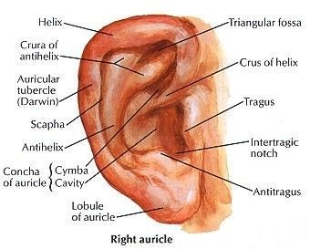
The auricle projects laterally from the side of the head, presenting a characteristic corrugated contour designed to collect acoustic waves and provide spatial sound localization along the vertical plane.
- Structural Framework: Consists of a pliable, thin plate of elastic cartilage covered tightly by thin, vascularized skin. The cartilage is continuous medially with the cartilaginous framework of the external acoustic meatus. The posteroinferior lobule (earlobe) is unique: it contains dense fibrofatty tissue and blood vessels but completely lacks cartilage.
- Surface Topography & Landmarks:
- Helix: The prominent outer curved cartilaginous rim of the auricle. Near its posterosuperior aspect, it occasionally features a small cartilaginous thickening termed the Darwinian tubercle (tubercle of Darwin). The anterior extremity curving into the concha is the crus of the helix.
- Antihelix: An inner curved elevation running parallel to the helix. Superiorly, it bifurcates into two limbs—the crura of the antihelix—which bound a shallow depression termed the triangular fossa. The elongated groove separating the helix from the antihelix is the scaphoid fossa (scapha).
- Concha of the Auricle: The deep central cavity leading directly into the external acoustic meatus. The crus of the helix divides the concha into a superior cymba conchae (overlying the suprameatal triangle of Macewen on the temporal bone) and an inferior cavum conchae.
- Tragus, Antitragus & Intertragic Notch: The tragus is a small, backward-curving cartilaginous flap situated anterior to the external meatus. Facing it across the deep intertragic notch is the posteroinferior tuberculum termed the antitragus.
- Motor Innervation: The intrinsic and extrinsic auricular muscles (anterior, superior, and posterior auricular muscles) are striated branchial muscles derived from the second pharyngeal arch and are innervated by the facial nerve (CN VII) via its posterior auricular branch and temporal branches.
- Sensory Innervation: Highly complex, reflecting multi-nerve embryological convergence:
- Great Auricular Nerve (C2, C3): Cervical plexus; supplies the lower half of both cranial and lateral surfaces, including the lobule and the angle of the jaw.
- Auriculotemporal Nerve (V3): Mandibular division of CN V; supplies the tragus, crus of the helix, and anterior upper lateral rim.
- Lesser Occipital Nerve (C2): Supplies the cranial surface of the superior auricle.
- Auricular Branch of Vagus (CN X, Arnold’s Nerve) & CN VII: Supplies the deep floor of the concha and antihelix. Irritation here can trigger a reflex cough (vagal cough reflex) or cardiac bradycardia during instrumentation.
2. The External Acoustic Meatus (EAM)
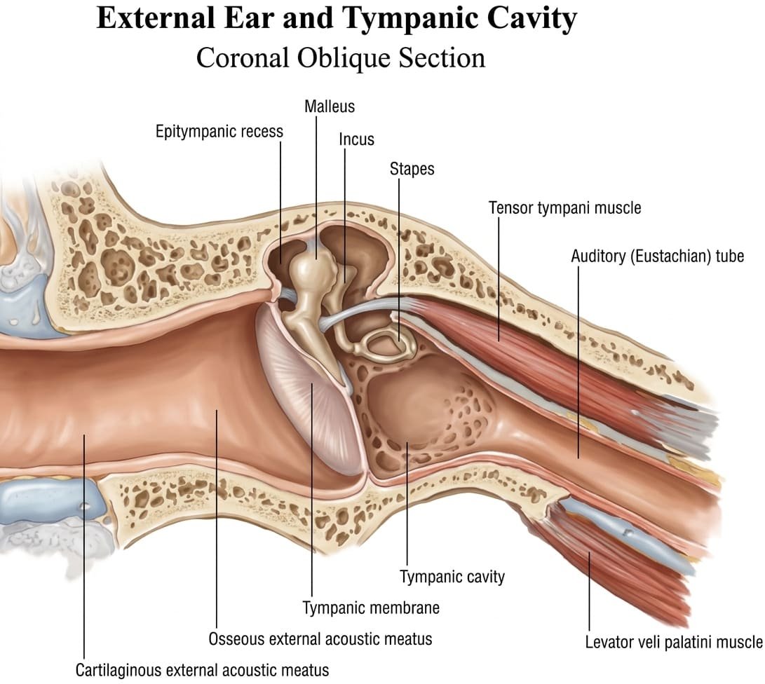
The external acoustic meatus is an S-shaped sinuous canal approximately 24 mm (2.4 cm) in length in adults, extending from the bottom of the concha medially to the tympanic membrane.
Structural Subdivisions of the Meatus
- Cartilaginous Outer One-Third (8 mm): Formed by a U-shaped trough of elastic cartilage continuous with the auricle, closed posterosuperiorly by dense fibrous tissue. The lining skin is thick and contains protective hairs (tragi), sebaceous glands, and modified apocrine ceruminous glands that secrete cerumen (earwax). Contains transverse cartilaginous fissures (fissures of Santorini) through which parotid infections or tumors can track into the canal.
- Bony Inner Two-Thirds (16 mm): Formed predominantly by the curved tympanic plate of the temporal bone anteriorly, inferiorly, and posteroinferiorly, and by the squamous temporal bone posterosuperiorly. The skin lining this section is exceptionally thin, devoid of hair follicles and ceruminous glands, and adheres tightly to the underlying periosteum, rendering infections here exquisitely painful.
- The Isthmus: A physiological constriction located approximately 5 mm lateral to the tympanic membrane. Foreign bodies pushed past the isthmus become firmly lodged and difficult to extract.
- Canal Curvature & Otoscopic Traction: The meatus is curved like an elongated “S”: it courses initially medially, anteriorly, and superiorly; then medially and posteriorly; and finally medially, anteriorly, and inferiorly.
Clinical Rule: To straighten the meatus during otoscopic examination:- In adults: Pull the auricle upward, backward, and slightly outward.
- In infants and neonates (who lack a fully ossified bony canal): Pull the auricle straight backward and downward.
The tympanic membrane (eardrum)
The tympanic membrane is a thin, semitransparent, pearly-gray fibrous partition separating the external acoustic meatus from the tympanic cavity of the middle ear. It sits obliquely at an angle of roughly 55 degrees to the horizontal canal floor, facing laterally, downward, and forward.

Anatomical Structure & Divisions
- Three Histological Layers:
- Cuticular Layer (Lateral): Stratified squamous epithelium continuous with the skin of the external auditory meatus.
- Fibrous Intermediate Layer (Lamina Propria): Composed of organized outer radial fibers and inner circular fibers, providing mechanical tensile strength.
- Mucosal Layer (Medial): Simple cuboidal epithelium continuous with the mucous lining of the middle ear cavity.
- Pars Tensa: The large inferior portion of the membrane. Its thickened fibrocartilaginous border (annulus fibrocartilagineus) slots firmly into the bony sulcus tympanicus of the temporal bone. It is held taut by the inward traction of the tensor tympani muscle acting on the handle of the malleus.
- Pars Flaccida (Shrapnell’s Membrane): A small, thin triangular area situated superiorly above the anterior and posterior malleolar folds, where the bony sulcus is deficient (the notch of Rivinus). It completely lacks the intermediate fibrous layer, making it mechanically flaccid and susceptible to retraction pocket formation and cholesteatoma.
- Umbo: The central, deepest point of depression on the lateral concave surface, produced by the firmly attached tip of the handle (manubrium) of the malleus.
- Cone of Light (Light Reflex): When illuminated during otoscopy, the downward and outward concavity of the membrane reflects a bright, triangular wedge of light projecting anteroinferiorly from the umbo toward the periphery (at approximately the 5 o’clock position in the right ear and 7 o’clock in the left ear). Distortion or loss of this cone signifies increased middle ear pressure, effusion, or retraction.
Sensory Innervation of the Tympanic Membrane
The nerve supply of the eardrum mirrors its embryological trilaminar derivation:
- Lateral (External) Cuticular Surface: Innervated mainly by the auriculotemporal nerve (CN V3) over its anterior and superior two-thirds, with the posterior and inferior one-third supplied by the auricular branch of the vagus nerve (CN X). Minor twigs from CN VII can also contribute.
- Medial (Internal) Mucosal Surface: Exclusively innervated by the tympanic branch of the glossopharyngeal nerve (CN IX, Jacobson’s nerve) through the submucosal tympanic plexus.
Middle ear: walls & boundaries
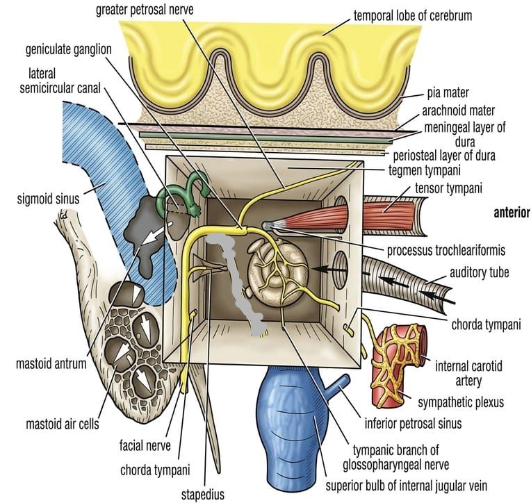
The middle ear (tympanic cavity) is an irregular, laterally compressed, air-filled space carved into the petrous part of the temporal bone. Lined by a vascular mucous membrane (ciliated columnar to cuboidal epithelium), it functions to convey mechanical vibrations from the tympanic membrane across its cavity via the auditory ossicles to the perilymph of the internal ear.
Primary Communications
- Anterior Communication: Communicates directly with the nasopharynx via the auditory (pharyngotympanic / Eustachian) tube, which equalizes intratympanic pressure with atmospheric pressure.
- Posterior Communication: Communicates with the mastoid antrum (and through it, the mastoid air cell system) via the aditus ad antrum.
The Six Anatomical Walls (The Tympanic “Box”)
1. Roof (Tegmental Wall / Paries Tegmentalis)
Formed by a paper-thin plate of bone from the petrous temporal bone called the tegmen tympani. It separates the middle ear cavity and epitympanic recess from the temporal lobe of the brain and the meninges within the middle cranial fossa. In young children, unossified petrosquamous sutures can permit direct spread of otitis media into the cranial cavity, causing meningitis or temporal lobe cerebral abscess.
2. Floor (Jugular Wall / Paries Jugularis)
A thin plate of bone separating the cavity from the superior bulb of the internal jugular vein. Near the medial border, it is pierced by the tympanic branch of the glossopharyngeal nerve (CN IX). A dehiscent bony floor can allow a high-riding jugular bulb to protrude directly beneath the mucous membrane of the middle ear.
3. Anterior Wall (Carotid Wall / Paries Caroticus)
- Superior Portion: Features two parallel bony tunnels separated by a thin horizontal shelf called the processus cochleariformis:
- Upper canal: Contains the tensor tympani muscle.
- Lower canal: The bony opening for the auditory (Eustachian) tube.
- Inferior Portion: A thin vertical plate of bone separating the middle ear from the internal carotid artery within the carotid canal, perforated by caroticotympanic nerves carrying sympathetic fibers from the internal carotid plexus.
4. Posterior Wall (Mastoid Wall / Paries Mastoideus)
- Superior Opening: The large opening termed the aditus ad antrum, leading directly from the epitympanic recess backward into the mastoid antrum. Below the aditus lies the fossa incudis, which accommodates the short crus of the incus.
- Inferior Projection: A small, hollow, conical bony projection termed the pyramid (eminentia pyramidalis). Through a minute aperture at its summit emerges the delicate tendon of the stapedius muscle. Lateral to the pyramid lies the posterior canaliculus for the chorda tympani nerve.
5. Lateral Wall (Membranous Wall / Paries Membranaceus)
Formed almost entirely by the circular concave tympanic membrane, along with the bony outer wall of the epitympanic recess (attic) superiorly, which shelters the head of the malleus and the body of the incus.
6. Medial Wall (Labyrinthine Wall / Paries Labyrinthinus)
Forms the lateral wall of the internal ear. It presents four major anatomical landmarks:
- The Promontory: A rounded, smooth bony prominence occupying the center of the wall, produced by the projection of the basal turn of the cochlea. Grooved by the submucosal tympanic plexus.
- Fenestra Vestibuli (Oval Window): An oval opening situated above and behind the promontory, leading into the vestibule of the internal ear. In life, it is completely sealed by the base (footplate) of the stapes and its surrounding annular ligament.
- Fenestra Cochleae (Round Window): A round aperture located below and behind the promontory, leading into the scala tympani of the cochlea. Closed by the delicate secondary tympanic membrane, which flexes to dissipate hydraulic fluid shockwaves generated by stapedial displacement.
- Prominence of the Facial Nerve Canal: A smooth horizontal bony ridge passing backward immediately above the promontory and oval window, then curving sharply downward behind the pyramid on the posterior wall. Superior to it lies the prominence of the lateral semicircular canal.
Auditory ossicles & muscles
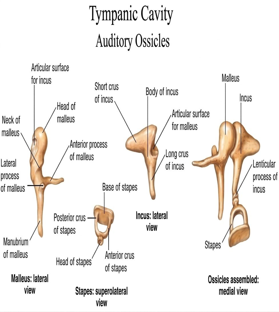
The middle ear houses three minute bones—the auditory ossicles: the malleus (hammer), incus (anvil), and stapes (stirrup). Linked by true synovial joints and suspended by delicate ligaments, they bridge the air gap from the tympanic membrane to the oval window.
1. The Malleus
- Head: Bulbous superior portion lying in the epitympanic recess; presents a posterior faceted articular surface for the body of the incus (incudomalleolar joint, a synovial saddle joint).
- Neck: Constricted segment beneath the head, related medially to the chorda tympani nerve.
- Handle (Manubrium): Long, tapered process directed downward, backward, and medially. Its lateral border is firmly attached along its length to the fibrous layer of the tympanic membrane, terminating at the umbo. Near its root, it receives the insertion of the tensor tympani tendon.
- Anterior Process: Slender bony spicule connected to the petrotympanic fissure by an anterior ligament.
- Lateral Process: Conical projection at the root of the manubrium, forming an obvious otoscopic bulge that anchors the anterior and posterior malleolar folds.
2. The Incus
- Body: Large central mass situated in the epitympanic recess, articulating anteriorly with the head of the malleus.
- Short Process (Crus Breve): Projects horizontally backward into the fossa incudis on the posterior tympanic wall, secured by the posterior incudal ligament.
- Long Process (Crus Longum): Descends vertically behind and parallel to the handle of the malleus. At its inferior tip, it turns sharply medially, terminating in a rounded knob called the lenticular process, which articulates with the head of the stapes (incudostapedial joint, a synovial ball-and-socket joint).
3. The Stapes
- Head (Capitulum): Articulates with the lenticular process of the incus.
- Neck: Constricted portion below the head, providing insertion for the delicate stapedius tendon.
- Crura: Two divergent hollow limbs (an anterior, straighter crus and a posterior, more curved crus) connecting the neck to the base.
- Base (Footplate): Flat oval plate that fits snugly into the fenestra vestibuli (oval window). Held in place by the fibroelastic annular ligament, forming a syndesmosis that seals the perilymph of the inner ear.
Intratympanic Muscles
Two striated muscles regulate ossicular tension to protect the inner ear from acoustic trauma:
Scroll the table sideways on a smaller screen.
| Muscle | Bony Origin | Point of Insertion | Innervation | Action & Reflex Role |
|---|---|---|---|---|
| Tensor Tympani | Cartilaginous part of auditory tube, canal wall above tube, and sphenoid bone | Handle (manubrium) of malleus, near its root | Nerve to tensor tympani (branch of Mandibular nerve, CN V3 via otic ganglion) | Pulls the handle of the malleus medially, tensing the tympanic membrane; damps down vibrations to reduce sound transmission; active during chewing to suppress self-generated noise. |
| Stapedius (Smallest skeletal muscle in the body) |
Interior hollow of the pyramid (eminentia pyramidalis) on the posterior wall | Posterior aspect of the neck of the stapes | Nerve to stapedius (branch of the Facial nerve, CN VII) | Pulls the stapes posteriorly, tilting the footplate and dampening excessive movement at the oval window (acoustic attenuation reflex); protects cochlea from loud acoustic injury (>80 dB). |
Intratympanic facial nerve
The facial nerve (CN VII) has an intimate anatomical course through the temporal bone and middle ear cavity, traveling inside the bony facial canal (Fallopian canal) before emerging at the base of the skull through the stylomastoid foramen.
Intratympanic Segments of the Facial Nerve
- Labyrinthine Segment: Traverses the petrous bone from the internal acoustic meatus to the geniculate ganglion, where the nerve turns sharply backward (first genu).
- Tympanic (Horizontal) Segment: Courses horizontally backward across the medial wall of the middle ear, situated directly above the promontory and fenestra vestibuli and below the lateral semicircular canal. The bony wall here is often thin and can be naturally dehiscent in up to 30% to 50% of individuals, exposing the nerve sheath directly to middle ear infections.
- Mastoid (Vertical) Segment: At the junction of the medial and posterior walls, behind the pyramid, the nerve turns sharply downward (second genu) through the posterior wall to exit the stylomastoid foramen.
Branches Arising Within the Temporal Bone
- 1. Greater Petrosal Nerve: Arises from the apex of the geniculate ganglion. Carries preganglionic parasympathetic secretomotor fibers destined for the pterygopalatine ganglion (supplying the lacrimal gland, nasal, and palatine mucosal glands) alongside special sensory taste fibers from the soft palate.
- 2. Nerve to Stapedius: Arises from the vertical mastoid segment behind the pyramid, entering the hollow of the pyramid to innervate the stapedius muscle.
- 3. Chorda Tympani Nerve: Arises from the facial nerve in the facial canal approximately 6 mm above the stylomastoid foramen.
- Intratympanic Trajectory: Enters the tympanic cavity through the posterior canaliculus, runs forward across the medial surface of the tympanic membrane (passing medial to the neck of the malleus and lateral to the long process of the incus), enveloped by mucous membrane. It exits anteriorly through the petrotympanic fissure (Glaserian fissure) to enter the infratemporal fossa.
- Functional Payload: Joins the lingual nerve (branch of CN V3) to carry taste fibers from the anterior two-thirds of the tongue and preganglionic parasympathetic fibers to the submandibular ganglion (innervating the submandibular and sublingual salivary glands).
The internal ear (labyrinth)
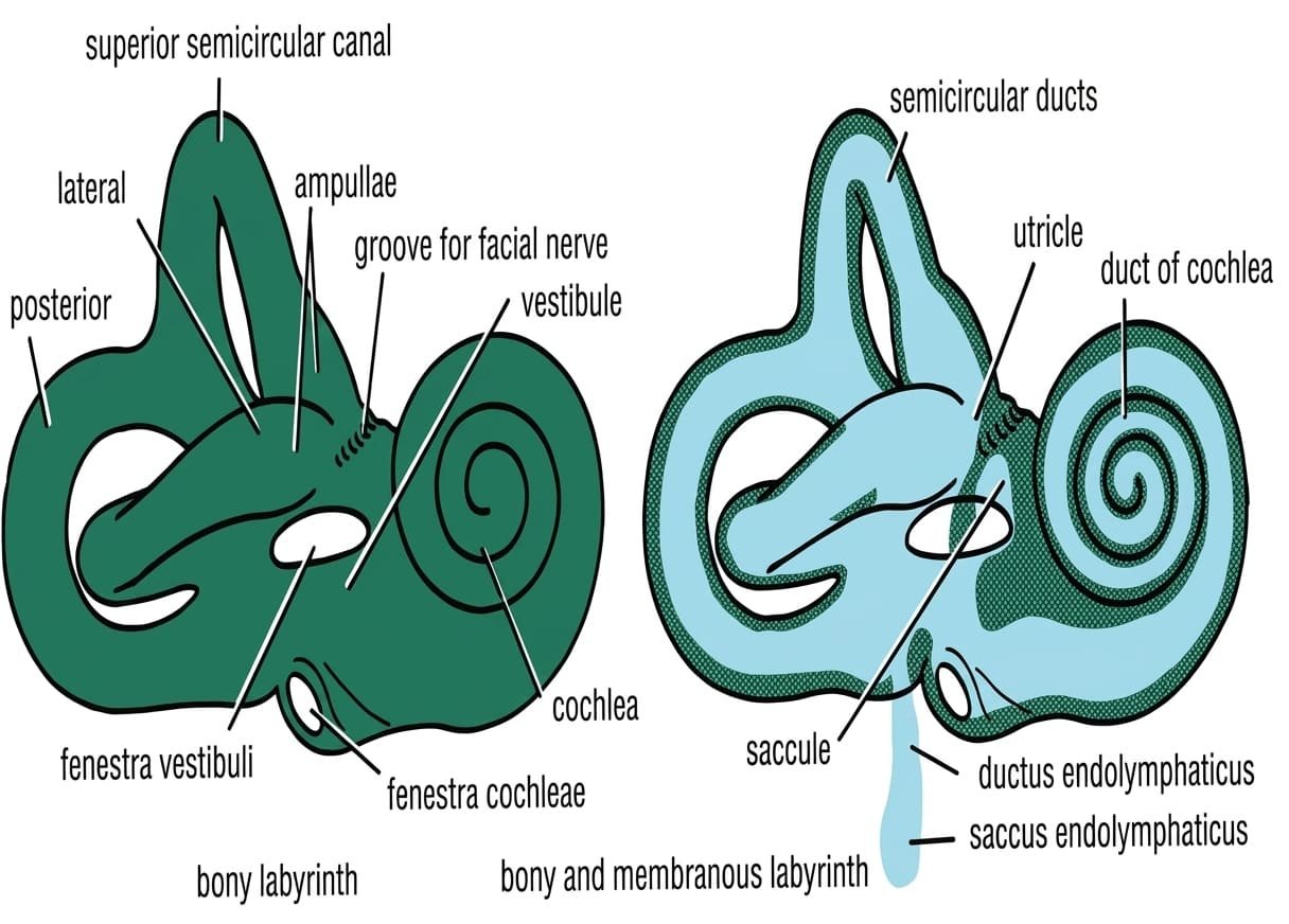
The internal ear (labyrinth) is embedded entirely within the petrous part of the temporal bone, situated medial to the middle ear cavity. It comprises two main structural systems: the rigid outer bony labyrinth and the delicate inner membranous labyrinth.
1. The Bony Labyrinth
A series of communicating cavities hollowed out of dense compact bone (the otic capsule), lined internally by endosteum and filled with perilymph (a fluid high in Na+ and low in K+, chemically similar to extracellular fluid and cerebrospinal fluid). It consists of three parts:
- Vestibule: The central, ovoid chamber (approx. 5 mm long). Its lateral wall contains the fenestra vestibuli (oval window). Medially, it is perforated by cribriform apertures for vestibular nerve fibers and features the opening of the vestibular aqueduct (transmitting the endolymphatic duct to the posterior cranial fossa). Houses the membranous utricle and saccule.
- Semicircular Canals: Three bony loops—anterior (superior), posterior, and lateral (horizontal)—situated posterosuperior to the vestibule, oriented at right angles (orthogonal) to one another in the three planes of space. Each canal presents a dilated terminal end called an ampulla. The anterior and posterior canals fuse medially to form the crus commune, so that the three canals open into the vestibule via five (rather than six) orifices.
- Cochlea: A conical, snail-shell-like bony structure consisting of a central conical bony axis, the modiolus, around which a spiral canal winds for 2.5 to 2.75 turns.
- A spiral shelf of bone, the osseous spiral lamina, projects outward from the modiolus like the thread of a screw.
- The membranous cochlear duct attaches to this lamina, dividing the perilymphatic bony canal into two separate channels: a superior scala vestibuli (continuous with the vestibule) and an inferior scala tympani (terminating at the round window).
- At the apex (cupula) of the cochlea, these two scale communicate via a narrow aperture termed the helicotrema.
- Near the round window, the cochlear aqueduct (perilymphatic duct) extends through the petrous bone to communicate directly with the subarachnoid space of the posterior cranial fossa.
2. The Membranous Labyrinth
A closed, continuous system of epithelial ducts and sacs floating entirely within the perilymph of the bony labyrinth. It is filled with endolymph (a unique extracellular fluid characterized by an extraordinarily high K+ concentration [~150 mmol/L] and very low Na+, actively secreted by the stria vascularis of the cochlear duct to maintain an endocochlear potential of +80 mV). It comprises:
- Utricle & Saccule: Delicate membranous sacs located in the bony vestibule.
- The utricle is the larger, oblong superior sac into which the three semicircular ducts open.
- The saccule is smaller and globular, situated anteroinferiorly; it communicates with the cochlear duct via the narrow ductus reuniens (of Hensen).
- The utriculosaccular duct connects the two sacs and gives off the endolymphatic duct, which traverses the vestibular aqueduct to expand into the blind endolymphatic sac beneath the dura mater on the posterior surface of the petrous temporal bone (mediating endolymph resorption).
- Both house a specialized neuroepithelial patch: the macula, covered by an otolithic membrane embedded with calcium carbonate crystals (otoliths / statoconia). The macula utriculi lies horizontally (detecting horizontal linear acceleration and head tilt), while the macula sacculi lies vertically (detecting vertical linear acceleration, such as gravity in an elevator).
- Semicircular Ducts: Three slender tubes lodged within the bony semicircular canals. Each presents a dilated ampulla housing a neuroepithelial ridge: the crista ampullaris. The hair cells are embedded in a gelatinous glycoprotein tower called the cupula. Fluid movement during angular (rotational) head acceleration deflects the cupula, stimulating or inhibiting hair cells to transduce dynamic rotational equilibrium.
- Cochlear Duct (Scala Media): A spiraling, blind-ended triangular tube lodged within the middle of the bony cochlea.
- Vestibular Membrane (Reissner’s Membrane): Forms the delicate roof, separating the endolymph of the cochlear duct from the perilymph of the scala vestibuli.
- Basilar Membrane: Forms the floor, spanning from the osseous spiral lamina to the spiral ligament, separating endolymph from the perilymph of the scala tympani. It supports the sensory neuroepithelium: the spiral organ of Corti.
- Organ of Corti: Contains sensory inner hair cells (primary auditory transducers) and outer hair cells (cochlear amplifiers), whose stereocilia embed into the overlying gelatinous tectorial membrane. Shear forces deflect the stereocilia, opening mechanically gated K+ channels that trigger depolarization and action potentials along the cochlear nerve.
Vestibulocochlear nerve (CN VIII)
The vestibulocochlear nerve (CN VIII) is a purely special sensory nerve dedicated to balance and audition. It exits the brainstem at the cerebellopontine angle, traverses the subarachnoid space, and enters the internal acoustic meatus alongside the facial nerve (CN VII), the nervus intermedius, and the labyrinthine artery.
Subdivisions & Peripheral Distribution of CN VIII
- 1. Vestibular Nerve (Nerve of Balance):
- Formed by the central axons of bipolar primary sensory neurons located in the vestibular ganglion (Scarpa’s ganglion) at the bottom of the internal acoustic meatus.
- Divides into a superior division (supplying the macula of the utricle and the ampullae of the anterior and lateral semicircular ducts) and an inferior division (supplying the macula of the saccule and the ampulla of the posterior semicircular duct).
- Transmits balance signals to the vestibular nuclei in the pontomedullary tegmentum and directly to the flocculonodular lobe of the cerebellum.
- 2. Cochlear Nerve (Nerve of Hearing):
- Formed by the central axons of bipolar sensory neurons located within the spiral ganglion, coiled within the modiolus of the cochlea.
- Peripheral processes synapse directly upon the base of the hair cells of the organ of Corti.
- Central processes bundle within the modiolus, pierce the cribriform tractus spiralis foraminosus in the fundus of the internal acoustic meatus, and enter the brainstem to terminate within the dorsal and ventral cochlear nuclei at the pontomedullary junction.
Clinical & surgical correlations
1. Acute Otitis Media & Mastoiditis
The middle ear mucosa is continuous with the nasopharynx via the Eustachian tube. Upper respiratory tract infections (especially in infants and young children, whose tubes are shorter, wider, and more horizontal) ascend readily into the middle ear, producing acute otitis media (AOM). Pus under tension bulges the tympanic membrane laterally, obliterating the light reflex. Untreated infections track through the aditus ad antrum into the mastoid air cells, causing acute mastoiditis, subperiosteal abscesses, or erosion through the tegmen tympani into the middle cranial fossa, resulting in meningitis or temporal lobe cerebral abscess.
2. Myringotomy & Tympanostomy Tube Placement
To relieve middle ear fluid or recurrent suppurative otitis media, an incision (myringotomy) is performed. Surgical safety dictates that incisions must be placed strictly in the posteroinferior or anteroinferior quadrant of the pars tensa. This avoids damaging the firmly attached handle of the malleus anteriorly and preserves the delicate chorda tympani nerve and incudostapedial joint, which lie in the posterosuperior quadrant.
3. Conductive vs. Sensorineural Hearing Loss
- Conductive Hearing Loss (CHL): Results from physical defects in the sound-conduction apparatus: external canal obstruction (impacted cerumen), tympanic membrane perforation, ossicular chain discontinuity, or fixative pathology like otosclerosis (fibrous/bony ankylosis of the stapes footplate in the oval window).
Tuning Fork Tests: Rinne test is negative (Bone conduction > Air conduction); Weber test lateralizes to the affected ear (due to absence of masking ambient background noise). - Sensorineural Hearing Loss (SNHL): Results from damage to the sensory neuroepithelium of the organ of Corti (ototoxic aminoglycosides, acoustic trauma, presbycusis) or lesions of the cochlear nerve/pathway (acoustic neuroma / vestibular schwannoma at the cerebellopontine angle).
Tuning Fork Tests: Rinne test is positive (Air conduction > Bone conduction, though both are reduced); Weber test lateralizes to the normal (unaffected) ear.
4. Ménière’s Disease (Endolymphatic Hydrops)
Characterized by impaired drainage or excess accumulation of endolymph within the membranous labyrinth, leading to elevated hydrostatic pressure and rupture of Reissner’s membrane. Clinically manifests as a classic tetrad: episodic vertigo, fluctuating low-frequency sensorineural hearing loss, tinnitus, and a sensation of aural fullness.
Quick recap
References
The following authoritative medical anatomy and clinical otorhinolaryngology textbooks support this curriculum note:
- Moore’s Clinically Oriented Anatomy: Dalley, A.F. and Agur, A.M.R. (2023) Moore’s Clinically Oriented Anatomy. 9th edn. Philadelphia: Wolters Kluwer. [Chapter 8: Head – Ear, pp. 880–905].
- Gray’s Anatomy: Standring, S. (2021) Gray’s Anatomy: The Anatomical Basis of Clinical Practice. 42nd edn. London: Elsevier. [Section 4: Head and Neck – Chapter 37: External and Middle Ear; Chapter 38: Inner Ear].
- Snell’s Clinical Anatomy by Regions: Waugh, A. and Grant, A. (2019) Snell’s Clinical Anatomy by Regions. 10th edn. Philadelphia: Wolters Kluwer. [Chapter 11: The Head and Neck – The Ear].
- Scott-Brown’s Otorhinolaryngology: Watkinson, J.C. and Clarke, R.W. (2018) Scott-Brown’s Otorhinolaryngology and Head and Neck Surgery. 8th edn. Boca Raton: CRC Press. [Volume 2: Head and Neck Surgery – Anatomy of the Ear and Temporal Bone].
- Guyton and Hall Textbook of Medical Physiology: Hall, J.E. and Hall, M.E. (2021) Guyton and Hall Textbook of Medical Physiology. 14th edn. Philadelphia: Elsevier. [Chapter 53: The Sense of Hearing; Chapter 56: The Vestibular Sensations and Maintenance of Equilibrium].