
Receptor theory & signal transduction: molecular mechanisms, second messengers & clinical pharmacology
The receptor concept
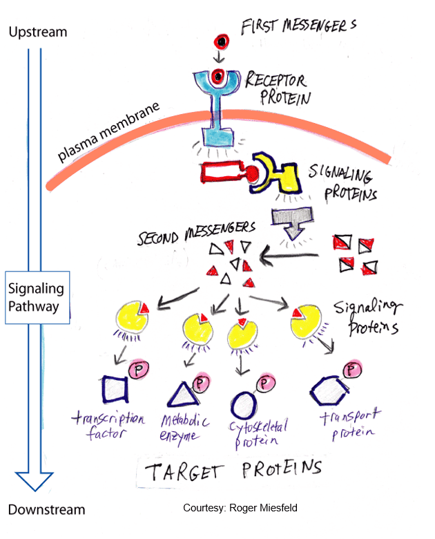
The receptor concept forms the central organizing paradigm of modern pharmacology and clinical therapeutics. A receptor is a specific biological macromolecule—most commonly a transmembrane or intracellular protein—that selectively binds an endogenous chemical messenger or exogenous xenobiotic to initiate a biochemical cascade culminating in a defined cellular response.
Fundamental Consequences in Clinical Medicine
- 1. Quantitative Dose-Response Relationships: Receptors determine the strict quantitative relationship between the concentration or dose of a drug and its therapeutic or toxic pharmacologic effect. The affinity of a drug for its binding site dictates the dose needed to form critical numbers of drug-receptor complexes, while the total receptor density limits the maximal achievable efficacy (Emax).
- 2. Selectivity of Drug Action: A drug’s molecular dimensions, geometric shape, stereochemical conformation, and electrical charge distribution determine whether and how tightly it fits within a specific receptor binding pocket among thousands of chemically distinct cellular proteins. Chemical modifications of a drug scaffold can dramatically alter its relative affinities across receptor subtypes, modulating its therapeutic-to-toxicity ratio.
- 3. Mediation of Agonists and Antagonists: Receptors act as biological molecular switches. Endogenous ligands and therapeutic agonists bind to and stabilize the active receptor state, directly driving downstream signal transduction. In contrast, pharmacologic antagonists bind without activating downstream signaling, occupying the receptor site and competitively preventing endogenous agonists from triggering responses.
Quantitative principles & receptor states
The Two-State Receptor Model
Classical receptor theory assumed that receptors exist in a quiescent, inactive ground state (R) until forced into an active conformation (R*) by ligand binding. Modern biophysical and structural evidence demonstrates that receptors fluctuate spontaneously between at least two thermodynamically reversible conformational states: the inactive state (R) and the active state (R*).
- Basal (Constitutive) Activity: In the absence of an agonist, the equilibrium strongly favors the inactive R state. However, a minute fraction of the receptor population spontaneously adopts the active R* state, generating a measurable baseline level of biological activity termed constitutive activity.
- Full Agonists: Possess high selective affinity for the R* state. By preferentially binding and stabilizing R*, full agonists shift the conformational equilibrium rapidly toward the active state, producing a maximal cellular response.
- Partial Agonists: Bind to both R and R* states with intermediate preference. Even at 100% receptor occupancy, they can only shift a fraction of the receptor pool into the R* conformation, resulting in a submaximal Emax compared to full agonists. In the presence of a full agonist, a partial agonist functions as a competitive antagonist.
- Neutral Competitive Antagonists: Exhibit equal affinity for both R and R* states. They do not alter the baseline equilibrium between R and R* (constitutive activity remains unchanged), but their physical occupation of the orthosteric binding site competitively blocks agonists from binding.
- Inverse Agonists: Possess selective affinity for the inactive R state. By binding and stabilizing R, they pull the equilibrium away from R*, suppressing baseline constitutive activity below resting levels. Classic examples include many β-blockers, antihistamines, and antimuscarinics previously misclassified as simple neutral antagonists.
The Phenomenon of “Spare Receptors”
In many pharmacological systems, a maximal biological response (Emax) can be elicited by an agonist at concentrations where only a tiny fraction of the total receptor population is occupied. The remaining unoccupied receptors are termed spare receptors.
- Biochemical Basis: Spareness is not due to physical hiding of receptors; rather, it reflects temporal and catalytic signal amplification downstream of receptor activation. For example, a single GTP-bound Gs protein can remain active for tens of seconds, keeping adenylyl cyclase active long after the initial agonist molecule has dissociated from the receptor.
- Mathematical Correlate: The concentration of drug required to produce half-maximal biological effect (EC50) is significantly lower than the concentration required to occupy 50% of the available receptors (KD):
EC50 < KD - Clinical Consequence: Spare receptors increase tissue sensitivity to low circulating hormone or drug concentrations. When an irreversible antagonist is introduced, low concentrations inactivate spare receptors without reducing the maximal agonist response (the curve shifts rightward). Only when the concentration of irreversible antagonist exceeds the spare receptor reserve does the maximal response (Emax) begin to depress.
Primary messengers & ligands
An extracellular chemical signaling molecule is termed a primary messenger (first messenger or ligand). Primary messengers are chemically diverse, ranging from small inorganic gases to complex polypeptides. Their physicochemical properties (polar vs. nonpolar, charge, molecular weight) dictate whether they require cell-surface transmembrane receptors or can passively permeate the lipid bilayer to bind intracellular targets.
| Signaling Molecule | Chemical Class | Primary Biological Source | Principal Physiological Function |
|---|---|---|---|
| Cortisol | Steroid (Lipophilic) | Adrenal cortex (zona fasciculata) | Suppresses inflammation, regulates gluconeogenesis, modulates stress responses |
| Epinephrine (Adrenaline) | Catecholamine / Amino acid derivative | Adrenal medulla & sympathetic nerves | Prepares the body for acute “fight-or-flight” autonomic mobilization |
| Erythropoietin (EPO) | Polypeptide glycoprotein (165 residues) | Renal peritubular interstitial cells | Stimulates erythropoiesis in the bone marrow under hypoxic conditions |
| Growth Hormone (Somatotropin) | Polypeptide hormone (191 residues) | Anterior pituitary gland (somatotrophs) | Stimulates linear body growth, chondrogenesis, protein synthesis, and lipolysis |
| Nitric Oxide (NO) | Inorganic free-radical gas (Lipophilic) | Vascular endothelial cells (via eNOS) | Diffuses into adjacent vascular smooth muscle to trigger cGMP-mediated vasodilation |
| Thromboxane A2 (TXA2) | Eicosanoid / Lipid derivative | Activated platelets (via COX-1) | Triggers platelet aggregation and potent local arteriolar vasoconstriction |
| Auxin (Indole-3-acetic acid) | Indole / Amino acid derivative | Apical botanical plant meristems | Promotes directional cell elongation, apical dominance, and plant phototropism |
Transmembrane signaling mechanisms
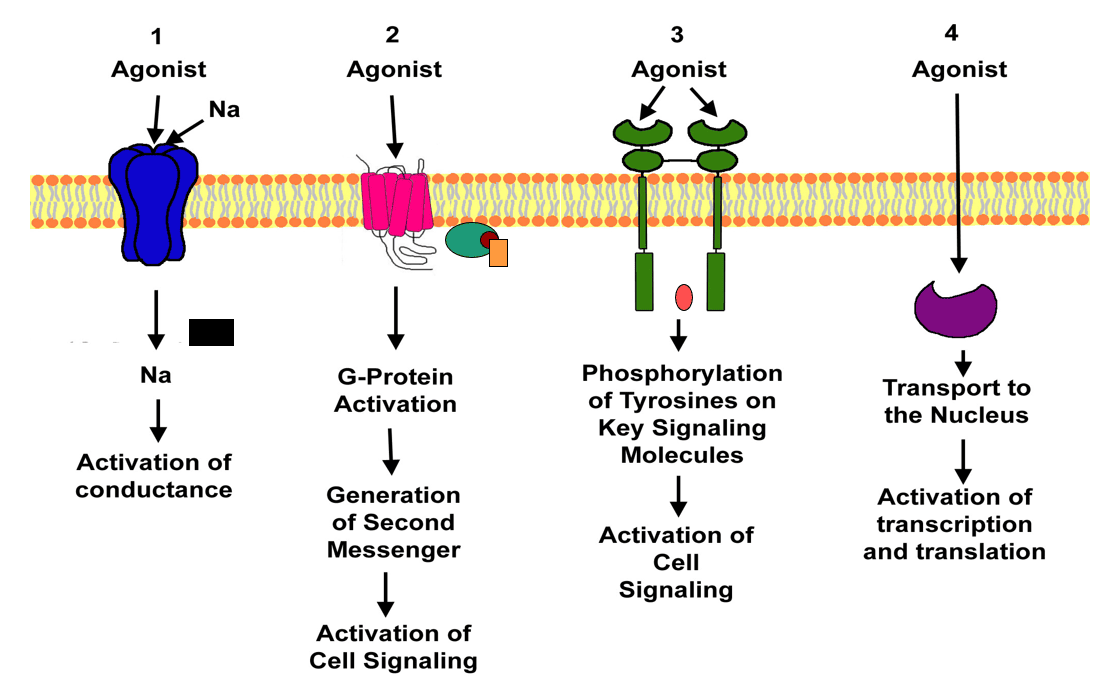
Cellular communication across biological membranes is governed by four major receptor superfamilies. These mechanisms allow water-soluble, polar ligands to transmit information without crossing the hydrophobic lipid bilayer, or enable lipophilic ligands to directly alter gene transcription.
1. Ligand-Gated Ion Channels (Ionotropic Receptors)
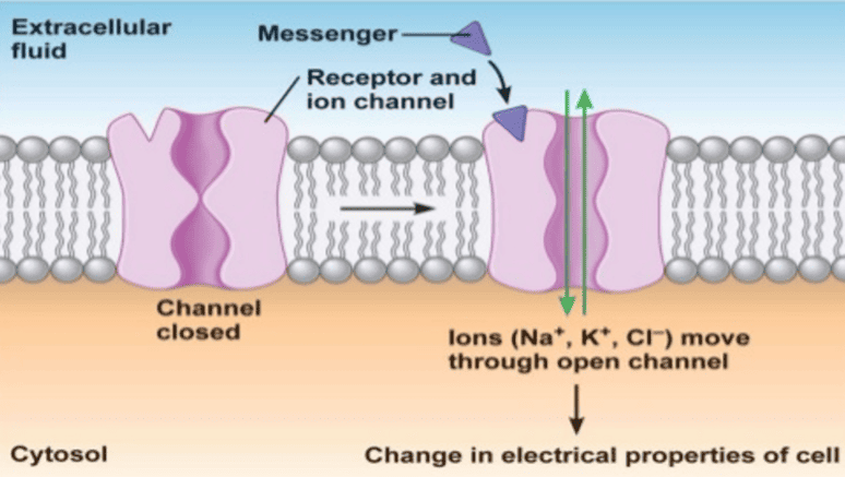
Cell-surface multimeric channel proteins that open or close directly upon binding an extracellular ligand, allowing selective inorganic ion flux (Na+, K+, Ca2+, or Cl−) down electrochemical gradients. Rapid signaling on a millisecond timescale drives fast synaptic transmission.
2. G-Protein Coupled Receptors (GPCRs / 7TM Receptors)
Heptaspanning serpentine transmembrane receptors that allosterically activate intracellular heterotrimeric guanine-nucleotide binding proteins (G-proteins). These G-proteins modulate downstream effector enzymes (adenylyl cyclase, phospholipase C) or ion channels to produce second messengers on a seconds timescale.
3. Enzyme-Linked Receptors (Transmembrane Receptor Kinases)
Single-pass transmembrane polypeptides containing an extracellular ligand-binding domain and an intracellular enzymatic domain (most commonly a protein tyrosine kinase, serine/threonine kinase, or guanylyl cyclase). Ligand binding drives non-covalent dimerization and trans-autophosphorylation, initiating downstream kinase cascades over minutes to hours.
4. Intracellular & Nuclear Receptors
Soluble cytoplasmic or nuclear proteins that bind lipid-soluble ligands (steroids, thyroid hormones, vitamin D) able to diffuse freely across the plasma membrane. Activated complexes bind specific DNA sequences (hormone response elements) to alter gene transcription and protein synthesis over a hours-to-days timescale.
Intracellular & nuclear receptors
Lipophilic ligands—including glucocorticoids, mineralocorticoids, sex steroids (estrogen, progesterone, testosterone), vitamin D, and thyroid hormone—cross the lipophilic plasma membrane via passive diffusion to engage intracellular receptor proteins.
Receptor Topography & Chaperone Complexes
- Compartmental Segregation: Unliganded steroid receptors occupy distinct subcellular compartments. The progesterone receptor and estrogen receptor reside predominantly within the nucleus in an unliganded state. In contrast, the unliganded glucocorticoid receptor (GR) is localized strictly within the cytoplasm.
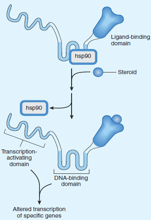
- The Inhibitory Heat-Shock Complex: In the absence of hormone, the glucocorticoid receptor polypeptide is trapped in an inactive conformation by a multi-protein chaperone complex containing heat-shock protein 90 (hsp90), hsp70, and immunophilin chaperones. The hsp90 dimer binds across the receptor’s hinge and ligand-binding domains, sterically preventing normal folding of the DNA-binding domain and obscuring its nuclear localization signal (NLS).
- Ligand-Induced Activation: Binding of a glucocorticoid molecule to the C-terminal ligand-binding domain induces a conformational change that relieves the inhibitory constraint, triggering dissociation of hsp90. This exposes the DNA-binding domain (containing two zinc-finger motifs) and unmasks the nuclear localization signal. The ligand-receptor complex forms a homodimer, translocates rapidly through the nuclear pore complex, and binds directly to specific palindromic DNA sequences termed Glucocorticoid Response Elements (GREs). By recruiting coactivator or corepressor complexes, the activated receptor modulates RNA polymerase II transcription of target genes.
Enzyme-linked receptors (RTKs)
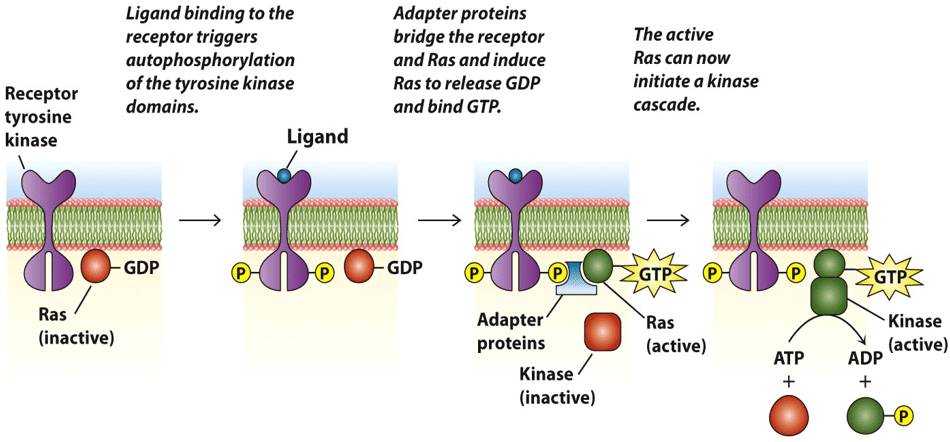
Receptor Tyrosine Kinases (RTKs) are single-pass transmembrane glycoproteins that transduce signals for polypeptide growth factors and trophic metabolic hormones, regulating cell growth, differentiation, survival, and intermediary metabolism.
Receptor Architecture & Activation Cascade
- Domain Organization: Composed of an extracellular ligand-binding domain (often enriched with cysteine-rich or immunoglobulin-like folds), a single hydrophobic transmembrane α-helix spanning the lipid bilayer, and an intracellular cytoplasmic tyrosine kinase catalytic domain.
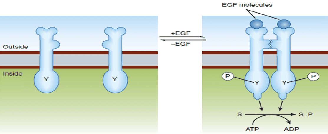
- Ligand-Induced Dimerization: In the resting state, RTKs (such as the epidermal growth factor receptor [EGFR]) exist as inactive monomers. Ligand binding alters the extracellular conformation, driving two receptor molecules to bind non-covalently into an active dimer. (The insulin receptor is an exception: it exists as a pre-formed disulfide-linked α2β2 heterotetramer).
- Trans-Autophosphorylation: Dimerization brings the cytoplasmic kinase domains into close apposition. Each kinase subunit phosphorylates specific tyrosine residues on the activation loop of its dimer partner. This autophosphorylation enhances intrinsic catalytic activity and creates high-affinity phosphotyrosine docking sites for intracellular signaling proteins containing Src-homology 2 (SH2) or phosphotyrosine-binding (PTB) domains.
The Ras-Raf-MEK-ERK (MAPK) Pathway
Following autophosphorylation, RTKs initiate the canonical mitogen-activated protein kinase (MAPK) cascade governing gene expression and mitosis:
- An adapter protein, Grb2 (Growth factor receptor-bound protein 2), docks onto receptor phosphotyrosine residues via its SH2 domain.
- Grb2 recruits SOS (Son of Sevenless), a guanine nucleotide exchange factor (GEF), via its SH3 domains.
- SOS interacts with membrane-anchored Ras, a monomeric small GTPase, stimulating the release of GDP and the binding of GTP.
- Active Ras-GTP recruits and activates Raf (a serine/threonine protein kinase), initiating sequential phosphorylation of MEK (MAPK/ERK kinase) and ERK (Extracellular signal-Regulated Kinase).
- Activated ERK translocates into the nucleus to phosphorylate transcription factors (e.g., c-Fos, c-Jun, c-Myc), driving cell-cycle progression.
Receptor Down-Regulation & Oncological Therapeutics
- Receptor Down-Regulation: The intensity and duration of RTK signaling are tightly controlled. Ligand binding triggers clathrin-mediated endocytosis of the receptor-ligand complex, followed by trafficking to late endosomes and complete degradation within lysosomes. If receptor endocytosis and proteolysis occur faster than de novo receptor synthesis, cell-surface receptor density declines, diminishing subsequent responsiveness. Mutations that disable endocytic down-regulation (e.g., loss of the Cbl ubiquitin ligase) lead to prolonged growth factor signaling and malignant transformation.
- Monoclonal Antibodies (Extracellular Targeters): Target the extracellular ligand-binding domain, sterically blocking growth factor engagement and preventing dimerization:
- Trastuzumab: Binds the extracellular domain of HER2/neu (ErbB2), an amplified RTK in aggressive invasive breast carcinomas.
- Cetuximab: Binds EGFR (HER1), inhibiting downstream signaling in colorectal carcinoma and head/neck squamous cell carcinoma.
- Small-Molecule Tyrosine Kinase Inhibitors (Intracellular Targeters): Cell-permeable, synthetic small molecules that competitively bind the intracellular ATP-binding pocket of the kinase domain:
- Gefitinib & Erlotinib: Inhibit EGFR tyrosine kinase activity, used in non-small cell lung cancer harboring activating EGFR mutations.
- Imatinib: Selectively inhibits the Bcr-Abl fusion tyrosine kinase in chronic myeloid leukemia (CML) and c-Kit in gastrointestinal stromal tumors (GIST).
Other Transmembrane Enzyme Receptors
- Transmembrane Serine/Threonine Kinases: Receptors for the Transforming Growth Factor-β (TGF-β) superfamily possess intracellular serine/threonine kinase domains. Ligand binding drives heterodimerization of Type I and Type II receptor subunits, phosphorylating cytosolic Smad proteins that translocate to the nucleus to regulate morphogenesis and tissue fibrosis.
- Membrane Guanylyl Cyclases: Receptors for Atrial Natriuretic Peptide (ANP) and B-type natriuretic peptide (BNP) contain an intrinsic intracellular catalytic domain that directly converts GTP into the second messenger cyclic GMP (cGMP), promoting renal natriuresis and vascular relaxation.
- Cytokine Receptors (JAK-STAT Pathway): Receptors for growth hormone, prolactin, erythropoietin, and interferons lack intrinsic catalytic enzymatic activity. Upon ligand-induced dimerization, they recruit non-receptor tyrosine kinases termed Janus Kinases (JAK1, JAK2, TYK2), which cross-phosphorylate the receptor and activate STAT (Signal Transducer and Activator of Transcription) transcription factors.
Ionotropic & voltage-gated channels
Ligand-Gated Ion Channels (Ionotropic Receptors)
Ionotropic receptors are multimeric transmembrane protein complexes enclosing a central aqueous ion conduction pore. The binding of a specific chemical neurotransmitter directly induces an allosteric conformational shift, opening or closing the gate on a millisecond timescale.
The Nicotinic Acetylcholine Receptor (nAChR)
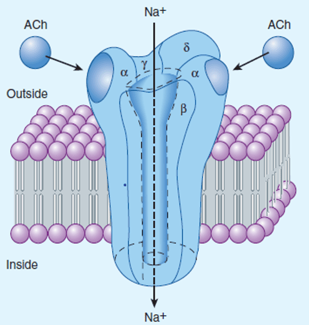
The muscle-type nicotinic acetylcholine receptor (NM) represents the prototype of the Cys-loop ligand-gated ion channel superfamily:
- Subunit Stoichiometry: A heteropentamer composed of five homologous transmembrane glycoprotein subunits arranged symmetrically around a central pseudo-pore: two α1, one β1, one γ (or ε in adult muscle), and one δ subunit (α2βγδ).
- Gating Mechanism: Each α-subunit contains an extracellular orthosteric binding pocket at the interface with adjacent subunits. Opening of the channel requires the simultaneous binding of two molecules of acetylcholine (one to each α-subunit). Ligand binding produces a rapid rotational twist in the pore-lining M2 α-helices, dilating the hydrophobic constriction gate.
- Electrochemical Consequence: The open pore is cation-selective, allowing rapid passive influx of Na+ down its steep inward electrochemical gradient (with minor K+ efflux). This net inward positive current generates a localized Excitatory Postsynaptic Potential (EPSP), known at the neuromuscular junction as the endplate potential (EPP), depolarizing the muscle membrane toward the threshold for voltage-gated Na+ channel activation.
- Other Primary Ionotropic Receptors:
- GABAA & Glycine Receptors: Selective for chloride (Cl−) anions; activation produces cellular hyperpolarization (Inhibitory Postsynaptic Potential, IPSP).
- 5-HT3 Serotonin Receptors: Cation-selective (Na+/K+); mediate rapid emesis in the chemoreceptor trigger zone and gut.
- Glutamate Receptors (NMDA, AMPA, Kainate): Excitatory cation channels; NMDA channels exhibit high Ca2+ permeability and require glycine as a co-agonist alongside membrane depolarization to relieve Mg2+ pore block.
Pharmacological Modulation: Digoxin & The Na+/K+-ATPase
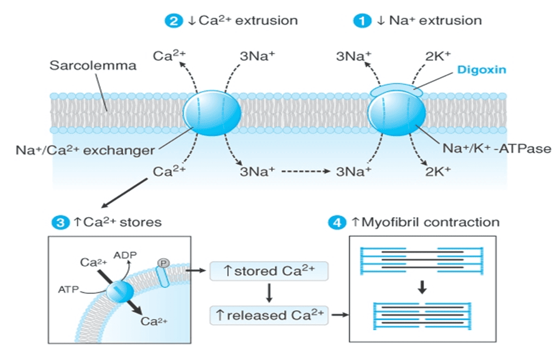
Therapeutic agents frequently alter ion gradients by inhibiting transport proteins rather than targeting ligand-gated receptors. A classic example is the cardiac glycoside Digoxin in congestive heart failure:
Voltage-Gated Ion Channels
Voltage-gated ion channels do not bind neurotransmitters directly; their gating is governed by changes in transmembrane electrical potential sensed by positively charged transmembrane segments (e.g., the S4 α-helix containing arginine and lysine residues).
- Presynaptic Voltage-Gated Ca2+ Channels (N-type, P/Q-type): Depolarization of the axon terminal activates these channels, permitting localized Ca2+ influx that triggers SNARE-dependent synaptic vesicle fusion and neurotransmitter release.
- L-Type Ca2+ Channels (Cav1.2): Predominate in cardiac nodal tissue, myocardium, and vascular smooth muscle.
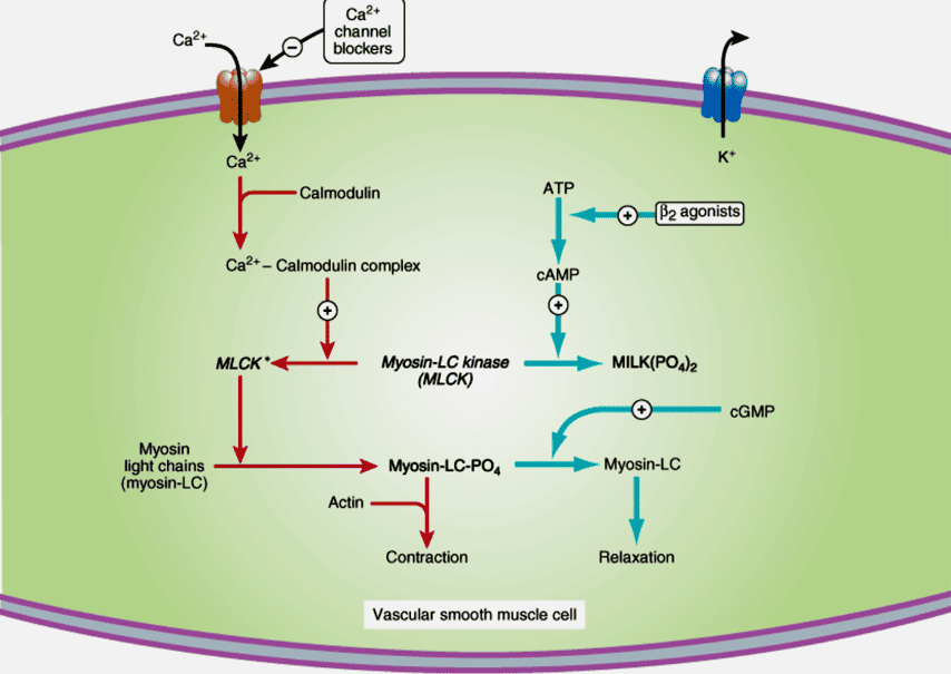
Calcium Channel Blockers (CCBs): Non-dihydropyridines (verapamil, diltiazem) and dihydropyridines (amlodipine, nifedipine) bind distinct allosteric sites on the α1 subunit of L-type Ca2+ channels, stabilizing the inactive closed state without mimicking endogenous transmitters. In vascular smooth muscle, blocking Ca2+ entry prevents calmodulin activation of Myosin Light Chain Kinase (MLCK), causing arterial vasodilation and lowering blood pressure.
GPCRs & heterotrimeric G-proteins
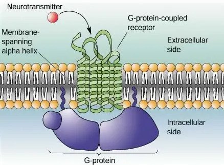
G-protein coupled receptors (GPCRs)—also termed seven-transmembrane (7TM) or serpentine receptors—comprise the largest single family of cell-surface signaling receptors in the human genome, representing the direct molecular targets for over 30% of approved pharmaceutical drugs.
The Heterotrimeric G-Protein Activation Cycle
Heterotrimeric G-proteins consist of three distinct subunits: a guanine nucleotide-binding Gα subunit and a stable, high-affinity Gβγ dimeric complex. In the basal inactive state, the Gα subunit binds guanosine diphosphate (GDP) and remains associated with the Gβγ dimer near the inner leaflet of the plasma membrane.
Stepwise G-Protein Cycle
- Ligand Engagement: An extracellular agonist binds the GPCR, causing a conformational shift in the transmembrane helices that is transmitted to the intracellular loops.
- Guanine Nucleotide Exchange: The activated receptor acts as a Guanine Nucleotide Exchange Factor (GEF), interacting with Gα to induce an open conformation that expels bound GDP. Because intracellular GTP concentrations exceed GDP concentrations, GTP immediately binds to Gα.
- Subunit Dissociation: GTP binding alters the switch regions of Gα, destabilizing the heterotrimer. Gα-GTP dissociates from both the Gβγ dimer and the receptor.
- Effector Modulation: Both the liberated, active Gα-GTP monomer and the freed Gβγ dimer diffuse laterally along the membrane inner surface to interact allosterically with specific effector enzymes (adenylyl cyclase, phospholipase C) or ion channels (opening K+ channels, inhibiting Ca2+ channels).
- Signal Inactivation (GTP Hydrolysis): The Gα subunit possesses intrinsic GTPase activity that slowly hydrolyzes bound GTP into GDP and inorganic phosphate (Pi). This hydrolysis acts as an internal molecular timer, returning Gα to its basal GDP-bound state. Acceleration of this GTPase clock is mediated by Regulators of G-protein Signaling (RGS proteins), which act as GTPase-Activating Proteins (GAPs).
- Reassociation: Upon GDP reconstitution, Gα-GDP reassociates with the Gβγ dimer, reforming the inactive heterotrimer ready for another cycle. The freed receptor can re-engage additional G-protein heterotrimers as long as agonist remains bound.
The adenylyl cyclase–cAMP pathway
The cyclic adenosine-3′,5′-monophosphate (cAMP) pathway regulates glycogen metabolism, cardiac inotropy and chronotropy, smooth muscle tone, and renal water excretion.
Enzymatic Synthesis & PKA Activation
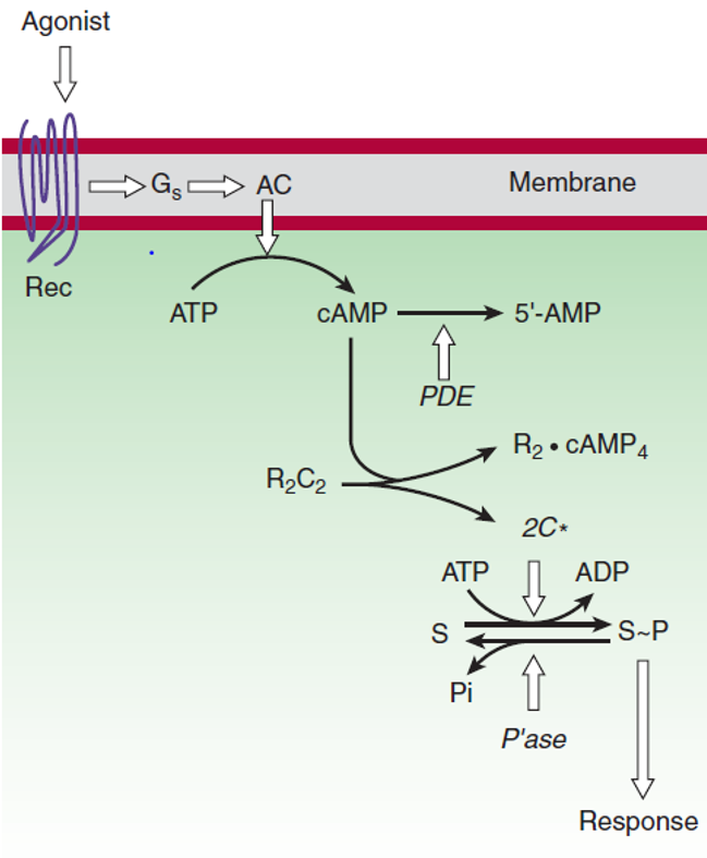
- Synthesis via Adenylyl Cyclase (AC): Activated Gαs-GTP binds to the catalytic core of adenylyl cyclase, a 12-transmembrane enzyme, stimulating the cyclization of cytosolic ATP into cyclic AMP (cAMP) with the release of inorganic pyrophosphate. Conversely, Gαi-GTP directly inhibits adenylyl cyclase, lowering cAMP production.
- Allosteric Activation of Protein Kinase A (PKA): In the basal state, PKA is an inactive heterotetramer composed of two regulatory (R) subunits and two catalytic (C) subunits (R2C2). The regulatory subunits occupy the active sites of the catalytic subunits, inhibiting their function. Cooperatively, four molecules of cAMP bind (two per R subunit), inducing a conformational release of the two active, free catalytic subunits (2C*).
- Substrate Phosphorylation: Free catalytic subunits phosphorylate serine and threonine residues on specific cellular enzymes:
- Glycogenolysis: PKA phosphorylates and activates phosphorylase kinase, which in turn phosphorylates and activates glycogen phosphorylase, driving glycogen breakdown into glucose-1-phosphate. Concurrently, PKA phosphorylates and inactivates glycogen synthase, shutting down glycogen storage.
- Cardiac Myocytes: PKA phosphorylates L-type Ca2+ channels on the sarcolemma (increasing Ca2+ influx) and phospholamban on the sarcoplasmic reticulum (accelerating SERCA Ca2+ re-uptake), increasing both the force (inotropy) and rate of relaxation (lusitropy) of contraction.
- Smooth Muscle Relaxation: In vascular and airway smooth muscle, PKA phosphorylates and inhibits Myosin Light Chain Kinase (MLCK), reducing actin-myosin cross-bridging and producing bronchodilation and vasodilation.
- Transcriptional Regulation: PKA translocates into the nucleus and phosphorylates the transcription factor CREB (cAMP Response Element-Binding protein), which recruits coactivators (CBP/p300) to bind CRE consensus sequences and drive gene expression.
Termination of cAMP Signaling & Therapeutic PDE Inhibition
- Degradation via Phosphodiesterases (PDEs): Cytosolic cAMP is converted into inactive 5′-AMP via hydrolysis by cyclic nucleotide phosphodiesterases, terminating PKA activation. Dephosphorylation of phosphorylated substrate proteins is executed by serine/threonine protein phosphatases (PP1, PP2A).
- Milrinone: A selective inhibitor of PDE-3, the primary isoform expressed in cardiac myocytes and vascular smooth muscle. In acute decompensated heart failure, milrinone prevents cAMP degradation, increasing cardiac contractility (positive inotropy) while inducing systemic arteriolar dilation (afterload reduction)—a clinical profile termed an inodilator.
- Methylxanthines (Theophylline, Caffeine): Non-selective competitive inhibitors of phosphodiesterases that block cAMP breakdown, producing bronchodilation in asthma/COPD and cortical CNS stimulation.
The PLC, IP3/DAG & Ca2+ cascade
The phosphoinositide (PI) cascade couples cell-surface Gq/11 receptors to the generation of two complementary second messengers: Inositol-1,4,5-trisphosphate (IP3) and Diacylglycerol (DAG).
Generation and Functional Actions of IP3 and DAG
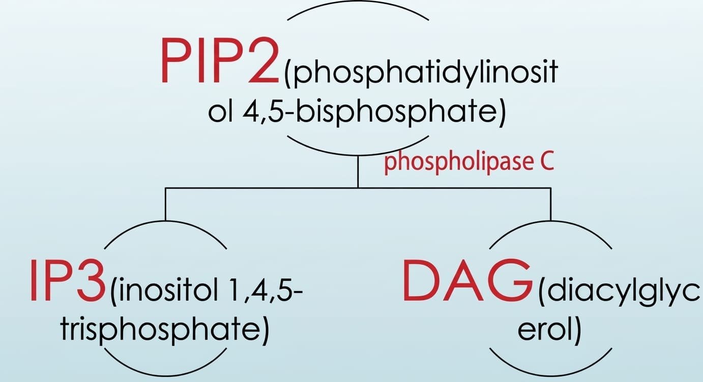
- 1. Phospholipase C Activation: Agonist binding to Gq-coupled receptors (α1-adrenoceptors, M1/M3 muscarinic receptors, V1 vasopressin receptors) releases Gαq-GTP, which activates membrane-bound phospholipase C-β (PLCβ). PLCβ hydrolyzes a minor membrane phospholipid, phosphatidylinositol-4,5-bisphosphate (PIP2).
- 2. Actions of Inositol-1,4,5-Trisphosphate (IP3): IP3 is small and water-soluble, diffusing rapidly away from the membrane through the cytoplasm to bind IP3 receptors (IP3R)—tetrameric ligand-gated Ca2+ channels—situated on the membrane of the smooth endoplasmic reticulum (and sarcoplasmic reticulum). This triggers the rapid release of sequestered Ca2+ into the cytoplasm, elevating free cytosolic Ca2+ concentrations from basal resting levels (~100 nmol/L) up to micromolar levels (>1 μmol/L).
- 3. Actions of Diacylglycerol (DAG): DAG is highly hydrophobic and remains confined to the inner leaflet of the plasma membrane. In the presence of membrane phosphatidylserine and elevated cytosolic Ca2+, DAG allosterically binds and activates Protein Kinase C (PKC). Active PKC translocates to the cell membrane and phosphorylates target enzymes that regulate inflammation, cellular proliferation, tumor promotion, smooth muscle contraction, and exocytosis.
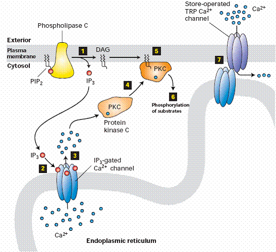
Downstream Effects of Elevated Cytosolic Calcium (Ca2+)
Free Ca2+ functions as a universal second messenger, cooperating with DAG to activate PKC and binding to the ubiquitous intracellular calcium sensor protein calmodulin (CaM):
- Calmodulin Activation: Calmodulin possesses four high-affinity EF-hand binding sites for Ca2+. Binding induces a conformational change that exposes a hydrophobic interaction surface, allowing the Ca2+-CaM complex to bind and activate Ca2+/calmodulin-dependent protein kinases (CaMKs) and Myosin Light Chain Kinase (MLCK), driving smooth muscle contraction and glycogenolysis.
- Cross-Talk with cAMP Pathways: Ca2+-CaM binds to and regulates specific adenylyl cyclase isoforms (AC1, AC8) and calcium-activated phosphodiesterases (PDE1), linking calcium and cyclic nucleotide cascades.
- Exocytosis & Secretion: Triggers synaptotagmin-mediated SNARE vesicle fusion, driving neurotransmitter release at synapses and hormone exocytosis across endocrine glands.
Pathway Termination & The Mechanism of Lithium
- Signal Termination: IP3 is rapidly deactivated by sequential enzymatic dephosphorylation via inositol polyphosphate phosphatases to produce free inositol, or phosphorylated into IP4. DAG is either phosphorylated by DAG kinase into phosphatidic acid (re-entering the phosphoinositide resynthesis cycle) or deacylated by DAG lipase to yield arachidonic acid (the substrate for prostaglandin and leukotriene synthesis). Cytosolic Ca2+ is cleared back into the ER/SR by ATP-dependent SERCA pumps and extruded across the plasma membrane by PMCA pumps and the Na+/Ca2+ exchanger (NCX).
- Clinical Action of Lithium: In the central nervous system, inositol is recycled from IP1 to free inositol via the enzyme inositol monophosphatase (IMPase). Lithium ion (Li+), used as a mood stabilizer in bipolar disorder, uncompetitively inhibits IMPase. In hyperactive cerebral neurons with excessive Gq signaling, lithium depletes intracellular free inositol reserves (the “inositol depletion hypothesis”), selectively suppressing runaway phosphoinositide signaling without impairing quiescent neural circuits.
The guanylyl cyclase–cGMP pathway
The cyclic guanosine-3′,5′-monophosphate (cGMP) pathway plays a central role in vascular smooth muscle tone, penile erectile function, intestinal fluid secretion, and retinal phototransduction.
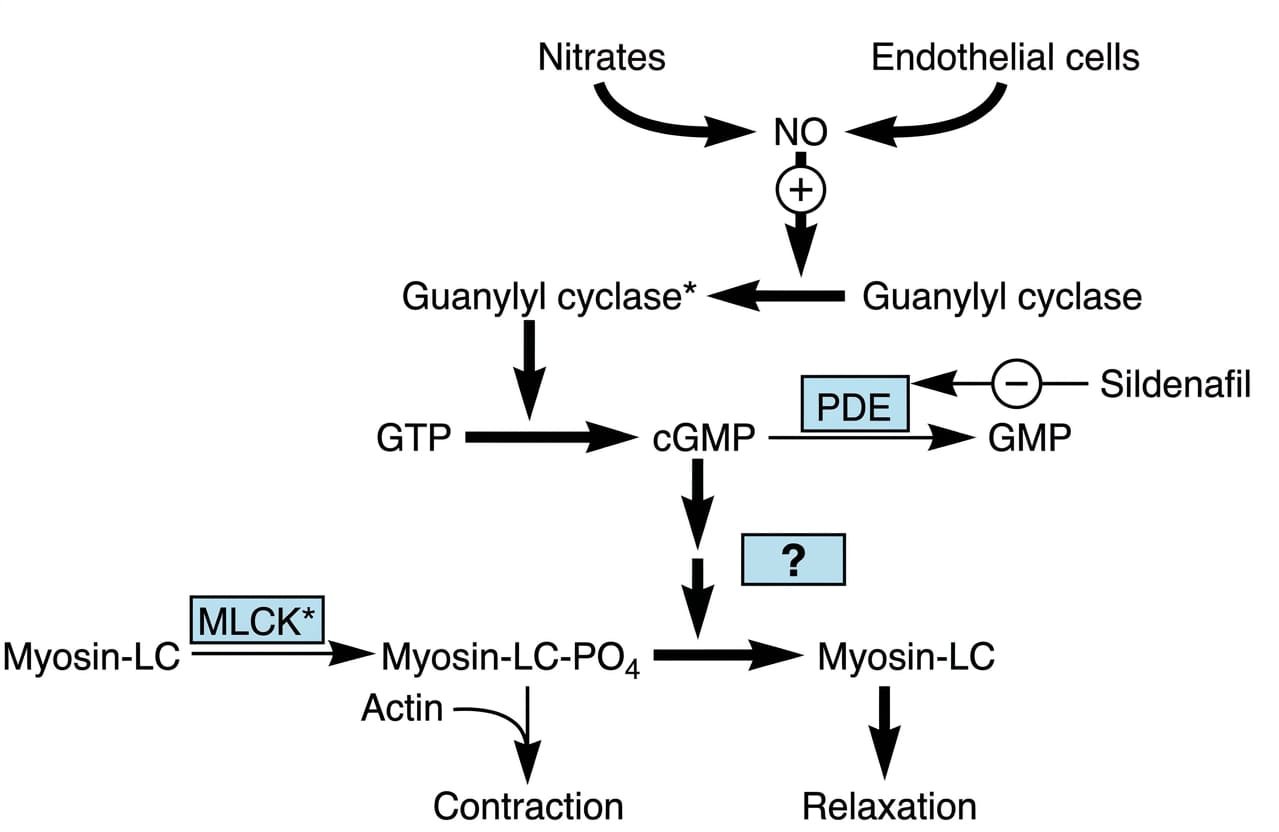
Mechanisms of cGMP Generation
Intracellular cGMP synthesis is catalyzed by two distinct forms of guanylyl cyclase:
- 1. Particulate / Membrane-Bound Guanylyl Cyclase (pGC): Transmembrane enzyme receptors whose extracellular domains bind natriuretic peptides:
- Atrial Natriuretic Peptide (ANP): Secreted by atrial myocytes in response to hypervolemic stretch; binds GC-A to generate cGMP, causing renal glomerular afferent arteriolar vasodilation, natriuresis, and systemic blood pressure reduction.
- Enterotoxins & Guanylin: Bacterial heat-stable enterotoxin (STa of enterotoxigenic E. coli) and the drug linaclotide bind GC-C on intestinal enterocytes, elevating cGMP to stimulate cystic fibrosis transmembrane conductance regulator (CFTR) chloride channels, driving secretory diarrhea.
- 2. Soluble / Cytoplasmic Guanylyl Cyclase (sGC): An intracellular heterodimeric hemoprotein that functions as a high-affinity receptor for nitric oxide (NO) gas. Endogenous vasodilators (acetylcholine, bradykinin) stimulate endothelial NO synthase (eNOS) to synthesize NO from L-arginine. Lipophilic NO gas diffuses across the inter-cellular space into adjacent vascular smooth muscle cells, binding to the heme iron of sGC to increase cGMP synthesis.
Vascular Smooth Muscle Relaxation & Pharmacology
- Dephosphorylation of Myosin Light Chains: cGMP allosterically activates Protein Kinase G (PKG). PKG phosphorylates and activates Myosin Light Chain Phosphatase (MLCP). Activated MLCP rapidly dephosphorylates myosin light chain phosphate (Myosin-LC-PO4 → Myosin-LC), terminating actin-myosin cross-bridge cycling and inducing profound vascular smooth muscle relaxation. PKG also lowers intracellular Ca2+ by inhibiting voltage-gated Ca2+ channels and stimulating Ca2+-ATPase pumps.
- Organic Nitrates: Vasodilator drugs such as nitroglycerin, isosorbide dinitrate, and sodium nitroprusside undergo bioactivation to release exogenous nitric oxide, stimulating sGC to produce selective venodilation (reducing cardiac preload in angina) and arteriolar dilation (reducing afterload in hypertensive crises).
- Phosphodiesterase-5 (PDE-5) Inhibitors: In the penile corpus cavernosum and pulmonary vasculature, cGMP is cleared by the PDE-5 enzyme. Sildenafil, tadalafil, and vardenafil competitively inhibit PDE-5, preventing cGMP degradation. This prolongs smooth muscle relaxation, maintaining penile erection in erectile dysfunction and dilating pulmonary arteries in pulmonary arterial hypertension (PAH).
Signal amplification & cross-talk
Exponential Catalytic Amplification
Signal transduction cascades do not operate in linear 1:1 stoichiometry. Each successive catalytic tier activates multiple downstream substrate molecules, generating an exponential cascade that allows minute nanomolar (10−9 M) concentrations of a circulating first messenger to rapidly mobilize millions of product molecules within the target cell.
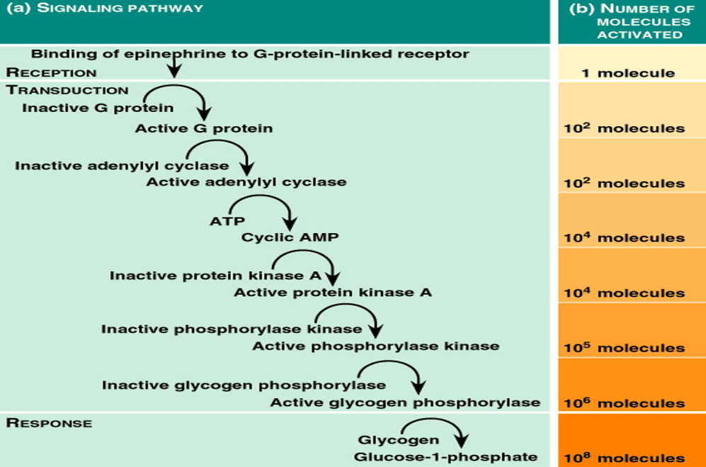
The 108-Fold Epinephrine Cascade
- Reception: Binding of 1 molecule of epinephrine to a β2-adrenoceptor.
- G-Protein Activation: The single occupied receptor sequentially encounters and activates 102 molecules of Gs before ligand dissociation.
- Enzyme Activation: 102 molecules of active adenylyl cyclase are stimulated.
- Second Messenger Generation: Each adenylyl cyclase converts thousands of ATP molecules, synthesizing 104 molecules of cAMP.
- Kinase Activation: cAMP allosterically activates 104 active PKA catalytic subunits.
- Intermediate Kinase Cascade: PKA phosphorylates and activates 105 molecules of phosphorylase kinase.
- Terminal Enzyme Activation: Phosphorylase kinase activates 106 molecules of glycogen phosphorylase.
- Final Biological Output: Glycogen phosphorylase cleaves stored glycogen to liberate 108 molecules of glucose-1-phosphate—a net 100-million-fold biological amplification.
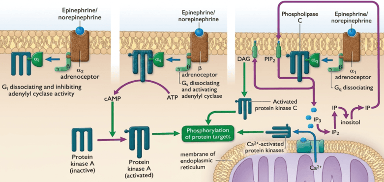
Receptor Signaling Cross-Talk
Target cells rarely respond to a single isolated signal. Multiple pathways converge, diverge, and cross-regulate to fine-tune homeostatic responses:
- Dual Adrenergic Fine-Tuning in Hepatocytes: Epinephrine stimulates β2 receptors (coupled via Gs to activate adenylyl cyclase and elevate cAMP) while simultaneously stimulating α1 receptors (coupled via Gq to activate PLC, generating IP3/Ca2+ and DAG). Both pathways converge synergistically to activate glycogen phosphorylase through independent kinase phosphorylation sites, maximizing hepatic glucose release during systemic hypoglycemia.
- Opposing Regulation of Vascular Smooth Muscle: Intracellular calcium (via Gq-coupled α1 receptors) drives MLCK and promotes vasoconstriction. Concurrently, Gs-coupled β2 receptors (via cAMP/PKA) and NO-stimulated sGC (via cGMP/PKG) phosphorylate and inactivate MLCK while activating MLCP, promoting vasodilation. The net vascular tone represents the integration of these opposing inputs.
Desensitization, down-regulation & tolerance
Prolonged or repeated stimulation of a receptor by an agonist frequently produces an adaptive attenuation of responsiveness, a homeostatic defense mechanism termed desensitization, tachyphylaxis, or tolerance.
Mechanisms of Receptor Desensitization
- 1. Homologous Desensitization (Receptor Phosphorylation):
Occurs within seconds to minutes of agonist occupancy. Agonist-activated GPCRs are recognized by specialized serine/threonine kinases termed G-protein-coupled Receptor Kinases (GRKs). GRKs selectively phosphorylate the cytoplasmic C-terminal tail and third intracellular loop of the occupied receptor.
This phosphorylation recruits the cytosolic adaptor protein β-arrestin. Binding of β-arrestin sterically blocks the receptor from coupling to heterotrimeric G-proteins, immediately extinguishing downstream signaling even while the agonist remains bound—a process termed receptor uncoupling.
- 2. Receptor Internalization (Endocytosis):
β-Arrestin functions as a clathrin adaptor, targeting the uncoupled receptor to clathrin-coated pits for dynamin-dependent endocytosis into early endosomes. Once internalized, receptors follow one of two pathways:
- Resensitization (Recycling): The endosomal proton pump acidifies the vesicle, causing the agonist to dissociate. Endosomal phosphatases dephosphorylate the receptor, allowing it to be recycled back to the plasma membrane fully resensitized.
- Down-Regulation (Lysosomal Degradation): Under prolonged, chronic agonist exposure, endosomes are targeted to lysosomes, where the receptor protein is degraded by acid hydrolases. Down-regulation reduces the total cell-surface receptor density (Bmax), and recovery requires de novo protein synthesis over days to weeks.
- 3. Heterologous Desensitization:
Second-messenger-activated kinases (PKA or PKC) activated by one receptor pathway can phosphorylate non-specific serine/threonine residues on unrelated, unliganded receptors. This broad feedback desensitizes multiple distinct receptor classes simultaneously.
Comparative tables
| Receptor Superfamily | Membrane Architecture | Coupling / Effector System | Signaling Scale | Representative Prototypes |
|---|---|---|---|---|
| 1. Ligand-Gated Ion Channels (Ionotropic) | Oligomeric (pentamers/tetramers) with central aqueous pore | Direct intrinsic ion channel flux (Na+, K+, Ca2+, Cl−) | Milliseconds | Nicotinic ACh (NM, NN), GABAA, Glycine, 5-HT3, NMDA, AMPA |
| 2. G-Protein Coupled Receptors (Metabotropic) | Seven-transmembrane (7TM) serpentine α-helical monomer | Heterotrimeric G-proteins (Gs, Gi, Gq, Go, Gt) modulating enzymes/channels | Seconds to minutes | Adrenoceptors (α, β), Muscarinic ACh (M1–M5), Opioid (μ, δ, κ), Rhodopsin |
| 3. Enzyme-Linked Receptors | Single-pass transmembrane α-helix; active as dimers | Intrinsic protein kinase (tyrosine, serine/threonine) or guanylyl cyclase | Minutes to hours | Insulin receptor, EGFR, PDGFR, VEGFR, TGF-β receptor, ANP receptor (GC-A) |
| 4. Intracellular & Nuclear Receptors | Soluble monomeric proteins; zinc-finger DNA-binding domains | Ligand-activated transcription factors binding Hormone Response Elements | Hours to days | Glucocorticoid (GR), Mineralocorticoid (MR), Estrogen (ER), Thyroid (TR), Vitamin D |
| Gα Class | Coupled Receptors | Primary Effector Enzyme | Second Messenger Produced | Key Downstream Physiological Action |
|---|---|---|---|---|
| Gαs (Stimulatory) | β1, β2, β3, Glucagon, Histamine H2, TSH, PTH, V2 | ↑ Adenylyl Cyclase; ↑ L-type Ca2+ channels | ↑ Cyclic AMP (cAMP) | PKA activation: cardiac inotropy/chronotropy (β1), bronchodilation (β2), renal water reabsorption (V2) |
| Gαi (Inhibitory) | α2, Muscarinic M2, M4, Opioid (μ, δ, κ), Dopamine D2 | ↓ Adenylyl Cyclase; ↑ KAch channels; ↓ Ca2+ channels | ↓ Cyclic AMP (cAMP) | Reduced PKA activity: decreased SA nodal firing, slowed AV nodal conduction, presynaptic inhibition of transmitter release |
| Gαq (PLC-Activating) | α1, Muscarinic M1, M3, M5, 5-HT2, Angiotensin AT1, V1a | ↑ Phospholipase C-β (PLCβ) | ↑ IP3, ↑ DAG, ↑ Cytosolic Ca2+ | PKC and CaM-kinase activation: smooth muscle contraction (α1, M3), exocrine glandular secretion (M3) |
| Gαo (Other/Neural) | Opioid, α2, Cannabinoid CB1 | ↓ Presynaptic Ca2+ currents | ↓ Intracellular Ca2+ | Inhibition of neurotransmitter release in central and peripheral nerves |
| Gαt (Transducin) | Rhodopsin (photons) in rod and cone cells | ↑ cGMP Phosphodiesterase (PDE6) | ↓ Cyclic GMP (cGMP) | Closure of cGMP-gated Na+ channels, membrane hyperpolarization, and visual phototransduction |
| Gαolf (Olfactory) | Odorant receptors in nasal olfactory neuroepithelium | ↑ Adenylyl Cyclase (AC3) | ↑ Cyclic AMP (cAMP) | Opening of cAMP-gated cation channels, membrane depolarization, and perception of smell |
| Second Messenger | Generating Effector Enzyme | Degrading / Inactivating Mechanism | Direct Intracellular Target | Major Physiological Actions |
|---|---|---|---|---|
| cAMP | Adenylyl Cyclase (from ATP) | Phosphodiesterases (PDE3, PDE4) to 5′-AMP | Protein Kinase A (PKA); EPAC; cyclic nucleotide-gated channels | Glycogenolysis, lipolysis, cardiac inotropy, smooth muscle relaxation, water conservation |
| cGMP | Guanylyl Cyclase (soluble or membrane-bound from GTP) | Phosphodiesterase-5 (PDE5) to 5′-GMP | Protein Kinase G (PKG); PDE3 modulation; cGMP-gated channels | Vascular smooth muscle relaxation (dephosphorylates MLC), natriuresis, penile erection |
| IP3 | Phospholipase C-β (cleaves PIP2) | Inositol polyphosphate phosphatases to inositol | IP3 receptor (ligand-gated Ca2+ channel on ER) | Rapid release of stored Ca2+ from the endoplasmic reticulum into the cytosol |
| DAG | Phospholipase C-β (cleaves PIP2) | DAG kinase (to phosphatidic acid); DAG lipase (to arachidonic acid) | Protein Kinase C (PKC) | Enzyme phosphorylation, cellular proliferation, inflammation, smooth muscle contraction |
| Ca2+ | IP3-gated channels; L-type Ca2+ channels; RyR | SERCA pumps (into ER); PMCA pumps & NCX (extrusion) | Calmodulin; PKC; troponin C; calpain | Myocardial and smooth muscle contraction, exocytosis, glycogenolysis, gene regulation |
| Nitric Oxide (NO) | Nitric Oxide Synthase (eNOS, nNOS, iNOS from L-arginine) | Spontaneous oxidation to nitrite and nitrate; scavenging by hemoglobin | Soluble Guanylyl Cyclase (heme iron) | Stimulates cGMP production, producing profound arteriolar and venous vasodilation |
Quick recap
References
The following authoritative medical textbooks support this curriculum study note:
- Katzung’s Basic & Clinical Pharmacology: Katzung, B.G. and Vanderah, T.W. (2021) Basic & Clinical Pharmacology. 15th edn. New York: McGraw-Hill Education. [Chapter 1: Introduction: The Nature of Drugs & Drug Development and Regulation; Chapter 2: Drug Receptors & Pharmacodynamics].
- Goodman & Gilman’s Pharmacological Basis of Therapeutics: Brunton, L.L., Hilal-Dandan, R. and Knollmann, B.C. (2018) Goodman & Gilman’s The Pharmacological Basis of Therapeutics. 13th edn. New York: McGraw-Hill Education. [Chapter 3: Mechanisms of Drug Action and the Relationship Between Drug Concentration and Effect].
- Rang & Dale’s Pharmacology: Ritter, J.M., Flower, R., Henderson, G., Loke, Y.K., MacEwan, D. and Rang, H.P. (2020) Rang & Dale’s Pharmacology. 9th edn. Edinburgh: Elsevier. [Chapter 3: How Drugs Act: Molecular Aspects].
- Guyton and Hall Textbook of Medical Physiology: Hall, J.E. and Hall, M.E. (2021) Guyton and Hall Textbook of Medical Physiology. 14th edn. Philadelphia: Elsevier. [Chapter 75: Pituitary Hormones and Their Control by the Hypothalamus; Section XIII: Endocrinology and Reproduction].