
Diuretic agents: nephron physiology, transport inhibitors & clinical therapeutics
On this page
Overview & core definitions
A diuretic (colloquially termed a “water pill”) is any pharmacological agent that promotes diuresis—an elevation in the rate of urine volume formation by the kidneys. These agents are central to managing expanded extracellular fluid (ECF) volume states and regulating systemic arterial hemodynamics.

Fundamental Pharmacological Definitions
- Diuresis: The physiological or drug-induced increase in total urine volume output. When achieved via pharmacological intervention, it is frequently described as forced diuresis.
- Natriuresis: The drug-induced increase in renal sodium (Na+) excretion. Because osmotic forces mandate that water follows sodium (“where sodium goes, water follows”), almost all clinically useful diuretics operate primarily as natriuretics.
- Antidiuretic: An agent that reduces urinary water excretion. The physiological prototype is vasopressin (antidiuretic hormone [ADH]), an octapeptide synthesized by the hypothalamus and secreted by the posterior pituitary that promotes water reabsorption through aquaporin-2 channels in the renal collecting duct.
- Primary Indications: Diuretics are deployed to treat volume overload in congestive heart failure (CHF), hepatic cirrhosis with ascites, nephrotic syndrome, acute and chronic renal failure, systemic hypertension, gestational edema, hypercalcemia, and acute intoxication (via forced alkaline or saline diuresis).
- Eating Disorders & Surreptitious Abuse: Diuretics are frequently abused by patients suffering from bulimia nervosa and anorexia nervosa. Patients misuse these agents to induce rapid weight loss through intravascular and interstitial volume depletion, frequently precipitating severe hypokalemic hypochloremic metabolic alkalosis, prerenal azotemia, and cardiac arrhythmias.
Renal hemodynamics & GFR formation
Urine formation requires three sequential processes: ultrafiltration at the glomerulus, selective tubular reabsorption from the luminal fluid back into peritubular capillaries, and active tubular secretion from peritubular blood into the tubular lumen.
Renal Hemodynamics & Filtration Fractions
The kidneys receive disproportionate perfusion relative to their physical mass, accounting for approximately 20% to 25% of resting cardiac output:
- Cardiac Output (CO): Standard physiological resting cardiac output is approximately 5.0 L/min (measured baseline ≈ 4.7 to 5.0 L/min).
- Renal Blood Flow (RBF): Twenty percent of cardiac output is distributed to the kidneys, yielding an RBF of 1,000 to 1,200 mL/min through both kidneys.
- Renal Plasma Flow (RPF): Assuming a standard hematocrit of 40% (40% cellular elements, 60% liquid plasma), actual renal plasma flow is:
RPF = RBF × (1 − Hematocrit) = 1,000 mL/min × 0.60 = 600 mL/min. - Glomerular Filtration Rate (GFR): Of the 600 mL/min of plasma traversing glomerular capillaries, only approximately 20% is filtered across the glomerular filtration barrier into Bowman’s space:
GFR = RPF × Filtration Fraction (0.20) = 120 mL/min (1.5 to 2.0 mL/sec).
This produces an ultrafiltrate volume of approximately 180 liters per day. The remaining 80% (480 mL/min) exits via efferent arterioles to perfuse peritubular capillaries and vasa recta.
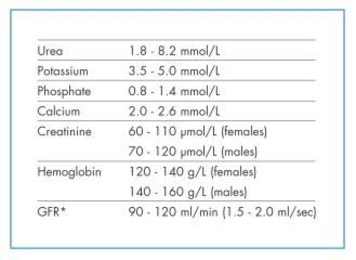
Scroll the table sideways on a smaller screen.
| Laboratory Parameter | SI Units (Standard Range) | Physiological Relevance in Diuretic Therapy |
|---|---|---|
| Serum Urea | 1.8 – 8.2 mmol/L | Marker of nitrogenous retention; fractional excretion altered by loop diuretics |
| Serum Potassium (K+) | 3.5 – 5.0 mmol/L | Critical electrolyte; depleted by loop/thiazides, retained by K+-sparers |
| Serum Phosphate | 0.8 – 1.4 mmol/L | Proximal tubular reabsorption altered by carbonic anhydrase inhibitors |
| Total Calcium (Ca2+) | 2.0 – 2.6 mmol/L | Excreted by loop diuretics (hypocalcemia); spared by thiazides (hypercalcemia) |
| Serum Creatinine (Females) | 60 – 110 μmol/L | Surrogate marker for GFR estimation; rises during diuretic-induced prerenal azotemia |
| Serum Creatinine (Males) | 70 – 120 μmol/L | Elevated baseline reflects higher average skeletal muscle mass |
| Hemoglobin (Females) | 120 – 140 g/L | Monitors hemoconcentration during aggressive volume depletion |
| Hemoglobin (Males) | 140 – 160 g/L | Monitors hemoconcentration during aggressive volume depletion |
| Glomerular Filtration Rate (GFR) | 90 – 120 mL/min (1.5 – 2.0 mL/s) | Defines functional renal capacity; thiazides lose efficacy when GFR <30 mL/min |
Clinical Classification of Daily Urine Output
- Normal Physiological Output: 800 to 2,000 mL/day (reflecting normal homeostatic balancing of fluid intake, insensible perspiration, and respiratory losses).
- Oliguria: 400 to 500 mL/day (<0.5 mL/kg/h in adults). Represents the absolute minimum urinary volume required to excrete the obligatory daily solute load (~600 mOsm/day) at maximal urinary concentration (~1,200 mOsm/L). Output below this threshold causes solute retention and uremia.
- Polyuria: >2,500 to 3,000 mL/day. Observed in diabetes mellitus (solute diuresis), central/nephrogenic diabetes insipidus (water diuresis), primary psychogenic polydipsia, and during the recovery phase of acute tubular necrosis.
- Anuria: <100 mL/day. Indicates complete bilateral urinary tract obstruction, catastrophic renal cortical necrosis, or end-stage vascular collapse.
Nephron sites & classification
Each human kidney contains approximately 1,000,000 functional nephrons. Diuretics are classified according to their primary anatomical site of action along the nephron, their specific membrane transport target, and their maximal natriuretic efficacy.

Segmental Tubular Sodium Handling
- Proximal Convoluted Tubule (PCT): Reabsorbs 65% of all filtered sodium, water, and chloride, alongside 100% of filtered glucose and amino acids, and 85% to 90% of filtered bicarbonate (HCO3−). Diuretics acting here (carbonic anhydrase inhibitors) display weak natriuretic efficacy because downstream nephron segments possess high compensatory reabsorptive capacity.
- Thick Ascending Limb of Henle’s Loop (TAL): Reabsorbs 20% to 25% of filtered sodium via the electroneutral Na+/K+/2Cl− cotransporter. Because the TAL is water-impermeable and downstream segments have limited capacity to reabsorb large delivered solute loads, inhibitors acting here are termed high-ceiling diuretics.
- Distal Convoluted Tubule (DCT): Reabsorbs 5% to 10% of filtered sodium via the electroneutral Na+/Cl− symporter. Inhibitors acting here are termed low-ceiling diuretics (thiazides).
- Cortical Collecting Tubule (CCT): Reabsorbs 2% to 3% of filtered sodium via epithelial sodium channels (ENaC) on principal cells, coupled to potassium (K+) and proton (H+) secretion. Inhibitors acting here function as potassium-sparing diuretics.
Taxonomic Classes of Diuretic Drugs
- 1. High-Ceiling / Loop Diuretics: Furosemide, bumetanide, torsemide, ethacrynic acid. Target: luminal Na+/K+/2Cl− (NKCC2) cotransporter in the thick ascending limb.
- 2. Thiazide & Thiazide-Like Diuretics: Hydrochlorothiazide, chlorothiazide, trichlormethiazide, chlorthalidone, indapamide, metolazone. Target: luminal Na+/Cl− (NCC) symporter in the early distal convoluted tubule.
- 3. Carbonic Anhydrase Inhibitors (CAIs): Acetazolamide, methazolamide, dorzolamide. Target: membrane-bound (CA-IV) and cytoplasmic (CA-II) carbonic anhydrase in the proximal convoluted tubule.
- 4. Potassium-Sparing Diuretics:
- Direct ENaC Blockers: Amiloride, triamterene.
- Mineralocorticoid Receptor Antagonists (MRAs): Spironolactone, eplerenone. Target: late distal tubule and cortical collecting duct principal cells.
- 5. Osmotic Diuretics: Mannitol, isosorbide, hypertonic urea, hypertonic glucose. Target: proximal tubule and thin descending limb of Henle (luminal osmotic water retention).
- 6. Calcium-Sparing Diuretics: Thiazides and potassium-sparing diuretics (reduce urinary Ca2+ clearance).
High-ceiling loop diuretics
Loop diuretics are the most efficacious natriuretic agents available. They produce prompt, profound diuresis and are capable of excreting up to 20% to 25% of the filtered sodium load.
Mechanism of Action
Loop diuretics bind reversibly to the chloride-binding site of the luminal Na+/K+/2Cl− cotransporter (NKCC2) in the medullary and cortical thick ascending limb of Henle’s loop, selectively inhibiting its function:
- Abolition of the Lumen-Positive Transepithelial Potential: Under normal conditions, physiological back-leak of K+ through apical ROMK channels creates a +8 to +10 mV lumen-positive potential that drives paracellular reabsorption of divalent cations: calcium (Ca2+) and magnesium (Mg2+). By halting NKCC2 activity and potassium recycling, loop diuretics abolish this electrical gradient, inducing profound urinary wasting of Ca2+ and Mg2+.
- Disruption of Medullary Hypertonicity: NKCC2 operates as the primary engine of the countercurrent multiplier system. By blocking solute transport into the medullary interstitium, loop diuretics wash out the corticomedullary hyperosmolar gradient. This eliminates the osmotic driving force required for ADH-mediated water reabsorption in the collecting duct, leaving the kidney unable to concentrate or dilute urine.
- Renal Prostaglandin Induction & Hemodynamics: Loop diuretics stimulate renal synthesis of vasodilator prostaglandins (PGE2 and PGI2). This dilates renal afferent arterioles, increases total renal blood flow, redistributes flow to the midcortex, and increases venous capacitance (systemic venodilation). In acute pulmonary edema, intravenous furosemide relieves left ventricular preload and orthopnea within 5 to 15 minutes, long before urinary diuresis begins. Co-administration of non-steroidal anti-inflammatory drugs (NSAIDs) blocks cyclooxygenase (COX), abolishes prostaglandin synthesis, and blunts loop diuretic efficacy.
- Secondary Pharmacological Actions: Loop diuretics increase proximal tubular reabsorption of uric acid (causing hyperuricemia). Furosemide possesses weak carbonic anhydrase inhibitory activity, slightly increasing urinary excretion of HCO3− and titratable phosphate.
Individual Agents & Formulations
- Furosemide (Lasix): The clinical prototype sulfonamide derivative. Oral dosage ranges from 20 to 80 mg once or twice daily (up to 500–1,000 mg/day in severe chronic kidney disease); IV doses range from 20 to 40 mg bolus up to high-dose continuous infusions. Bioavailability is erratic (~50%, range 10–90%). Oral onset is 30 to 60 minutes; duration is 4 to 6 hours (“La-six” = lasts six hours). Half-life is ≈ 1.5 hours (extended in renal or hepatic failure).
- Bumetanide (Bumex): Highly potent sulfonamide loop diuretic (1 mg bumetanide ≈ 40 mg furosemide; 40-fold potency difference). Clinical dosage ranges from 0.5 to 2.0 mg (up to 10–20 mg/day in refractory states). Exhibits high, predictable oral bioavailability (80% to 100%).
- Torsemide (Demadex): Sulfonylurea-derived loop diuretic with superior oral bioavailability (80% to 90%) and a longer terminal elimination half-life (≈ 3.5 hours), providing stable once-daily diuresis (10 to 20 mg/day).
- Ethacrynic Acid (Edecrin): A phenoxyacetic acid derivative containing no sulfonamide moiety. Reserved strictly for patients with documented, severe hypersensitivity/allergy to sulfonamide drugs. Carries the highest risk of irreversible ototoxicity. Clinical dosage: 25 to 50 mg orally.
Therapeutic Indications & Clinical Use
- Acute Pulmonary Edema & Decompensated CHF: Rapid intravenous administration unloads the failing left ventricle through prompt venodilation, followed by profound natriuresis that clears pulmonary vascular congestion.
- Hepatic Cirrhosis & Nephrotic Edema: Mobilizes massive ascites and peripheral edema (usually paired with spironolactone to counter secondary hyperaldosteronism).
- Acute Oliguric Renal Failure: Used in volume-overloaded acute kidney injury to convert an oliguric state to a non-oliguric state, simplifying fluid and electrolyte management (though without changing overall mortality).
- Severe Hypercalcemia: Administered alongside aggressive isotonic saline volume resuscitation to safely accelerate renal calcium excretion.
- Severe Hyperkalemia: Accelerates renal potassium wasting, lowering life-threatening serum potassium levels.
Adverse Reactions & Clinical Toxicities
- Ototoxicity: Manifests as tinnitus, vertigo, and transient or permanent sensorineural hearing loss. Caused by alteration of the electrolyte composition of endolymph via inhibition of NKCC1 cotransporters in the stria vascularis of the inner ear. Risk increases with rapid intravenous bolus injection, high cumulative doses, renal impairment, and concurrent administration of ototoxic drugs (e.g., aminoglycosides like gentamicin, amikacin).
- Electrolyte & Metabolic Derangements:
- Hypokalemic Hypochloremic Metabolic Alkalosis: Massive sodium delivery to the late nephron accelerates K+ and H+ excretion, while extracellular volume contraction triggers aldosterone-mediated hydrogen secretion.
- Hypovolemia & Hypotension: Overly rapid fluid mobilization triggers intravascular volume depletion, orthostatic hypotension, hemoconcentration, and prerenal azotemia.
- Hypomagnesemia & Hypocalcemia: Inability to generate the lumen-positive potential causes progressive divalent cation wasting. Hypomagnesemia can trigger refractory cardiac arrhythmias.
- Hyperuricemia: Volume contraction enhances proximal tubular uric acid reabsorption, which may precipitate acute gouty arthritis.
- Hyperglycemia: Hypokalemia impairs insulin release from pancreatic beta-cells, destabilizing glycemic control in diabetes mellitus.
- Immunologic & Hematologic Reactions: Sulfonamide-associated photosensitivity, skin rashes, urticaria, and rare bone marrow suppression (agranulocytosis, neutropenia, thrombocytopenia).
Thiazide & thiazide-like agents
Thiazide diuretics are sulfonamide derivatives that function as moderate-efficacy, low-ceiling natriuretics. They represent cornerstone first-line agents in managing primary essential hypertension.
Mechanism of Action & Hemodynamics
- Inhibition of the Na+/Cl− Symporter: Thiazides act on the luminal membrane of the early distal convoluted tubule (DCT), competing for the chloride-binding site of the electroneutral Na+/Cl− cotransporter (NCCT/SLC12A3). This inhibits the reabsorption of 5% to 10% of filtered sodium and chloride, producing mild-to-moderate natriuresis.
- Calcium-Sparing Mechanism: Unlike loop diuretics, thiazides decrease urinary calcium excretion, elevating serum calcium. Blocking Na+ entry via NCCT lowers intracellular Na+ in DCT epithelial cells. This accelerates basolateral Na+/Ca2+ exchanger (NCX1) activity, which increases sodium entry in exchange for extruding calcium into the interstitial space. The resulting drop in intracellular Ca2+ enhances apical calcium reabsorption through TRPV5 calcium channels, which are stimulated by parathyroid hormone (PTH).
- Biphasic Antihypertensive Mechanism:
- Short-Term Action (1 to 4 Weeks): The initial drop in blood pressure results from mild natriuresis, which contracts plasma volume and lowers cardiac preload and cardiac output.
- Long-Term Action (>4 to 6 Weeks): Plasma volume and cardiac output normalize due to compensatory activation of the renin-angiotensin-aldosterone system (RAAS). However, blood pressure remains reduced because thiazides induce direct, sustained peripheral vasodilation. This reduction in systemic vascular resistance is mediated by opening calcium-activated potassium channels (KCa) in vascular smooth muscle and desensitizing vascular smooth muscle to circulating vasoconstrictors (norepinephrine, angiotensin II).
Key Agents & Pharmacological Profiles
- Hydrochlorothiazide (HCTZ / Esidrix / HydroDIURIL): The most widely prescribed thiazide. Standard dosage: 12.5 to 25 mg once daily (doses >50 mg increase metabolic adverse effects without adding clinical efficacy). Half-life is 6 to 12 hours.
- Chlorothiazide (Diuril): The original thiazide; the only member of the class available in an intravenous formulation. Low lipid solubility requires higher oral dosing (500 to 1,000 mg/day).
- Trichlormethiazide (Metahydrin): Highly potent thiazide administered at 1 to 4 mg once daily.
- Chlorthalidone (Hygroton): A phthalimidine-derivative thiazide-like diuretic. Possesses an exceptionally long elimination half-life (t1/2 ≈ 40 to 60 hours) due to extensive partitioning into erythrocytes and binding to red cell carbonic anhydrase. Provides sustained 24-hour ambulatory blood pressure reduction and superior cardiovascular event reduction in landmark trials (e.g., ALLHAT). Dosage: 12.5 to 25 mg once daily.
- Indapamide (Lozol): An indolyl-chlorosulfonamide thiazide-like diuretic that undergoes primarily hepatic clearance. Exerts potent direct vasodilatory effects alongside modest natriuresis; considered metabolically neutral regarding glucose and lipid profiles at lower doses (1.25 to 2.5 mg/day).
- Metolazone (Zaroxolyn): A quinazoline-derivative thiazide-like diuretic of exceptional potency. Unlike standard thiazides (which lose efficacy when GFR falls below 30 mL/min), metolazone remains effective in advanced renal failure (GFR <20 mL/min). Frequently combined with loop diuretics to achieve sequential nephron blockade in refractory edema.
Therapeutic Indications
- Primary Essential Hypertension: First-line monotherapy or combination therapy across national and international guidelines.
- Congestive Heart Failure & Mild Edematous States: Used in mild volume expansion, or synergistically with loop diuretics to overcome diuretic resistance.
- Idiopathic Hypercalciuria & Calcium Nephrolithiasis: By lowering urinary calcium excretion, thiazides prevent recurrent calcium oxalate and calcium phosphate kidney stones. Frequently combined with amiloride and allopurinol.
- Nephrogenic Diabetes Insipidus: Exerts a paradoxical antidiuretic effect. By inducing mild extracellular volume depletion, thiazides trigger compensatory proximal tubular reabsorption of sodium and water, reducing solute and water delivery to downstream collecting ducts and decreasing total urine volume by up to 50%.
- Senile & Postmenopausal Osteoporosis: Long-term thiazide therapy preserves bone mineral density and reduces hip fracture incidence through sustained renal calcium retention.
Adverse Reactions & Metabolic Complications
- The “Hyper” Metabolic Complications:
- Hyperglycemia: Inhibits insulin secretion from pancreatic beta-cells (mediated by opening ATP-sensitive K+ channels via hypokalemia) and worsens peripheral insulin resistance, destabilizing glycemic control.
- Hyperuricemia: Competes with uric acid for organic acid transporters (OAT1/OAT3) in the proximal tubule; combined with volume contraction, this elevates plasma uric acid and may precipitate acute gout.
- Hypercalcemia: Enhanced DCT reabsorption can unmask subclinical primary hyperparathyroidism.
- Hyperlipidemia: Causes modest increases (5% to 15%) in total serum cholesterol, low-density lipoproteins (LDL), and triglycerides (often resolving during long-term maintenance).
- The “Hypo” Electrolyte Complications:
- Hypokalemia & Hypomagnesemia: Distal sodium delivery accelerates potassium and magnesium excretion, predisposing patients to muscle cramps, fatigue, and ventricular dysrhythmias (especially in patients taking cardiac glycosides like digoxin).
- Hyponatremia: Impaired diluting capacity in the DCT combined with volume-stimulated ADH secretion can induce severe, symptomatic dilutional hyponatremia.
- Neurological, Urogenital & Cutaneous Effects: Dizziness, headache, paresthesias, erectile dysfunction / loss of libido (a common cause of non-compliance in males), allergic skin eruptions, urticaria, and sulfonamide photosensitivity.
Carbonic anhydrase inhibitors
Carbonic anhydrase inhibitors (CAIs) are non-bacteriostatic sulfonamide derivatives that target bicarbonate reabsorption in the proximal nephron. They function as weak diuretics with specialized clinical utility in ophthalmology, neurology, and high-altitude medicine.
Biochemical Mechanism in the Proximal Convoluted Tubule
Under physiological conditions, filtered Na+ is reabsorbed across the apical PCT membrane by the Na+/H+ exchanger (NHE3), which secretes an intracellular proton (H+) into the lumen. In the tubular lumen, secreted H+ combines with filtered HCO3− to form carbonic acid (H2CO3):
- Enzymatic Dehydration: Membrane-bound carbonic anhydrase type IV (CA-IV) anchored to the apical brush border rapidly dehydrates luminal carbonic acid into carbon dioxide and water:
H+ + HCO3− ↔ H2CO3 → CO2 + H2O. - Intracellular Rehydration: Lipophilic CO2 diffuses passively across the apical membrane into the PCT epithelial cell. Cytoplasmic carbonic anhydrase type II (CA-II) rehydrates CO2 back into carbonic acid, which spontaneously dissociates into H+ and HCO3−:
CO2 + H2O → H2CO3 ↔ H+ + HCO3−. - Basolateral Extrusion: Intracellular H+ is recycled back to the luminal fluid via NHE3, while intracellular HCO3− is transported into the interstitial blood alongside Na+ via the electrogenic Na+/HCO3− cotransporter (NBCe1).
- Consequences of CA Inhibition: Drugs like acetazolamide inhibit both CA-IV and CA-II, halting luminal dehydration and intracellular rehydration. This prevents the reabsorption of up to 85% of filtered bicarbonate. Sodium reabsorption via NHE3 is indirectly blunted due to intracellular H+ depletion.
Self-Limiting Diuretic Action & Urinary Alkalinization
CAIs produce a prompt, marked increase in urinary excretion of HCO3−, Na+, and K+, driving the urine pH up to alkaline levels (urine pH ≈ 8.0). However, their natriuretic efficacy is self-limiting: as plasma bicarbonate drops and systemic hyperchloremic metabolic acidosis develops, the filtered load of bicarbonate falls, leaving less substrate for the drug to block. Diuretic tolerance typically develops within 48 to 72 hours of continuous therapy.
Clinical Indications
- Glaucoma: Ciliary body epithelial cells utilize carbonic anhydrase to produce bicarbonate-rich aqueous humor. Oral acetazolamide, oral methazolamide, or topical agents (dorzolamide, brinzolamide) inhibit ciliary CA-II, reducing aqueous humor secretion and lowering intraocular pressure in open-angle and secondary glaucomas. Used preoperatively with topical miotics to manage acute angle-closure crises.
- Acute Mountain Sickness (High-Altitude Prophylaxis): Rapid ascent above 3,000 meters causes hypobaric hypoxia, which stimulates peripheral chemoreceptor-driven hyperventilation. The resulting respiratory alkalosis depresses central medullary chemoreceptors, triggering periodic Cheyne-Stokes breathing and hypoxemia. Acetazolamide induces mild renal bicarbonate wasting and metabolic acidosis, acidifying the cerebrospinal fluid. This stimulates central respiratory drive, prevents nocturnal hypoxemia, and accelerates acclimatization.
- Urinary Alkalinization: By elevating urine pH to ≥7.5, acetazolamide increases the ionization and solubility of weakly acidic compounds. It accelerates renal clearance of salicylates in acute aspirin toxicity and prevents renal tubular precipitation of cystine and uric acid stones.
- Epilepsy & Idiopathic Intracranial Hypertension (Pseudotumor Cerebri): CAIs reduce cerebrospinal fluid (CSF) secretion by the choroid plexus, lowering intracranial pressure in pseudotumor cerebri. In epilepsy, local metabolic acidosis suppresses abnormal neuronal firing in catamenial and absence seizures.
Adverse Reactions & Clinical Complications
- Hyperchloremic Normal Anion Gap Metabolic Acidosis: Caused by continuous urinary loss of bicarbonate buffer.
- Hypokalemia: Increased luminal sodium and non-reabsorbable bicarbonate delivery to the cortical collecting tubule elevates negative luminal potential, driving potassium excretion.
- Calcium Phosphate Nephrolithiasis: Calcium salts precipitate in an alkaline urinary environment, while metabolic acidosis promotes tubular reabsorption of citrate (a natural inhibitor of calcium stone formation), causing hypocitraturia.
- Hepatic Encephalopathy Precipitation: Urinary alkalinization converts charged ammonium (NH4+) back into lipid-soluble, uncharged ammonia (NH3), which is reabsorbed into the circulation. In patients with severe hepatic cirrhosis, this can trigger acute hyperammonemic coma.
- Neurological Effects: Drowsiness, fatigue, anorexia, perioral and acral paresthesias (“pins and needles” in fingers and toes), and altered taste sensation (carbonated beverages taste flat due to inhibition of salivary and lingual carbonic anhydrase).
Potassium-sparing diuretics
Potassium-sparing diuretics act on the principal and intercalated cells of the late distal convoluted tubule and the cortical collecting tubule (CCT). While possessing modest natriuretic efficacy (blocking only 2% to 3% of filtered sodium), their critical clinical value lies in their ability to prevent or reverse hypokalemia induced by loop and thiazide diuretics.
1. Direct ENaC Channel Inhibitors: Amiloride & Triamterene
- Mechanism of Action: Weak organic bases that act from the luminal tubular fluid to directly block the pore of epithelial sodium channels (ENaC) in the apical membrane of principal cells, independent of aldosterone:
- Under normal conditions, electrogenic sodium entry via ENaC leaves chloride behind in the lumen, generating a −10 to −20 mV lumen-negative transepithelial potential.
- This negative luminal potential drives potassium secretion through apical ROMK channels and proton secretion through α-intercalated cell H+-ATPase pumps.
- By blocking ENaC, amiloride and triamterene abolish the lumen-negative charge, eliminating the electrical driving force for potassium and hydrogen secretion.
- Specific Clinical Indications:
- Liddle’s Syndrome (Pseudo-Hyperaldosteronism): An autosomal dominant disorder caused by gain-of-function mutations in ENaC β- or γ-subunits that prevent channel ubiquitination and degradation. Channels remain constitutively active, causing severe hypertension, hypokalemia, and metabolic alkalosis with suppressed aldosterone and renin. Amiloride directly blocks these mutated channels, serving as the definitive targeted therapy (whereas aldosterone antagonists are ineffective).
- Lithium-Induced Nephrogenic Diabetes Insipidus: Lithium enters principal cells via ENaC, accumulating intracellularly and uncoupling V2 receptors from adenylyl cyclase. Amiloride competitively blocks lithium entry through ENaC, preventing and treating lithium nephrotoxicity.
- Aerosolized Amiloride in Cystic Fibrosis: Investigated to block airway epithelial ENaC, preserving surface liquid hydration in bronchial secretions.
2. Mineralocorticoid Receptor Antagonists (MRAs): Spironolactone & Eplerenone
- Mechanism of Action: Aldosterone diffuses across the basolateral membrane of principal cells, binding to cytoplasmic mineralocorticoid receptors (MR). The ligand-receptor complex translocates into the nucleus, binding hormone response elements to induce the transcription of Aldosterone-Induced Proteins (AIPs). AIPs upregulate apical ENaC expression, increase open channel probability, and accelerate basolateral Na+/K+-ATPase synthesis. Spironolactone and eplerenone act as competitive antagonists at intracellular mineralocorticoid receptors, preventing AIP transcription.
- Spironolactone (Aldactone): A non-selective synthetic steroid antagonist. Extensively metabolized in the liver to active metabolites, primarily canrenone (which contributes to its prolonged 16- to 24-hour biological duration). Standard dosage: 25 to 100 mg/day (up to 200–400 mg/day in hepatic ascites).
Endocrine Side Effects: Non-specifically binds and blocks androgen receptors and inhibits 17α-hydroxylase, while binding progesterone receptors. In males, this produces dose-dependent painful gynecomastia, erectile dysfunction, and decreased libido; in females, it causes menstrual irregularities, amenorrhea, and breast tenderness. - Eplerenone (Inspra): A second-generation spironolactone derivative containing an epoxy bridge. Highly selective for the mineralocorticoid receptor, displaying >100-fold lower affinity for androgen and progesterone receptors. Eliminates anti-androgenic side effects (gynecomastia rate <1%).
- Cardiovascular Mortality Benefit: Aldosterone promotes myocardial hypertrophy, coronary endothelial dysfunction, and perivascular myocardial fibrosis. In landmark clinical trials (RALES with spironolactone; EPHESUS and EMPHASIS-HF with eplerenone), low-dose MRA therapy (12.5 to 50 mg/day) reduced all-cause mortality and heart failure hospitalizations by up to 30% in patients with heart failure with reduced ejection fraction (HFrEF).
Osmotic diuretics (mannitol)
Osmotic diuretics are low-molecular-weight, pharmacologically inert compounds that are freely filtered at the glomerulus, undergo negligible tubular reabsorption, and are excreted intact into the urine.
Mechanism of Action
The clinical prototype is mannitol (Osmitrol / Resectisol), an uncharged six-carbon sugar alcohol:
- Luminal Osmotic Water Retention: Filtered mannitol remains trapped within the tubular lumen, raising the osmolarity of the tubular fluid. In segments that are freely permeable to water—primarily the proximal convoluted tubule and the thin descending limb of Henle’s loop—mannitol exerts an opposing osmotic force that prevents water from following reabsorbed sodium into peritubular capillaries.
- Sodium Dilution & Back-Diffusion: Retained luminal water dilutes the luminal sodium concentration. When luminal Na+ drops below interstitial levels, passive paracellular sodium reabsorption ceases, and sodium may leak back into the lumen down its concentration gradient, producing secondary natriuresis.
- Medullary Washout: Intravenous mannitol expands intravascular volume, increasing renal blood flow and dilating peritubular capillaries and vasa recta. This washes out the hypertonic medullary solute gradient, reducing the concentrating capacity of the loop of Henle.
Clinical Indications
- Acute Intracranial Pressure (ICP) Reduction: The primary indication. Mannitol does not cross an intact blood-brain barrier. Intravenous administration rapidly elevates plasma osmolarity, creating an osmotic gradient that extracts water from parenchymal brain tissue into the cerebral microcirculation, lowering ICP and brain mass in acute cerebral edema, traumatic brain injury, and prior to neurosurgery.
- Acute Intraocular Pressure (IOP) Reduction: Rapidly lowers IOP in acute refractory or angle-closure glaucoma before definitive surgical iridectomy.
- Maintenance of Urine Flow in Acute Renal Insufficiency: Used during the early oliguric phase of acute tubular necrosis (ATN), following severe crush injury (rhabdomyolysis), or during complex aortic aneurysm surgeries to flush out cellular debris, myoglobin casts, and hemoglobin casts.
- Forced Diuresis: Accelerates the renal clearance of dialyzable, water-soluble toxins and overdosed drugs.
Adverse Effects & Clinical Hazards
- Transient Intravascular Volume Expansion: Before producing diuresis, hypertonic mannitol rapidly draws water from the intracellular fluid into the intravascular compartment. In patients with compromised cardiac reserve, this volume expansion can precipitate acute pulmonary edema and decompensated heart failure.
- Dehydration & Hypernatremia: Excessive free water diuresis without adequate electrolyte replacement causes dehydration and severe hypernatremia.
- Osmotic Nephrosis (Acute Kidney Injury): High cumulative doses of mannitol (>200 g/day or serum osmolar gap >55 mOsm/kg) can cause intracellular vacuolation of proximal tubular cells and acute oliguric renal failure.
- Injection Site Complications: Extravasation causes local tissue necrosis, and peripheral administration can trigger thrombophlebitis. Manifests systemically with headache, chills, fever, and nausea.
Electrolyte & acid-base dynamics
Mastering the comparative urinary electrolyte excretion patterns and systemic acid-base derangements induced by each diuretic class is essential for safe prescribing and clinical problem-solving.
Scroll the table sideways on a smaller screen.
| Diuretic Class & Prototypes | Nephron Site & Primary Molecular Target | Maximal FeNa (%) | Urinary Electrolyte Impact (Na+, K+, Cl−, HCO3−) | Divalent Cation Impact (Ca2+, Mg2+) | Systemic Acid-Base Consequence |
|---|---|---|---|---|---|
| Carbonic Anhydrase Inhibitors (Acetazolamide) |
Proximal Convoluted Tubule • Apical CA-IV & Cytosolic CA-II |
≈ 2% – 4% (Weak, self-limiting) |
• Na+: ↑ • K+: ↑↑ • Cl−: ↓ (retained) • HCO3−: ↑↑↑ (alkaline urine) |
• Ca2+: No direct effect (promotes calcium phosphate stone precipitation) • Mg2+: Negligible |
Hyperchloremic Normal Anion Gap Metabolic Acidosis |
| High-Ceiling Loop Diuretics (Furosemide, Bumetanide) |
Thick Ascending Limb • Luminal Na+/K+/2Cl− (NKCC2) |
≈ 20% – 25% (High ceiling) |
• Na+: ↑↑↑ • K+: ↑↑ • Cl−: ↑↑↑ • HCO3−: ↑ (weak CAI action) |
• Ca2+: ↑↑ Wasting (hypocalcemia) • Mg2+: ↑↑ Wasting (hypomagnesemia) |
Hypokalemic Hypochloremic Metabolic Alkalosis (Contraction alkalosis) |
| Thiazide Diuretics (Hydrochlorothiazide, Chlorthalidone) |
Early Distal Convoluted Tubule • Luminal Na+/Cl− (NCCT) |
≈ 5% – 10% (Low ceiling) |
• Na+: ↑↑ • K+: ↑↑ • Cl−: ↑↑ • HCO3−: ↔ / slight ↑ |
• Ca2+: ↓↓ Excretion (calcium-sparing / hypercalcemia) • Mg2+: ↑ Wasting |
Hypokalemic Hypochloremic Metabolic Alkalosis |
| Potassium-Sparing: ENaC Blockers (Amiloride, Triamterene) |
Late Distal & Cortical Collecting Tubule • Apical Epithelial Na+ Channel (ENaC) |
≈ 2% – 3% (Mild natriuresis) |
• Na+: ↑ • K+: ↓↓ Sparing • Cl−: ↑ • HCO3−: ↓ (acid retention) |
• Ca2+: ↓ Excretion (mild sparing) • Mg2+: ↓ Excretion (spares magnesium) |
Hyperkalemic Normal Anion Gap Metabolic Acidosis |
| Potassium-Sparing: MRAs (Spironolactone, Eplerenone) |
Late Distal & Cortical Collecting Tubule • Intracellular Mineralocorticoid Receptor |
≈ 2% – 3% (Aldosterone-dependent) |
• Na+: ↑ • K+: ↓↓ Sparing • Cl−: ↑ • HCO3−: ↓ (acid retention) |
• Ca2+: ↔ / mild sparing • Mg2+: ↓ Excretion (spares magnesium) |
Hyperkalemic Normal Anion Gap Metabolic Acidosis |
| Osmotic Diuretics (Mannitol) |
PCT & Thin Descending Limb of Henle • Luminal Osmotic Particle Trapping |
Variable / High (Water diuresis > natriuresis) |
• Na+: ↑ (dilutional washout) • K+: ↑ • Cl−: ↑ • Water: ↑↑↑↑ |
• Ca2+: ↑ Wasting • Mg2+: ↑ Wasting |
Variable (Initial hypervolemic hyponatremia transitioning to hypernatremic dehydration) |
Doping & forensic toxicology
Diuretics are explicitly banned both in-competition and out-of-competition by the World Anti-Doping Agency (WADA) under Category S5 (Diuretics and Other Masking Agents). Their misuse in athletic contexts falls into two distinct categories:
1. The Masking Agent Mechanism
Athletes who ingest performance-enhancing substances—such as anabolic-androgenic steroids (e.g., stanozolol, nandrolone), recombinant human erythropoietin (EPO), or stimulants—consume loop or thiazide diuretics immediately before mandatory drug testing. The resulting rapid expansion of urinary output produces dilution of urinary solute concentration:
- Massive free water and sodium excretion lowers the specific gravity of the urine and dilutes banned xenobiotics and their metabolic breakdown products below established liquid chromatography-mass spectrometry (LC-MS) detection thresholds.
- Alkalinizing diuretics like acetazolamide accelerate the clearance of acidic doping agents, shortening their systemic half-lives.
- To counteract this manipulation, WADA testing protocols automatically flag urine samples with abnormally low specific gravity (<1.005) or dilute urinary creatinine, requiring re-testing.
2. Rapid Acute Weight Reduction
In weight-category sports (boxing, wrestling, judo, mixed martial arts, weightlifting, and horse racing jockeys), athletes use diuretics to shed 2 to 5 kg of body mass within 12 to 24 hours to qualify for a lower weight division (“making weight”):
- This rapid weight loss reflects acute extracellular fluid depletion rather than reduction of adipose tissue.
- Following the official pre-competition weigh-in, athletes aggressively rehydrate with oral fluids and electrolytes before competing.
- Severe Physiological Dangers: Rapid dehydration diminishes stroke volume, reduces cardiac output, impairs thermoregulatory sweating (accelerating exertional heat stroke), precipitates severe muscle cramping, and increases the risk of intracranial hemorrhage due to volume-depleted brain shrinkage tearing bridging subdural veins.
Clinical trials & recent advances
Recent high-impact clinical trials have identified novel applications for diuretic agents outside traditional nephrology and challenged established diagnostic algorithms in intensive care medicine.
1. Diuretic Efficacy in Obstructive Sleep Apnea (OSA)
A propensity score-matched cohort analysis by B. Revol et al. (2020) investigated the therapeutic impact of diuretic administration on the severity of obstructive sleep apnea:
- Pathophysiological Rationale: In patients with fluid retention (hypertension, mild heart failure), shifting from an upright posture to a recumbent position during sleep causes gravity-dependent lower-extremity interstitial fluid to migrate cephalad (rostral fluid shift). This fluid accumulates in the peripharyngeal soft tissues of the neck, increasing upper airway resistance, narrowing the pharyngeal lumen, and worsening airway collapse.
- Key Trial Results: The study demonstrated that diuretic therapy significantly reduced the apnea-hypopnea index (AHI) in overweight or moderately obese patients (p = 0.03) and in hypertensive patients (p < 0.01), particularly those with a body mass index (BMI) between 25 and 35 kg/m2 (p < 0.01).
- Clinical Implication: Diuretics had no significant impact on OSA severity in patients with self-reported low physical activity or severe, refractory heart failure. However, they offer a valuable adjunctive therapeutic option to reduce airway collapsibility in hypertensive patients with mild-to-moderate obesity and sleep apnea.
2. Loop Diuretic Impact on Fractional Excretion of Urea (FEUrea) in Acute Kidney Injury
A prospective cohort study by Zachary L. Cox et al. (2020) examined the diagnostic reliability of the fractional excretion of urea in patients presenting with decompensated heart failure and acute kidney injury (AKI):
- Clinical Background: Fractional excretion of sodium (FENa <1%) is the classical metric used to distinguish prerenal azotemia from intrinsic acute tubular necrosis (ATN). However, when patients receive loop diuretics, pharmacological natriuresis elevates FENa >1% even in the presence of true prerenal hypoperfusion, rendering FENa unreliable. Clinicians have traditionally substituted the fractional excretion of urea (FEUrea <35% = prerenal), assuming that urea handling is unaffected by loop diuretics because urea reabsorption occurs primarily in the proximal tubule and collecting duct.
- Trial Findings: Following administration of loop diuretics (median dose 80 mg furosemide equivalents, range 40–160 mg), mean baseline FEUrea was 35.2% ± 10.5% and increased significantly by a mean of 5.6% ± 10.5% (p < 0.001).
- Clinical Impact: Loop diuretics significantly alter FEUrea. The magnitude of change is highly variable and can cause false reclassification of prerenal AKI as intrinsic renal injury. Clinicians cannot assume that FEUrea remains an unconfounded diagnostic tool in patients actively receiving loop diuretic therapy.
Clinical practice & guidelines
Uganda Clinical Guidelines (UCG 2023) Therapeutic Recommendations
- Hypertension Management: Thiazide-like and thiazide diuretics remain first-line options for primary hypertension. Under the UCG protocols:
- Step 1 Monotherapy: Recommended in black African patients (who predominantly display low-renin, volume-sensitive hypertension) as either a calcium channel blocker (CCB, e.g., amlodipine 5–10 mg daily) or a thiazide/thiazide-like diuretic (e.g., hydrochlorothiazide 12.5–25 mg daily or bendroflumethiazide 2.5 mg daily).
- Step 2 Combination Therapy: A thiazide diuretic is combined with a CCB or an angiotensin-converting enzyme inhibitor (ACEI, e.g., enalapril 5–20 mg daily) to provide complementary vasodilation and volume reduction while blunting reactive RAAS activation.
- Heart Failure Management (HFrEF):
- Congestion Relief: Intravenous or oral furosemide (20 to 80 mg daily) is titrated to relieve pulmonary rales, elevated jugular venous pressure (JVP), and peripheral pitting edema. Once euvolemia is attained, the dose is titrated to the minimum effective maintenance level to avoid neurohormonal activation.
- Disease Modification & Mortality Reduction: Low-dose spironolactone (25 mg daily) is added for all patients with symptomatic NYHA Class II–IV HFrEF to suppress myocardial fibrosis and reduce mortality, provided serum potassium is <5.0 mmol/L and estimated GFR is >30 mL/min.
Quick recap
References
The following authoritative medical textbooks, national guidelines, and peer-reviewed studies support this curriculum study note:
- Katzung’s Basic & Clinical Pharmacology: Katzung, B.G. and Vanderah, T.W. (2021) Basic & Clinical Pharmacology. 15th edn. New York: McGraw-Hill Education. [Chapter 15: Diuretic Agents].
- Goodman & Gilman’s The Pharmacological Basis of Therapeutics: Brunton, L.L., Hilal-Dandan, R. and Knollmann, B.C. (2018) Goodman & Gilman’s The Pharmacological Basis of Therapeutics. 13th edn. New York: McGraw-Hill Education. [Chapter 25: Regulation of Renal Function and Vascular Volume].
- Guyton and Hall Textbook of Medical Physiology: Hall, J.E. and Hall, M.E. (2021) Guyton and Hall Textbook of Medical Physiology. 14th edn. Philadelphia: Elsevier. [Chapter 28: Urine Formation by the Kidneys: II. Tubular Processing of the Glomerular Filtrate; Chapter 32: Diuretics and Kidney Diseases].
- Essential of Medical Pharmacology (Tripathi): Tripathi, K.D. (2019) Essentials of Medical Pharmacology. 8th edn. New Delhi: Jaypee Brothers Medical Publishers. [Chapter 41: Diuretics and Antidiuretics].
- Uganda Clinical Guidelines: Ministry of Health Uganda (2023) Uganda Clinical Guidelines: National Guidelines for the Management of Common Conditions. Kampala: Ministry of Health. [Section 6: Cardiovascular Conditions – Hypertension and Heart Failure Protocols].
- Obstructive Sleep Apnea Diuretic Trial: Revol, B., Jullian-Desayes, I., Tamisier, R., et al. (2020) ‘Who may benefit from diuretics in OSA? A propensity score-matched observational study’, Thorax, 75(9), pp. 798–801.
- Fractional Excretion of Urea Study: Cox, Z.L., Rao, V.S., Ivey-Miranda, J.B., et al. (2020) ‘Effect of loop diuretics on the fractional excretion of urea in decompensated heart failure’, Journal of the American Heart Association, 9(19), e017059.