Doctors Revision

Physiology

The steeplechase exam in Human Anatomy practical 1 2048
Physiology

Anatomy steeplechase questions pdf

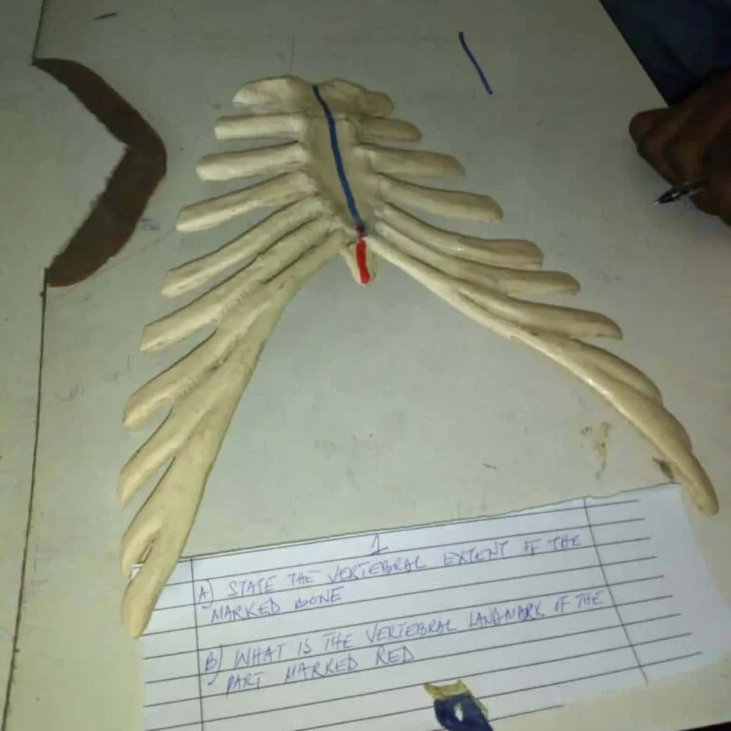
Anatomy Steeplechase Anatomy Steeplechase: Embryology, Histology & Limbs Anatomy Steeplechase Embryology, Histology, Upper & Lower Limb Exam Rules: Be Specific: Don’t just identify the bone; identify the landmark. Side Matters: In a real exam, always specify Left/Right. Clinical Correlation: Think about nerve supplies and injuries. Start Spotter Exam Station 1 Histology Check Answer Next Station Steeplechase Completed! Great job, . Score 0/0 Accuracy 0% Download PDF Report (with Images) Processing images and generating PDF…

Respiratory Function Tests
Physiology

Respiratory Function Tests

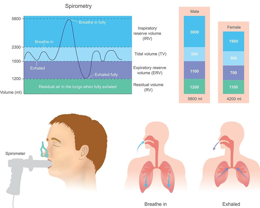
Respiratory Function Tests Respiratory Function Tests Respiratory Function Tests (RFTs), or lung function tests, are painless breathing evaluations measuring how well your lungs take in air, move it in and out, and transfer oxygen to blood, using tools like spirometry (how fast you breathe out) and plethysmography (total lung capacity in a booth) to diagnose breathing issues, monitor lung diseases, and assess lung health before surgery. These tests provide crucial data for managing asthma, COPD, and other respiratory conditions. Overall Objective To understand the principles, methodologies, and clinical significance of various tests used to assess pulmonary function, differentiate between obstructive and restrictive lung diseases, and monitor disease progression. Objective 1: Describe the principles and interpretation of spirometry, including FEV1, FVC, and FEV1/FVC ratio. Spirometry is the most common and fundamental pulmonary function test. It measures how much air a person can inhale and exhale, and how quickly they can exhale it. It’s an indispensable tool for diagnosing and managing a wide range of respiratory conditions. A. Spirometry Basics Definition and Purpose Definition: Spirometry is a simple, non-invasive test that measures the volume and flow of air that can be inhaled and exhaled. Purpose: Diagnose respiratory diseases (e.g., asthma, COPD). Monitor disease progression and response to treatment. Assess severity of lung impairment. Evaluate disability for legal or insurance purposes. Pre-operative assessment of respiratory risk. How Spirometry is Performed (Forced Exhalation Maneuver) The patient takes the deepest breath possible (maximal inspiration) to reach Total Lung Capacity (TLC). Then, they forcefully and rapidly exhale all the air they can, for as long as they can (at least 6 seconds, or until no more air can be exhaled) into a mouthpiece connected to a spirometer. It requires good patient cooperation and effort to obtain reliable and reproducible results. Parameters Measured Spirometry primarily measures two key volumes, from which a crucial ratio is derived: FVC Forced Vital Capacity (Total Volume Exhaled) Definition: The total volume of air exhaled during a maximal forced expiration, starting from a maximal inspiration. Represents: Total “usable” air. Reflects overall size/elasticity of lungs and chest wall. Normal Value: ~4-6 liters (varies by age/height). FEV1 Volume in 1st Second Definition: The volume of air exhaled in the first second of the FVC maneuver. Represents: Speed/ease of expulsion. Indicator of airway patency/resistance. Normal Value: 75-85% of FVC. FEV1/FVC Ratio The Critical Ratio Definition: Ratio of FEV1 to FVC, expressed as percentage. Represents: Most important parameter for differentiating obstructive vs. restrictive disease. Normal Value: ≥ 70-75% (or ratio ≥ 0.70-0.75). Flow-Volume Loops Description: A graphical representation generated during spirometry that plots instantaneous expiratory flow rate (y-axis) against lung volume (x-axis). Normal Loop: A rapid rise to peak expiratory flow, followed by a linear decrease in flow as lung volume decreases, forming a triangular or “sail-like” shape. Inspiratory limb is a smooth, concave curve. Obstructive Pattern: Characterized by a “scooped-out” or concave shape of the expiratory limb, reflecting significant airflow limitation. Peak flow may be reduced. Restrictive Pattern: Characterized by a “witch’s hat” appearance – smaller loop overall (reduced FVC) but with a relatively normal, preserved flow rate shape (proportional but scaled down). Fixed Airway Obstruction: Both inspiratory and expiratory limbs are flattened. B. Interpretation of Spirometry Results Interpretation involves comparing measured values to predicted normal values (based on age, sex, height, ethnicity). Normal Ratio: ≥ 70-75% FEV1: ≥ 80% predicted FVC: ≥ 80% predicted Suggests healthy lung function. Obstructive Example: COPD, Asthma Ratio: < 70-75% (KEY DIAGNOSTIC) FEV1: Reduced (< 80%) FVC: Normal or slightly reduced Loop: “Scooped-out” Increased resistance makes exhalation difficult. Restrictive Example: Fibrosis, Scoliosis Ratio: Normal/Increased (≥ 70%) FEV1: Reduced (< 80%) FVC: Reduced (< 80%) Loop: “Witch’s hat” (small) Reduced compliance/volume; both reduced proportionally. Severity Grading (e.g., COPD GOLD) Based on FEV1 % predicted: Mild: ≥ 80% Moderate: 50-79% Severe: 30-49% Very Severe: < 30% Bronchodilator Reversibility Purpose: Differentiate Asthma vs. COPD. Significant Reversibility: Increase in FEV1/FVC of >12% AND >200 mL. Asthma: Typically significant reversibility. COPD: Often less pronounced/consistent; obstruction is largely fixed. Objective 2: Understand additional lung volumes and capacities measured by methods other than spirometry, particularly Residual Volume (RV) and Total Lung Capacity (TLC). While spirometry is excellent for measuring dynamic lung function (how much air can be quickly moved), it has limitations. Specifically, it cannot measure volumes of air that cannot be exhaled from the lungs. This necessitates other techniques to determine the complete picture of lung volumes. A. Limitations of Spirometry Spirometry directly measures vital capacity (VC or FVC) and its components (IRV, TV, ERV). However, it cannot measure: Residual Volume (RV): The volume of air remaining in the lungs after a maximal forced expiration. Functional Residual Capacity (FRC): The volume of air remaining in the lungs after a normal tidal expiration (ERV + RV). Total Lung Capacity (TLC): The total volume of air in the lungs after a maximal inspiration (VC + RV, or FRC + IC). These volumes are essential for diagnosing and characterizing certain lung conditions, particularly restrictive lung diseases (where TLC is reduced) and obstructive diseases with air trapping (where RV and TLC might be increased). B. Methods for Measuring RV, FRC, and TLC Since RV, FRC, and TLC all include the residual volume, which cannot be exhaled, specialized techniques are required to measure them. 1. Helium Dilution Method Principle: Inert gas dilution. If a known quantity of a tracer gas (helium, insoluble in blood) is introduced into a closed system, it distributes throughout the available lung volume until equilibrium is reached. The extent of dilution is used to calculate the unknown volume. Procedure: Patient connected to spirometer with known volume/concentration of helium (C1). Patient breathes normally (tidal breathing) from closed system. Exhales to FRC, then circuit opens. Helium mixes with air in lungs (FRC volume). Breathing continues until equilibrium is reached (C2). Usually 5-7 minutes. Patient performs maximal expiration (ERV) and maximal inspiration (VC). Calculation: (Vspirometer * C1) = (Vspirometer + FRC) * C2 FRC = ( (Vspirometer * C1) / C2 ) – Vspirometer Once FRC is known:

Control of Respiration (Neural and Chemical Regulation)
Physiology

Control of Respiration (Neural and Chemical Regulation)

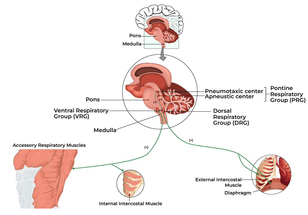
Control of Respiration: Neural and Chemical Regulation Control of Respiration (Neural and Chemical Regulation) Respiration is controlled by both the involuntary and voluntary nervous systems. Involuntary control, which governs automatic breathing, is managed by the respiratory centers in the brainstem (medulla oblongata and pons), which respond to blood levels of oxygen, carbon dioxide, and pH. Voluntary control comes from the cerebral cortex and allows you to control your breathing for activities like speaking or holding your breath. Overall Objective: To understand the neural pathways and chemical factors that regulate the rate and depth of breathing, ensuring appropriate gas exchange to meet metabolic demands and maintain blood gas homeostasis. Objective 1: Identify and describe the key neural control centers for respiration in the brainstem. The control of breathing is a complex process involving both voluntary and involuntary mechanisms. The involuntary, rhythmic control of breathing primarily originates in the brainstem, specifically in the medulla oblongata and the pons. These areas contain specialized groups of neurons that generate and modulate the respiratory rhythm. I. Medullary Respiratory Centers The medulla oblongata houses the most crucial respiratory control centers, responsible for setting the basic rhythm of breathing. These are broadly divided into two main groups: the Dorsal Respiratory Group (DRG) and the Ventral Respiratory Group (VRG). A. Dorsal Respiratory Group (DRG) Location: Located in the posterior portion of the medulla, near the nucleus of the tractus solitarius. Primary Function: The DRG is the most fundamental and active group involved in controlling the basic rhythm of breathing, especially during quiet (eupneic) respiration. It primarily controls inspiration. Neuronal Activity: Contains inspiratory neurons that fire rhythmically. These neurons generate a ramp-like signal: they start weakly and increase in intensity over approximately 2 seconds, then abruptly cease for about 3 seconds, allowing for elastic recoil and exhalation. This gradual increase helps to ensure a smooth, progressive filling of the lungs. Innervation: Sends efferent (motor) signals via the phrenic nerves to the diaphragm. Sends signals via the intercostal nerves to the external intercostal muscles. Activation of these muscles causes the diaphragm to contract and flatten, and the rib cage to expand, leading to inspiration. Afferent Input: Receives sensory input (afferent signals) from: Peripheral chemoreceptors: via glossopharyngeal (CN IX) and vagus (CN X) nerves, detecting changes in PO2, PCO2, and pH. Lung receptors: via vagus (CN X) nerve, detecting stretch and irritation in the lungs and airways. This sensory input allows the DRG to modify the basic respiratory rhythm in response to physiological demands. B. Ventral Respiratory Group (VRG) Location: Located in the anterior portion of the medulla, extending from the brainstem to the upper spinal cord, including the pre-Bötzinger complex. Primary Function: The VRG is largely inactive during quiet breathing. It becomes active and crucial for generating the respiratory rhythm only when there is an increased ventilatory demand, such as during forceful (active) inspiration and expiration. Neuronal Activity: Contains both inspiratory and expiratory neurons. Inspiratory neurons: When stimulated (e.g., by intense DRG signals or strong chemoreceptor input), they send signals to accessory muscles of inspiration (e.g., sternocleidomastoid, scalenes). Expiratory neurons: When stimulated, they send signals to the internal intercostals and abdominal muscles, which are primarily active during forceful exhalation. Rhythm Generation (Pre-Bötzinger Complex): Current research suggests that a small area within the VRG, known as the pre-Bötzinger complex, is the primary site responsible for generating the basic respiratory rhythm. It acts as the pacemaker for breathing, relaying signals to the DRG. Innervation: Inspiratory neurons: Innervate accessory muscles of inspiration. Expiratory neurons: Innervate internal intercostal and abdominal muscles (for active expiration). Role in Forced Breathing: During exercise or respiratory distress, the DRG activates the VRG. The VRG then significantly increases the strength of both inspiratory and expiratory signals, leading to a deeper and more rapid breathing pattern. II. Pontine Respiratory Centers (Pontine Respiratory Group – PRG) The pons contains centers that modify and fine-tune the activity of the medullary respiratory centers, ensuring smooth transitions between inspiration and expiration. These are often collectively referred to as the Pontine Respiratory Group (PRG) and include the Pneumotaxic and Apneustic centers. A. Pneumotaxic Center Upper Pons (Nucleus Parabrachialis) Primary Function: Primarily acts to limit inspiration and fine-tune the respiratory rate. It essentially “switches off” the inspiratory ramp signal from the DRG. Effect: By shortening the inspiratory phase, it leads to: Decreased tidal volume (shallower breaths). Increased respiratory rate. Analogy: Think of it as an “off switch” or a “brake” for inspiration. A strong pneumotaxic signal reduces the duration of inspiration. Clinical Significance: Damage to this center can lead to prolonged inspiration and decreased respiratory rate. B. Apneustic Center Lower Pons Primary Function: Has an excitatory effect on the medullary inspiratory neurons, particularly the DRG. It essentially prolongs inspiration. Effect: If unopposed by the pneumotaxic center, it would lead to: Prolonged, gasping inspirations followed by brief, insufficient expirations (a breathing pattern called apneusis). Interaction with Pneumotaxic Center: Normally, the pneumotaxic center overrides the apneustic center, preventing prolonged inspiration and ensuring rhythmic breathing. Clinical Significance: Damage to the pneumotaxic center or vagal nerves (which also inhibit inspiration) can allow the apneustic center to dominate, leading to apneustic breathing. Summary of Brainstem Control Medulla (DRG & VRG): Generates the basic rhythm of breathing. DRG for quiet inspiration; VRG for forceful inspiration/expiration and contains the pacemaker (pre-Bötzinger complex). Pons (Pneumotaxic & Apneustic): Modulates the medullary centers. Pneumotaxic center limits inspiration and increases rate; Apneustic center prolongs inspiration. This intricate interplay of neural centers ensures that breathing is a continuous, rhythmic process that can be finely adjusted to meet the body’s changing metabolic demands. Objective 2: Explain the roles of central chemoreceptors in regulating breathing. The chemical control of respiration is paramount for maintaining arterial blood gas homeostasis (PCO2, PO2, and pH). Chemoreceptors are specialized sensory receptors that detect changes in the chemical composition of the blood and cerebrospinal fluid (CSF) and send signals to the respiratory centers in the brainstem to adjust ventilation accordingly. Central chemoreceptors are the most potent and important regulators of ventilation under normal physiological

Gas Exchange and Transport
Physiology

Gas Exchange and Transport

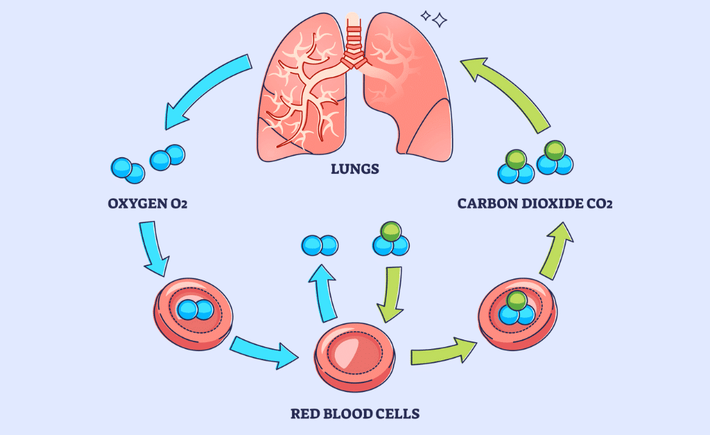
Gas Exchange &: Transport Gas Exchange and Transport Gas exchange is the process by which oxygen and carbon dioxide move between the lungs and the bloodstream, driven by simple diffusion along partial pressure gradients. This is coupled with the transport of these gases throughout the body via the circulatory system, primarily using hemoglobin in red blood cells. The entire process involves two main stages: I. External Respiration (in the Lungs) This is the exchange of gases between the air in the alveoli and the blood in the pulmonary capillaries. Oxygen uptake: Inhaled air has a high partial pressure of oxygen (PO2 ≈ 100 mmHg) in the alveoli, while the deoxygenated blood in the capillaries has a low PO2 ≈ 40 mmHg. This gradient causes oxygen to diffuse rapidly from the alveoli into the blood. Carbon dioxide release: The deoxygenated blood in the capillaries has a higher partial pressure of carbon dioxide (PCO2 ≈ 45 mmHg) compared to the air in the alveoli (PCO2 ≈ 40 mmHg). This causes carbon dioxide to diffuse from the blood into the alveoli to be exhaled. II. Internal Respiration (in the Tissues) This is the exchange of gases between the blood in systemic capillaries and the body’s tissue cells. Oxygen release: Oxygenated blood arriving at the tissues has a high PO2 ≈ 100 mmHg, while the metabolizing tissue cells have a low PO2 < 40 mmHg due to continuous consumption for cellular respiration. This gradient causes oxygen to dissociate from hemoglobin and diffuse into the cells. Carbon dioxide uptake: Tissue cells produce carbon dioxide as a waste product, resulting in a high PCO2 > 45 mmHg compared to the blood in the capillaries (PCO2 ≈ 40 mmHg). Carbon dioxide diffuses from the cells into the blood. Objective 1: Describe the partial pressures of oxygen and carbon dioxide in atmospheric air, alveoli, arterial blood, and venous blood. To understand gas exchange, we first need to grasp the concept of partial pressure. 1. Dalton’s Law of Partial Pressures Dalton’s Law states that in a mixture of non-reacting gases, the total pressure exerted is equal to the sum of the partial pressures of the individual gases. Ptotal = P1 + P2 + P3 + … Pn Where Ptotal is the total pressure of the gas mixture and P1, P2, etc. are the partial pressures of each individual gas. The partial pressure of an individual gas in a mixture is the pressure that gas would exert if it alone occupied the volume. It is directly proportional to its percentage concentration in the mixture. Partial Pressure of Gas (Px) = % Concentration of Gas x Total Pressure Example Calculation (Atmospheric Air at Sea Level): Total Pressure = 760 mmHg. Composition: Nitrogen (N2): ~79% Oxygen (O2): ~21% Carbon Dioxide (CO2): ~0.04% PO2 = 0.21 x 760 mmHg = ~160 mmHg 2. Partial Pressures in Different Locations Gases always diffuse down their partial pressure gradients from an area of higher partial pressure to an area of lower partial pressure. This is the driving force for gas exchange. Let’s examine the typical partial pressures of Oxygen (O2) and Carbon Dioxide (CO2) in four key locations: A. Atmospheric (Inspired) Air (at sea level, dry) This is the air we breathe in. PO2 (Atmospheric): Percentage: ~21% 0.21 x 760 = ~160 mmHg PCO2 (Atmospheric): Percentage: ~0.04% 0.0004 x 760 = ~0.3 mmHg (often rounded to 0 mmHg) B. Alveolar Air As atmospheric air enters the lungs, it mixes with the air already present in the dead space and alveoli, and it becomes saturated with water vapor. This significantly alters the partial pressures. Influencing Factors: Water Vapor: At 37°C, water vapor pressure is 47 mmHg. This dilutes other gases (Effective pressure: 760 – 47 = 713 mmHg). Gas Diffusion: CO2 continuously enters from blood; O2 continuously leaves into blood. PO2 (Alveolar – PAO2): ~104 mmHg Lower than atmospheric due to water vapor dilution and O2 diffusion into blood. PCO2 (Alveolar – PACO2): ~40 mmHg Higher than atmospheric due to CO2 diffusion from blood. C. Arterial Blood This is the blood leaving the pulmonary capillaries (oxygenated blood) and traveling to the systemic tissues. PO2 (Arterial – PaO2): ~95-100 mmHg Slightly lower than alveolar PO2 due to physiological shunts (bronchial circulation). PCO2 (Arterial – PaCO2): ~40 mmHg Same as alveolar PCO2; high solubility allows rapid equilibration. D. Venous Blood (Mixed Venous Blood) This is the blood returning to the lungs from the systemic tissues, carrying metabolic waste products. PO2 (Mixed Venous – PvO2): ~40 mmHg Lower than arterial because O2 was delivered to tissues. PCO2 (Mixed Venous – PvCO2): ~45 mmHg Higher than arterial because CO2 was picked up from tissues. Summary of Partial Pressures (Approximate Values at Sea Level) Location PO2 (mmHg) PCO2 (mmHg) Atmospheric Air 160 0.3 Alveolar Air 104 40 Arterial Blood 95-100 40 Mixed Venous Blood 40 45 Key Gradients for Gas Exchange O2 gradient for diffusion (Alveoli to Pulmonary Capillaries): 104 mmHg (alveolar) – 40 mmHg (venous) = 64 mmHg CO2 gradient for diffusion (Pulmonary Capillaries to Alveoli): 45 mmHg (venous) – 40 mmHg (alveolar) = 5 mmHg Note: Notice the much larger gradient for O2 compared to CO2. This is important because CO2 is much more soluble than O2, allowing it to diffuse efficiently even with a smaller pressure gradient. Checkpoint Question: Why is the partial pressure of oxygen in the alveoli (PAO2) significantly lower than the partial pressure of oxygen in atmospheric air (PO2)? Objective 2: Explain the principles governing gas exchange across the alveolar-capillary membrane (e.g., Dalton’s Law, Henry’s Law, Fick’s Law of Diffusion). Gas exchange, both between the alveoli and pulmonary capillaries, and between systemic capillaries and tissues, is driven by fundamental physical laws. We’ve already touched upon Dalton’s Law of Partial Pressures, which establishes the pressure gradient for individual gases. Now let’s integrate Henry’s Law and Fick’s Law of Diffusion to understand how these gases actually move and dissolve. 1. Dalton’s Law of Partial Pressures (Recap) Principle: The total pressure exerted by a mixture of gases is

Mechanics of Breathing (Pulmonary Ventilation)
Physiology

Mechanics of Breathing (Pulmonary Ventilation)

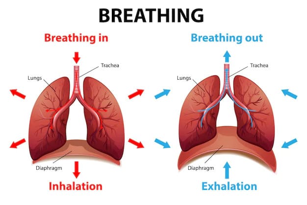
Mechanics of Breathing : Pulmonary Ventilation Mechanics of Breathing (Pulmonary Ventilation) The mechanics of breathing (pulmonary ventilation) involve creating pressure changes in the thoracic cavity to move air in (inspiration) and out (expiration) of the lungs, driven by muscle contraction and elastic recoil. During inhalation, the diaphragm and intercostal muscles contract, expanding the chest cavity, which lowers pressure, causing air to flow in (Boyle’s Law). Exhalation is usually passive, as muscles relax, the cavity shrinks, pressure increases, and air is pushed out, relying on lung elasticity. Precisely, Pulmonary ventilation, also known as breathing, involves the movement of air into and out of the lungs. Inspiration (inhalation) is an active process primarily driven by muscle contractions, such as the diaphragm. Objective 1: Identify and define the key pressures involved in the respiratory cycle, and explain the significance of each pressure, particularly the negative intrapleural pressure and positive transpulmonary pressure, in maintaining lung expansion. To understand how air moves into and out of the lungs, it’s crucial to grasp the various pressure gradients that drive this process. These pressures are always described relative to each other and, often, relative to atmospheric pressure. 1. Atmospheric Pressure (Patm) Definition: Atmospheric pressure is the pressure exerted by the column of air surrounding the Earth’s surface. It’s the “weight” of the air above us. Typical Value: At sea level, Patm is approximately 760 millimeters of mercury (mmHg) or 1 atmosphere (atm). This is equivalent to about 1033 cm H2O. Reference Point: In respiratory physiology, atmospheric pressure is often set as the reference point (0 mmHg or 0 cm H2O). This simplification allows us to discuss other pressures as positive (higher than atmosphere) or negative (lower than atmosphere) values. Significance: Air, like any fluid, moves from an area of higher pressure to an area of lower pressure. Therefore, differences between atmospheric pressure and pressures within the respiratory system are what ultimately drive the bulk flow of air during breathing. 2. Intrapulmonary Pressure (Ppul) / Alveolar Pressure (Palv) Definition: This is the pressure within the alveoli, the air sacs deep within the lungs where gas exchange occurs. It represents the pressure of the air inside the lungs. Characteristics and Changes during Breathing: Between Breaths (End-Expiration or End-Inspiration): At the end of a normal breath, when airflow momentarily ceases, Ppul equilibrates with Patm. Therefore, Ppul = 0 mmHg (relative to atmospheric). During Inspiration: For air to flow into the lungs from the atmosphere, Ppul must become lower than Patm. As the thoracic cavity expands (due to muscle contraction), the lung volume increases. According to Boyle’s Law (Volume and Pressure are inversely proportional), this increase in volume causes the pressure within the alveoli to drop slightly below atmospheric pressure (e.g., Ppul = -1 to -3 mmHg). This negative pressure gradient (Patm > Ppul) draws air into the lungs. During Expiration: For air to flow out of the lungs into the atmosphere, Ppul must become higher than Patm. As the thoracic cavity decreases in volume (due to elastic recoil of the lungs and chest wall), the lung volume decreases. This decrease in volume causes the pressure within the alveoli to rise slightly above atmospheric pressure (e.g., Ppul = +1 to +3 mmHg). This positive pressure gradient (Ppul > Patm) pushes air out of the lungs. Significance: Ppul is the direct driving force for airflow. Its fluctuations above and below atmospheric pressure determine the direction of air movement. 3. Intrapleural Pressure (Pip) Definition: This is the pressure within the pleural cavity—the narrow, fluid-filled space between the visceral pleura (lining the lungs) and the parietal pleura (lining the thoracic wall). Key Characteristic: Always Negative During normal breathing, Pip is always negative relative to both Patm and Ppul. At rest (between breaths), Pip is typically around -4 mmHg (-5 cm H2O). During inspiration, as the chest expands, it becomes even more negative (e.g., -6 to -8 mmHg). During expiration, it becomes less negative, returning towards -4 mmHg. Why is Pip always negative? This is a critical concept and results from two opposing elastic forces: 1. Lungs’ Natural Tendency to Recoil The elastic connective tissue within the lung parenchyma (especially around the alveoli) constantly pulls the lungs inward, trying to collapse them down to their smallest possible size. This creates an outward-pulling force on the visceral pleura. 2. Chest Wall’s Natural Tendency to Expand The thoracic wall (ribs, sternum, diaphragm) has its own natural elasticity, tending to spring outwards, increasing the volume of the thoracic cavity. This creates an inward-pulling force on the parietal pleura. These two opposing forces pull on the fluid in the pleural cavity, creating a “suction” effect that results in a sub-atmospheric (negative) pressure. The thin layer of pleural fluid, due to its surface tension, also acts as an adhesive, effectively “sticking” the two pleurae together, preventing the lungs from pulling away from the chest wall. Significance: The persistent negative intrapleural pressure is essential for: Maintaining Lung Expansion: It acts as a “suction” that keeps the lungs inflated and prevents them from collapsing due to their natural elastic recoil. Without this negative pressure, the lungs would collapse (atelectasis). Coupling Lung and Chest Wall Movement: It ensures that as the chest wall expands and contracts, the lungs follow suit, enabling effective changes in lung volume. Clinical Relevance: If the integrity of the pleural cavity is compromised (e.g., by a puncture wound, ruptured bleb), air can enter the pleural space, causing the Pip to equalize with Patm. This condition is called a pneumothorax, and without the negative Pip, the lung will collapse due to its inherent elastic recoil. 4. Transpulmonary Pressure (Ptp) Definition: Transpulmonary pressure is the pressure difference between the intrapulmonary pressure (Ppul) and the intrapleural pressure (Pip). Ptp = Ppul – Pip Key Characteristic: Always Positive: Since Pip is always negative relative to Ppul (and Patm), the transpulmonary pressure is always a positive value during normal breathing. For example: If Ppul = 0 mmHg and Pip = -4 mmHg, then Ptp = 0 – (-4) = +4 mmHg. Significance: Transpulmonary

Platelets and Hemostasis
Physiology

Blood Related Pathophysiology

Blood : Related Physiologies Physiology of Red Blood Cells I. Erythropoiesis: The Journey of a Red Blood Cell Erythropoiesis is the highly regulated process of red blood cell (RBC) production, primarily occurring in the bone marrow in adults. It’s a continuous, dynamic process designed to maintain a stable red blood cell mass and oxygen-carrying capacity in the blood. A. Sites of Erythropoiesis Embryonic/Fetal Life Yolk Sac (0-3 mo): Initial primitive site. Liver (3-7 mo): Primary peak activity. Spleen (3-6 mo): Contributes lesser extent. Bone Marrow (5 mo+): Gradually takes over. Adult Life Red Bone Marrow: Exclusive site (Vertebrae, sternum, ribs, pelvis, proximal long bones). Extramedullary: Reversion to Liver/Spleen in severe pathology (e.g., myelofibrosis). B. Stages of Erythropoiesis Progresses from Stem Cell to Mature RBC through distinct morphological changes. 1. Pluripotent Hematopoietic Stem Cell (HSC) “Master cells” capable of self-renewal. Differentiate into Common Myeloid Progenitors (CMPs). 2. Erythroid Progenitors (BFU-E & CFU-E) BFU-E: Primitive, EPO-sensitive but not dependent. CFU-E: Mature, highly sensitive and dependent on EPO for survival. 3. Pronormoblast (Proerythroblast) First recognizable precursor. Large (20-25 µm), basophilic cytoplasm (ribosomes), prominent nucleoli. Begins globin synthesis. 4. Basophilic Normoblast Smaller, intensely basophilic cytoplasm. Active hemoglobin synthesis begins. 5. Polychromatophilic Normoblast Grayish-blue cytoplasm (mix of ribosomes + Hb). Most active stage of Hb synthesis. 6. Orthochromatophilic Normoblast Smallest nucleated precursor. Dense, pyknotic nucleus. Pink cytoplasm (massive Hb). Nucleus is extruded at this stage. 7. Reticulocyte (Polychromatophilic Erythrocyte) Anucleated, contains residual RNA network (reticulum). Released from marrow to blood. Constitutes 0.5-2.5% of circulating RBCs. Reticulocytosis: Indicates increased production (e.g., response to anemia). 8. Mature Erythrocyte Biconcave disc, anucleated, no organelles. Packed with Hemoglobin for O2 transport. Lifespan: ~120 days. C. Regulation of Erythropoiesis 1. Erythropoietin (EPO) – The Key Hormone Source: Kidneys (90%), Liver (10%). Stimulus: Renal Hypoxia (Low O2) due to anemia, altitude, lung disease. Action: Binds receptors on progenitors (CFU-E) → Promotes proliferation, survival, Hb synthesis, and early release. 2. Nutritional Requirements Iron: Essential for Heme. Deficiency = Anemia. B12 & Folate: DNA synthesis cofactors. Deficiency = Macrocytic Anemia. Protein/Vitamins: Globin synthesis, C, B6, Copper, Zinc. 3. Hormonal Influences Androgens (Testosterone): Stimulate EPO + direct marrow effect (Higher RBCs in males). Thyroid/Growth Hormone: Stimulatory effects. Hypothyroidism can cause mild anemia. II. Hemoglobin Synthesis Hemoglobin (Hb) is the primary protein within red blood cells, responsible for oxygen transport from the lungs to the tissues and carbon dioxide transport from the tissues back to the lungs. It is a complex molecule, and its synthesis is a highly coordinated process. A. Structure of Hemoglobin A mature hemoglobin molecule is a tetramer (four subunits). Each subunit has two parts: 1. Heme (Non-Protein) Porphyrin ring structure with a central Iron (Fe2+) atom. Function: Site where oxygen binds reversibly. Capacity: 4 Heme groups per Hb molecule = 4 O2 molecules. 2. Globin (Protein) Four polypeptide chains (typically 2 pairs). Adult Hb: Two alpha (α) + Two beta (β) chains. Each globin chain enfolds a heme group. Specific combination determines Hb type. B. The Synthesis Process Occurs primarily in the cytoplasm of developing RBCs (pronormoblasts through reticulocytes). 1. Globin Chain Synthesis Occurs on ribosomes in the cytoplasm. Alpha (α) chains: Encoded on Chromosome 16. Beta (β), Gamma (γ), Delta (δ), Epsilon (ε): Encoded on Chromosome 11. 2. Heme Synthesis Multi-step enzymatic pathway occurring in Mitochondria and Cytoplasm. Start: Succinyl CoA + Glycine. Rate-Limiting Step: Formation of delta-aminolevulinic acid (ALA) by ALA synthase. Intermediates: Porphobilinogen → Uroporphyrinogen → Coproporphyrinogen → Protoporphyrin. Final Step: Insertion of Ferrous Iron (Fe2+) into Protoporphyrin IX ring by Ferrochelatase (Heme synthase). Iron Delivery: Transported by Transferrin, taken up via Transferrin Receptors. 3. Assembly Heme + Globin rapidly combine. 1 Globin + 1 Heme = Globin-Heme Monomer. 4 Monomers assemble = Final Hemoglobin Tetramer. C. Types of Normal Hemoglobin & Developmental Changes Globin chain production changes to adapt to oxygen environments. 1. Embryonic Hemoglobins (First 8-10 weeks) Gower 1 (ζ2ε2): Zeta + Epsilon. Gower 2 (α2ε2): Alpha + Epsilon. Hb Portland (ζ2γ2): Zeta + Gamma. Very high O2 affinity for extraction from maternal blood. 2. Fetal Hemoglobin (HbF – α2γ2) Predominant from 10 weeks to birth. Composition: 2 Alpha (α) + 2 Gamma (γ). Function: Higher O2 affinity than adult Hb (HbA). Crucial for O2 transfer across placenta. Post-Birth: Constitutes 60-90% at birth; gradually declines and is replaced by HbA. 3. Adult Hemoglobins Hemoglobin A (HbA – α2β2): 95-97% of adult Hb. 2 Alpha (α) + 2 Beta (β). Affinity regulated by 2,3-BPG for efficient tissue release. Hemoglobin A2 (HbA2 – α2δ2): 1.5-3.5% (Minor). 2 Alpha (α) + 2 Delta (δ). Elevated in Beta-thalassemia trait. D. Regulation of Hemoglobin Synthesis Iron Availability: Most critical. Deficiency impairs heme synthesis → reduced Hb. Globin Chain Balance: Synthesis of alpha/non-alpha chains is tightly balanced. Imbalance (Thalassemias) causes unstable chains/ineffective erythropoiesis. Erythropoietin (EPO): Indirectly stimulates synthesis by promoting precursor proliferation/maturation. III. Red Blood Cell Metabolism Unlike most cells in the body, mature red blood cells (erythrocytes) are anucleated and lack mitochondria, endoplasmic reticulum, and other organelles. This means they cannot synthesize proteins or carry out oxidative phosphorylation. Their metabolism is highly specialized and focuses on two main goals: Generating energy (ATP): To maintain membrane integrity, ion gradients (Na+/K+ pump), and cell shape. Protecting hemoglobin from oxidative damage: Hemoglobin is susceptible to oxidation, which impairs function and damages the cell. A. Energy Production (ATP Generation) RBCs rely almost exclusively on Anaerobic Glycolysis (Embden-Meyerhof pathway). 1. Embden-Meyerhof Pathway Converts Glucose → Pyruvate → Lactate. Yield: Net 2 ATP per glucose. Key Functions of ATP: Ion Gradients: Powers Na+/K+ ATPase pump (prevents osmotic lysis). Cell Shape: Phosphorylation of cytoskeletal proteins maintains deformability. 2. Rapoport-Luebering Shunt Offshoot pathway producing 2,3-Bisphosphoglycerate (2,3-BPG). Significance: Binds deoxyhemoglobin, stabilizing T-state → Promotes O2 release. High BPG: Decreased affinity (Right shift) → Increased delivery. Low BPG: Increased affinity (Left shift) → Decreased delivery. Cost: Consumes 1 ATP otherwise generated by glycolysis. B. Protection Against Oxidative Damage RBCs have antioxidant systems to neutralize Reactive Oxygen Species (ROS) that cause Methemoglobin (Fe3+) or Heinz bodies (denatured Hb). 1. Hexose Monophosphate

Platelets and Hemostasis
Physiology

Platelets and Hemostasis

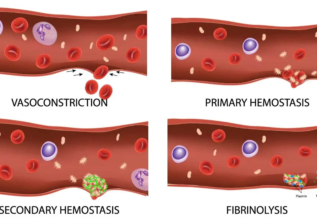
Platelets &: Hemostasis Introduction to Hemostasis Hemostasis is the physiological process that stops bleeding at the site of vascular injury while maintaining normal blood flow elsewhere. It involves interactions between blood vessels, platelets, and coagulation factors. Dysregulation leads to hemorrhage (excessive bleeding) or thrombosis (inappropriate clotting). Platelets (thrombocytes) are small, anucleated cell fragments that play a central role in primary hemostasis – the initial formation of a platelet plug at the site of injury. I. Morphology of Platelets Physical Traits Size/Shape: Tiny (2-4 µm), discoid (lens-shaped) when inactive. Upon activation, they become spherical with pseudopods (finger-like projections) to enhance adhesion. Anucleated: Lack a nucleus; cannot synthesize proteins. Limited lifespan (7-10 days). Membrane Rich in glycoproteins (e.g., GP Ib/IX/V, GP Ia/IIa, GP IIb/IIIa) acting as receptors for adhesion molecules (vWF, collagen, fibrinogen). Cytoplasmic Granules The cytoplasm contains critical granules and organelle systems. Alpha-granules Contain proteins for adhesion/coagulation: Fibrinogen von Willebrand factor (vWF) Platelet factor 4 (PF4) PDGF, P-selectin Dense (delta) granules Contain non-protein activators: ADP, ATP Serotonin Calcium Lysosomes Contain hydrolytic enzymes for digesting material. II. Formation of Platelets (Thrombopoiesis) Occurs in bone marrow, regulated by Thrombopoietin (TPO). 1. Origin: Hematopoietic Stem Cells (HSCs) → Common Myeloid Progenitor (CMP). 2. Megakaryoblast: Progenitor undergoes endoreduplication (DNA replication without division), becoming polyploid. 3. Megakaryocyte: Largest marrow cell (up to 100 µm). Highly lobulated nucleus. 4. Platelet Release: Megakaryocytes extend proplatelets into sinusoidal capillaries. Blood shear flow fragments these into thousands of platelets (1,000-3,000 per megakaryocyte). Regulation: TPO (from liver) stimulates megakaryocytes. Platelets bind and degrade TPO. High platelet mass = Low free TPO = Downregulated production (Negative Feedback). III. Function of Platelets in Hemostasis (Primary Hemostasis) 1. Adhesion Injury exposes subendothelial collagen. Platelets adhere via GP Ib receptor binding to von Willebrand factor (vWF) (bridge between platelet and collagen). Direct binding via GP Ia/IIa also occurs. Result: Anchors platelets to injury site. 2. Activation Triggered by adhesion, Thrombin, and ADP. Causes shape change (discoid → spherical + pseudopods) and granule release. Key Molecules Released: ADP: Potent activator, recruits more platelets. Thromboxane A2 (TxA2): Synthesized via COX-1; powerful vasoconstrictor and aggregator. Serotonin: Vasoconstriction. vWF/Fibrinogen: Aid further adhesion/aggregation. 3. Aggregation Activated platelets express GP IIb/IIIa receptor. Fibrinogen acts as a bridge, binding to GP IIb/IIIa on adjacent platelets. Links platelets together to form the primary hemostatic plug. 4. Procoagulant Activity Activated platelets provide a negatively charged phospholipid surface (phosphatidylserine). This surface concentrates coagulation factors (Tenase/Prothrombinase complexes), accelerating Thrombin generation to convert fibrinogen to fibrin, stabilizing the plug. Summary of Platelet Function When a vessel is damaged, platelets: Adhere to exposed matrix. Activate (shape change + release substances). Aggregate to form primary plug. Provide surface for Secondary Hemostasis (Coagulation Cascade). Steps and Components of the Coagulation Cascade While primary hemostasis (platelet plug formation) provides an initial, temporary seal at the site of vascular injury, it is not strong enough to withstand arterial pressure or provide long-term protection. Secondary hemostasis reinforces the platelet plug with a meshwork of fibrin, a strong, insoluble protein. This process is known as blood coagulation or the coagulation cascade, and it involves a series of enzymatic reactions involving plasma proteins called coagulation factors. I. Overview The coagulation cascade is traditionally described as having two main pathways that converge on a common pathway. However, a more modern and physiologically relevant view is the cell-based model. We will present both models for a comprehensive understanding. II. Traditional Model: Intrinsic, Extrinsic, and Common Pathways This model helps to understand individual factors and their interactions, especially in laboratory testing. 1. Extrinsic Pathway (Initiation) Initiated when blood is exposed to Tissue Factor (TF), expressed by subendothelial cells (fibroblasts, smooth muscle) upon injury. Step 1: TF binds to circulating Factor VII (VIIa) → Forms TF-VIIa complex. Step 2: TF-VIIa complex activates Factor X to Xa and Factor IX to IXa. Rapid pathway; primarily responsible for initiation. 2. Intrinsic Pathway (Amplification) Activated by contact of Factor XII with negatively charged surfaces (collagen, platelets) or by XIIa itself. Step 1: Factor XII → XIIa. Step 2: XIIa activates Factor XI → XIa. Step 3: XIa activates Factor IX → IXa. Step 4: IXa + Factor VIIIa (activated by thrombin) → Tenase Complex (IXa/VIIIa). The Tenase complex activates Factor X → Xa. Slower pathway; significant contribution to amplification. 3. Common Pathway Both pathways converge at the activation of Factor X. Step 1: Activated Factor X (Xa) + Factor Va (cofactor activated by thrombin) → Prothrombinase Complex (Xa/Va). Assembles on activated platelet surfaces. Step 2: Prothrombinase converts Prothrombin (Factor II) → Thrombin (Factor IIa). Central event of the cascade. Step 3: Roles of Thrombin: Converts Fibrinogen (I) → Fibrin monomers. Activates Factor XIII → XIIIa. Activates cofactors V and VIII. Activates Factor XI (feedback amplification). Activates platelets (positive feedback). Step 4: Fibrin monomers spontaneously polymerize → Unstable clot. Step 5: Factor XIIIa (transglutaminase) cross-links fibrin monomers → Stable, insoluble fibrin mesh. III. Cell-Based Model of Coagulation This model emphasizes the role of cellular surfaces (TF-bearing cells and activated platelets) and occurs in three overlapping phases. 1. Initiation Phase (on TF-bearing cells) Vascular injury exposes TF on subendothelial cells. TF binds VIIa → TF-VIIa. TF-VIIa activates small amounts of X to Xa and IX to IXa. Xa + Va generates a small “Thrombin Spurt”. Crucial for activating platelets/cofactors. 2. Amplification Phase (on Activated Platelets) The initial thrombin activates platelets (shape change, exposure of phosphatidylserine). Thrombin activates cofactors V, VIII, and Factor XI. Activated platelets provide negatively charged phospholipid surface. Factor IXa binds VIIIa on platelet surface → Tenase Complex. Tenase efficiently converts large amounts of X → Xa. 3. Propagation Phase (on Activated Platelets) Large amounts of Xa + Va assemble on platelet surface → Prothrombinase Complex. Converts massive amounts of prothrombin → thrombin. Result: “Thrombin Burst”. Thrombin burst rapidly converts fibrinogen to fibrin, activates XIIIa (cross-linking), and further activates platelets → Robust, stable clot. IV. Key Coagulation Factors & Components Plasma proteins, mostly synthesized in the liver. Vitamin K-Dependent Factors: Factors II, VII, IX, X, Protein C, Protein S. Require Vitamin

White Blood Cells Leukocytes Physiology
Physiology

White Blood Cells (Leukocytes) Physiology

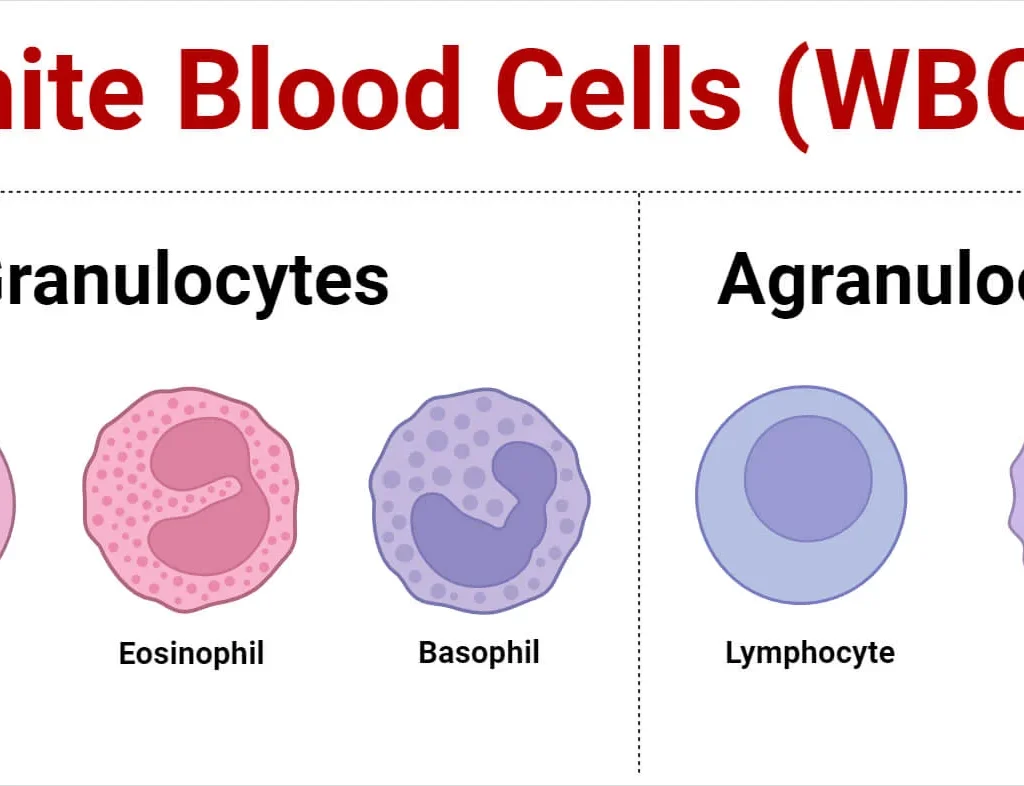
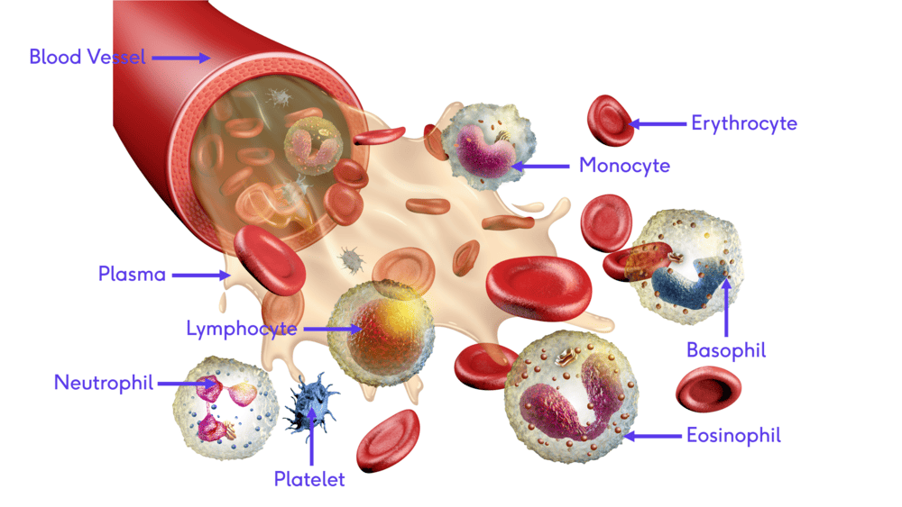
White blood Cell: Physiology White Blood Cells (Leukocytes) White blood cells (WBCs), also known as leukocytes, are a diverse group of immune cells that circulate in the blood and lymphatic system. Unlike red blood cells, they are complete cells, possessing a nucleus and other organelles, and their primary function is to defend the body against infection and disease. They are generally much less numerous than RBCs. Leukocytes are broadly classified into two main categories based on the presence or absence of visible granules in their cytoplasm when stained with Romanowsky stains (like Wright’s or Giemsa): I. Granulocytes These cells have prominent cytoplasmic granules that contain various enzymes and antimicrobial substances. They also have lobed nuclei. 1. Neutrophils Polymorphonuclear Leukocytes – PMNs Morphology Nucleus: Multi-lobed nucleus (usually 2-5 lobes) connected by thin strands of chromatin. Lobes increase with age. Cytoplasm: Fine, pale lilac or pinkish-tan granules; typically very faint. Size: 10-14 µm. Key Features Abundance: Most numerous (50-70%). Key Characteristic: “First responders” against bacteria. Primary Function: Phagocytosis: Rapid responders to bacterial/fungal infections. First to arrive at inflammation. Destroy Pathogens: Granules contain lysosomal enzymes, defensins, and antimicrobial agents. Formation of Pus: Dead neutrophils + debris + bacteria form pus. 2. Eosinophils Morphology Nucleus: Bi-lobed nucleus, resembling eyeglasses or headphones. Cytoplasm: Large, coarse, distinct red-orange granules. Size: 12-17 µm. Key Features Abundance: Relatively uncommon (1-4%). Key Characteristic: Associated with parasites and allergies. Primary Function: Parasitic Infections: Effective against multicellular parasites (worms) via toxic granule release. Allergic Reactions: Modulate responses by releasing antihistamines. Accumulate in asthma/hay fever. 3. Basophils Morphology Nucleus: Bi-lobed/S-shaped, often obscured by granules. Cytoplasm: Large, coarse, distinct dark blue-purple granules containing histamine and heparin. Size: 10-14 µm. Key Features Abundance: Rarest WBC (0.5-1%). Key Characteristic: Severe allergic reactions, histamine release. Primary Function: Allergic/Inflammatory Responses: Release histamine (vasodilator) and heparin (anticoagulant). Similar to Mast Cells: Share functional similarities but are distinct cells. II. Agranulocytes These cells have few or no visible granules in their cytoplasm. Their nuclei are typically non-lobed or kidney-shaped. 1. Lymphocytes Morphology Nucleus: Large, round, densely stained; occupies most of the cell. Cytoplasm: Scant, light blue rim; few/no granules. Size: Variable; small (7-9 µm) most common. Key Features Abundance: Second most numerous (20-40%). Key Characteristic: Immune “memory” and specific defense. Primary Function (Specific Immunity): T Lymphocytes (T cells): Cell-mediated immunity (attack virus-infected/cancer cells). B Lymphocytes (B cells): Humoral immunity (produce antibodies). Differentiate into plasma cells. NK Cells: Rapid response to infected/tumor cells (Innate immunity). 2. Monocytes Morphology Nucleus: Large, kidney or horse-shoe shaped; lighter stain. Cytoplasm: Abundant, pale gray-blue (“ground-glass”). Size: Largest WBC (14-20 µm). Key Features Abundance: 2-8%. Key Characteristic: Precursors to macrophages, “big eaters.” Primary Function: Macrophages: Circulate briefly then migrate to tissues to differentiate into macrophages. Phagocytosis: Engulf bacteria, debris, old RBCs (“clean-up crew”). Antigen Presentation: Present antigens to lymphocytes. Chronic Inflammation: Crucial role. Summary Table of WBC Types WBC Type Granules (Staining) Nucleus Morphology Abundance Primary Function Neutrophil Fine, pale lilac 2-5 lobes, polymorphous 50-70% Phagocytosis of bacteria/fungi (first responders) Eosinophil Large, red-orange Bi-lobed 1-4% Allergic reactions, parasitic infections Basophil Large, dark blue-purple Bi-lobed, often obscured 0.5-1% Allergic reactions (histamine), inflammation Lymphocyte None/scant Large, round, dense 20-40% Specific immunity (T/B cells), memory Monocyte None/fine dust-like Kidney-shaped, horse-shoe 2-8% Phagocytosis (macrophages), antigen presentation Process of Leukopoiesis Leukopoiesis is the process of white blood cell (WBC) production, occurring primarily in the red bone marrow. Unlike erythropoiesis, which is mainly stimulated by erythropoietin, leukopoiesis involves a broader array of growth factors called colony-stimulating factors (CSFs) and interleukins (ILs) that guide the differentiation of hematopoietic stem cells into the various leukocyte lineages. I. Hematopoietic Stem Cells (HSCs) and Lineage Commitment All blood cells originate from pluripotent Hematopoietic Stem Cells (HSCs) in the red bone marrow. These HSCs differentiate into two major progenitor cell lines: Common Myeloid Progenitor (CMP) Gives rise to granulocytes (neutrophils, eosinophils, basophils), monocytes, red blood cells, and platelets. Common Lymphoid Progenitor (CLP) Gives rise to lymphocytes (T cells, B cells, NK cells). II. Myelopoiesis (Granulocytes & Monocytes) This is the pathway from the CMP to mature granulocytes and monocytes. Pathway: Common Myeloid Progenitor (CMP) → Granulocyte-Monocyte Progenitor (GMP) (A bipotential progenitor). A. Granulocyte Development (Neutrophil, Eosinophil, Basophil) 1. Myeloblast: First morphologically recognizable precursor. Large cell, prominent nucleus, fine chromatin, basophilic cytoplasm, no granules. 2. Promyelocyte: Larger than myeloblast. Prominent primary (azurophilic) granules (dark purple). 3. Myelocyte: Beginning of specific granule synthesis (neutrophilic, eosinophilic, or basophilic). Nucleus becomes more kidney-shaped. Last stage capable of mitosis. 4. Metamyelocyte: Nucleus indented (kidney-bean shaped). No longer capable of mitosis. 5. Band Cell (Stab Cell): Nucleus elongated and curved (Band or “C” shape), not fully segmented. Released in infection (“left shift”). 6. Mature Granulocyte: Nucleus segmented (multi-lobed for neutrophils, bi-lobed for eosinophils/basophils). B. Monocyte Development Monoblast: Precursor, similar to myeloblast but committed to monocytic lineage. Promonocyte: Large cell with indented nucleus, somewhat basophilic cytoplasm. Monocyte: Mature cell released into bloodstream. Circulates briefly before migrating to tissues to become a macrophage or dendritic cell. III. Lymphopoiesis (Lymphocytes) Pathway from Common Lymphoid Progenitor (CLP) to mature lymphocytes. Stages: Lymphoblast: First recognizable precursor. Large nucleus, scant cytoplasm. Prolymphocyte: Slightly smaller, less prominent nucleolus. Lymphocyte: Mature cells released into circulation. Maturation Sites: B Lymphocytes: Mature in Bone Marrow → migrate to lymph nodes/spleen. T Lymphocytes: Migrate from marrow to Thymus to mature and undergo selection. NK Cells: Mature in marrow and secondary lymphoid organs. IV. Regulation: Colony-Stimulating Factors (CSFs) and Interleukins (ILs) Leukopoiesis is tightly regulated by a complex network of signaling molecules (glycoproteins) acting as growth factors. Colony-Stimulating Factors GM-CSF (Granulocyte-Macrophage CSF): Stimulates production of granulocytes and monocytes/macrophages from myeloid progenitors. G-CSF (Granulocyte CSF): Primarily stimulates production and maturation of neutrophils. Clinical: Used to boost neutrophil counts in neutropenic patients. M-CSF (Macrophage CSF): Promotes differentiation of monocytes into macrophages. Interleukins (ILs) & Others IL-3: Multilineage CSF; stimulates growth of various hematopoietic stem cells (myeloid & lymphoid). IL-5: Crucial for growth, differentiation, and activation of eosinophils. IL-7: Essential for development of B and T lymphocytes. IL-6: Involved in immune responses; stimulates HSCs. Stem Cell

Red Blood Cells (Erythrocytes) Physiology
Physiology

Red Blood Cells (Erythrocytes) Physiology

Red blood Cell: Physiology Red Blood Cells (Erythrocytes) Red blood cells (RBCs), also known as erythrocytes, are arguably the most crucial component of blood in terms of overall physiological function. Their primary role is to transport oxygen from the lungs to the body’s tissues and to transport carbon dioxide from the tissues back to the lungs. To efficiently carry out this vital function, RBCs possess a unique and highly specialized structure. I. Structure of Red Blood Cells 1. Biconcave Disc Shape Description: Mature RBCs are flexible, anucleated (lacking a nucleus), and lack most other organelles. Their most distinctive feature is their biconcave disc shape – a flattened disc with depressed centers on both sides. Functional Significance: Increased Surface Area to Volume Ratio: Maximizes the surface area available for gas exchange (O2 and CO2). A spherical cell would have a much smaller surface area. Flexibility and Deformability: The biconcave shape and flexible membrane (maintained by a spectrin protein network) allow RBCs to bend and squeeze through narrow capillaries (3-4 µm) despite being 7.5 µm wide. Essential for circulation. Rouleaux Formation: Allows RBCs to stack like coins in single file to pass through narrow vessels without jamming. 2. Anucleated & Lack of Organelles Description: Unlike most cells, mature RBCs extrude their nucleus and lose their mitochondria, endoplasmic reticulum, and Golgi apparatus during maturation. Functional Significance: Maximized Hemoglobin Content: Frees up space to be packed almost entirely with hemoglobin. Approx. 97% of the non-water content is hemoglobin. No Oxygen Consumption: Lacking mitochondria, RBCs do not consume the O2 they transport. They generate ATP primarily through anaerobic glycolysis. Limited Lifespan: Lack of protein synthesis machinery limits lifespan to approx. 100-120 days. 3. Plasma Membrane Description: A phospholipid bilayer highly specialized with a dense network of cytoskeletal proteins (spectrin, ankyrin, band 3) on its inner surface. Functional Significance: Maintain Shape and Flexibility: The spectrin-actin cytoskeleton provides structural integrity to withstand shear stress. Antigen Presentation: Displays glycoproteins and glycolipids (e.g., ABO and Rh antigens) important for blood typing. II. Function of Red Blood Cells 1. Oxygen Transport Mechanism: This is the primary function. Hemoglobin (Hb) binds reversibly to oxygen. Each Hb molecule binds up to four O2 molecules. Lungs: High O2 concentration → O2 loads onto Hb → Oxyhemoglobin (HbO2) (bright red). Tissues: Low O2 concentration → O2 unloads → diffuses into tissues. Efficiency: High Hb concentration + large surface area = highly efficient transport. 2. Carbon Dioxide Transport RBCs transport CO2 (waste product) via three methods: Bicarbonate Buffer System (~70%) Enzyme Carbonic Anhydrase converts CO2 + H2O → H2CO3 → H+ + HCO3-. HCO3- moves to plasma (chloride shift) and acts as a buffer. Carbaminohemoglobin (~20-23%) CO2 binds directly to the amino groups of the globin protein chains (not the heme iron). Forms HbCO2. This is reversible based on PCO2 levels. Dissolved in Plasma (~7-10%) A small amount of CO2 is simply dissolved in the fluid. 3. pH Regulation (Buffering) Mechanism: Hemoglobin acts as a buffer. When CO2 is converted to H+ and HCO3-, the free H+ ions are buffered by deoxyhemoglobin. This prevents significant drops in intracellular pH and helps maintain blood pH within the narrow physiological range (7.35-7.45). Summary of Specializations Biconcave Shape: Surface area & flexibility. Anucleated: Max space for Hb, no O2 consumption. Packed with Hb: Gas transport vehicle. Carbonic Anhydrase: Facilitates CO2 transport & pH regulation. Flexible Membrane: Passage through capillaries. Structure and Function of Hemoglobin Hemoglobin (Hb) is the specialized globular protein found within red blood cells responsible for their ability to transport oxygen and, to a lesser extent, carbon dioxide. It has a complex quaternary protein structure perfectly adapted for its vital role in gas exchange. I. Structure of Hemoglobin 1. Four Polypeptide Chains (Globins): A single hemoglobin molecule is composed of four protein subunits. In adults, the most common type (HbA) consists of two alpha (α) chains and two beta (β) chains. Each globin chain has a specific amino acid sequence and a characteristic folded structure. 2. Heme Groups: Each of the four globin chains is associated with a non-protein, iron-containing prosthetic group called a heme group. (Therefore: 1 Hemoglobin molecule = 4 Heme groups). Porphyrin Ring: A large organic molecule structure. Iron Ion (Fe2+): A central ferrous iron ion is chelated within the ring. Critical Function: This Fe2+ is the binding site for oxygen. Each iron can bind one O2 molecule. The bond is weak and reversible. Capacity: One Hb molecule can bind up to 4 O2 molecules. Important: Iron must be in the Ferrous (Fe2+) state. If oxidized to Ferric (Fe3+), it forms methemoglobin and cannot bind oxygen. II. Function of Hemoglobin A. Oxygen Transport (Primary Function) Oxygenation (Lungs): High PO2 → Oxygen diffuses into RBCs → Binds to Fe2+ in heme → Forms Oxyhemoglobin (HbO2). Appearance: Bright Red. Deoxygenation (Tissues): Low PO2 → Oxygen bond breaks → O2 released and diffuses into tissue cells → Becomes Deoxyhemoglobin (HHb) (or reduced hemoglobin). Appearance: Darker, dull red. Concept: Cooperative Binding: Hemoglobin exhibits a unique phenomenon where the binding of oxygen facilitates further binding. When the first O2 molecule binds to one heme group, it causes a conformational (shape) change in the entire hemoglobin molecule. This change increases the affinity of the remaining three heme groups for oxygen. Conversely, when one O2 is released, it decreases the affinity of the others, facilitating further release. Result: The characteristic S-shaped (sigmoidal) oxygen-hemoglobin dissociation curve, allowing for highly efficient loading in lungs and unloading in tissues. B. Carbon Dioxide Transport (Secondary Function) Carbaminohemoglobin (HbCO2): About 20-23% of blood CO2 binds directly to the amino groups of the globin chains (NOT the heme iron). This is reversible based on PCO2 levels. Role in Bicarbonate System (Haldane Effect): While Hb doesn’t transport bicarbonate directly, it buffers the Hydrogen ions (H+) produced during the conversion of CO2 to bicarbonate. Deoxyhemoglobin binds these H+ ions, preventing a pH drop and facilitating more CO2 uptake. Deoxyhemoglobin has a higher H+ affinity than oxyhemoglobin. C. Buffering Blood pH Deoxyhemoglobin acts as a stronger buffer for

Blood Physiology Introduction (1)
Physiology

Blood Physiology: Introduction

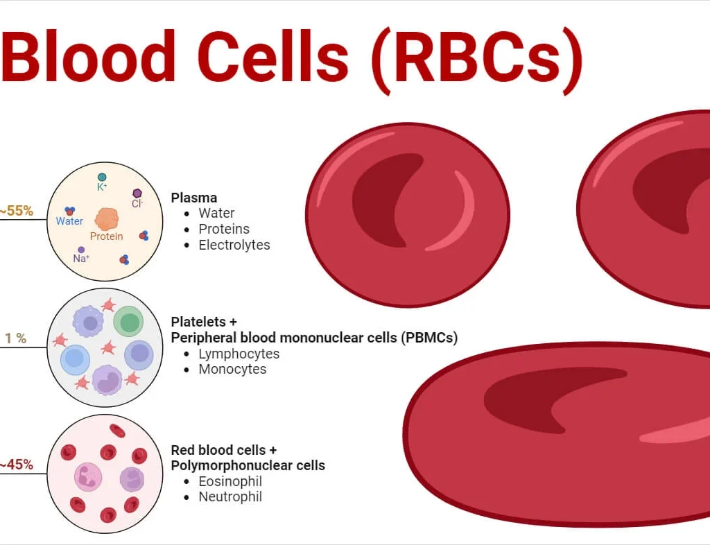
Blood Physiology: Introduction Introduction to Blood Blood is often described as a unique connective tissue, though it differs significantly from other connective tissues like bone or cartilage. Its uniqueness stems from its cellular components being suspended in a liquid extracellular matrix (plasma) rather than being anchored to solid fibers. This fluidity is crucial for its transport functions. It is the only fluid tissue in the body, continuously circulating within the closed system of the cardiovascular system (heart, blood vessels). It is a complex, viscous fluid that accounts for approximately 8% of total body weight in an average adult (e.g., about 5-6 liters in males, 4-5 liters in females). Origin: All blood cells originate from hematopoietic stem cells in the red bone marrow. Why is it essential for life? Blood serves as the body’s primary transport and communication medium, ensuring that all cells receive necessary resources and waste products are efficiently removed. Without its continuous circulation, cells would rapidly cease to function due to lack of oxygen and nutrients, and the accumulation of toxic metabolic byproducts. It acts as a dynamic internal environment, constantly adapting to the body’s changing needs and maintaining homeostasis (the stable internal conditions required for survival). Overview of Major Roles of Blood 1. Distribution/Transportation Blood acts as the delivery system for the body: Respiratory Gases: Carries oxygen from the lungs (where it’s loaded onto hemoglobin in red blood cells) to all body tissues and cells for cellular respiration. Transports carbon dioxide, a waste product of cellular respiration, from body cells back to the lungs for exhalation (dissolved in plasma, bound to hemoglobin, or as bicarbonate ions). Nutrients: Delivers absorbed nutrients (e.g., monosaccharides like glucose, amino acids, fatty acids, glycerol, vitamins, minerals) from the digestive tract to the liver first, and then to all body cells for energy, growth, and repair. Hormones: Acts as the “circulatory highway” for endocrine hormones, transporting them from their sites of production (endocrine glands) to their specific target organs or cells throughout the body, regulating diverse physiological processes. Metabolic Wastes: Collects and transports metabolic waste products, such as urea (from protein metabolism) and uric acid (from nucleic acid metabolism) to the kidneys for excretion in urine, and lactic acid (from anaerobic respiration) to the liver for conversion. 2. Regulation Blood plays a pivotal role in maintaining the stability of the interstitial fluid (homeostasis): Body Temperature: Blood possesses a high heat capacity due to its water content. It absorbs heat generated by metabolically active tissues (e.g., muscles) and distributes it throughout the body. By regulating blood flow to the skin, it can either dissipate excess heat (vasodilation) or conserve heat (vasoconstriction) to maintain a stable core body temperature. pH Levels: Crucial for maintaining the extremely narrow and vital physiological pH range of 7.35-7.45. It achieves this through various buffer systems present in plasma proteins and within red blood cells (e.g., bicarbonate buffer system, phosphate buffer system, protein buffer system). These buffers can accept or donate hydrogen ions to resist drastic changes in acidity or alkalinity. Fluid Volume and Blood Pressure: Blood plasma proteins, particularly albumin, exert significant osmotic pressure (colloid osmotic pressure). This pressure draws water from the interstitial fluid back into the capillaries, maintaining proper fluid volume within the circulatory system and helping to prevent edema (swelling of tissues). Maintaining adequate blood volume is directly linked to maintaining sufficient blood pressure for tissue perfusion. Electrolyte Balance: Transports various electrolytes (Na+, K+, Ca2+, Cl-, HCO3-) which are vital for nerve impulse transmission, muscle contraction, and fluid balance. 3. Protection Blood provides defense mechanisms against blood loss and foreign invaders: Prevention of Blood Loss (Hemostasis): Initiates a rapid and efficient series of events when a blood vessel is damaged. This process, called hemostasis, involves the aggregation of platelets (thrombocytes) and the activation of clotting factors (plasma proteins) to form a fibrin clot, sealing the injured vessel and preventing excessive hemorrhage. Prevention of Infection: Leukocytes (White Blood Cells): Are the mobile units of the immune system. They identify and destroy pathogens (bacteria, viruses, fungi, parasites) and remove damaged or abnormal cells (e.g., cancer cells, dead cells). Different types of leukocytes have specialized roles in this defense. Antibodies: Specific proteins (immunoglobulins) produced by certain lymphocytes that target and neutralize specific pathogens or toxins. Complement Proteins: A group of plasma proteins that, when activated, can lyse microorganisms, enhance phagocytosis, and contribute to inflammation. Physical Characteristics Appearance & Texture Color: Oxygen-rich blood: (typically arterial) is a bright, scarlet red. This vibrant color is due to hemoglobin picking up oxygen in the lungs (oxyhemoglobin). Oxygen-poor blood: (typically venous) is a darker, duller red, sometimes described as brick-red or maroon. This is because hemoglobin has released its oxygen (deoxyhemoglobin). Note: Venous blood is never blue, despite how veins appear through the skin. Viscosity (Thickness): Blood is about 5 times more viscous (thicker/stickier) than water, primarily due to RBCs and plasma proteins. Clinical Significance: Increased viscosity (e.g., polycythemia, severe dehydration) increases resistance to flow, straining the heart. Decreased viscosity (e.g., severe anemia) can lead to turbulent flow. Properties pH Level: Slightly alkaline (basic), maintained tightly between 7.35 and 7.45. Clinical Significance: pH < 7.35 = Acidosis pH > 7.45 = Alkalosis Both disrupt enzyme function and can be fatal. Temperature: Circulates at ~38°C (100.4°F), slightly higher than body temperature, to absorb and distribute metabolic heat. Taste/Odor: Metallic taste (iron content) and faint characteristic odor. Volume: The average adult has approximately 5-6 liters (1.5 gallons), constituting 7-8% of total body weight. Clinical Significance: Significant deviations (hemorrhage, fluid overload) severely compromise tissue perfusion. Composition of Blood: The Two Major Components When a sample of blood is collected and centrifuged (spun at high speed), its components separate into distinct layers due to differences in density. This separation reveals two main components: Plasma55% RBCs45% Buffy Coat 1. Plasma (Liquid Matrix) Constitutes ~55% of total volume. Least dense component; forms top, yellowish-straw colored layer. A sticky, non-living fluid matrix. (Detailed composition covered in Objective 1.3) 2. Formed Elements (Cellular) Constitutes ~45% of total volume (Hematocrit). Normal
Scroll to Top