Doctors Revision

Oral and Dental Health

Oral and Dental Health

Periodontal Diseases: Gingivitis and Periodontitis — Diagnosis and Management

Dental Health · Oral and Dental Conditions Periodontal Diseases: Gingivitis and Periodontitis — Diagnosis and Management Periodontal diseases affect the tissues that surround and support the teeth. They include gingival inflammation, most commonly plaque-induced gingivitis, and periodontitis, in which inflammation is associated with loss of the tooth’s supporting attachment and alveolar bone. Gingivitis is often reversible when the cause is controlled. Periodontitis can usually be stabilised and managed, but lost attachment and bone are not routinely restored by simple cleaning alone. Early recognition, accurate examination, effective daily plaque control, risk-factor management and professional follow-up help protect oral function and reduce the risk of tooth loss. This study guide expands the course slide resources and applies current periodontal terminology. It is written for health-science learners and does not replace supervised clinical training, a dental diagnosis or local Ugandan protocols. A trained dental professional should assess individual patients and decide treatment according to the findings, available facilities, patient preferences and referral pathways. Learning objectives By the end of this lesson, you should be able to: define the periodontium and common periodontal terms; distinguish gingivitis from periodontitis; describe causes, risk factors, clinical features and complications; take a focused periodontal history; outline screening, periodontal charting and indicated radiography; explain the stepwise principles of management; counsel patients on prevention and risk reduction; and recognise situations that need urgent dental or specialist referral. 1. The periodontium and key terms The periodontium is the group of tissues that support and attach a tooth. Its main parts are the gingiva, periodontal ligament, cementum covering the tooth root and alveolar bone. Disease may affect the gingiva alone or damage deeper supporting structures. Gingiva: the gum tissue surrounding the teeth and covering the alveolar processes. Healthy gingiva is generally firm and closely adapted to the tooth, though colour and appearance vary with pigmentation and individual anatomy. Dental plaque biofilm: a structured community of microorganisms in a matrix that forms on teeth and restorations. Persistent biofilm at the gingival margin can provoke inflammation in a susceptible host. Calculus (tartar): mineralised plaque. It is not itself the primary cause of periodontitis, but its rough surface can retain biofilm and it cannot be removed by ordinary brushing once firmly deposited. Gingivitis: inflammation confined to the gingiva, typically causing redness, swelling and bleeding on gentle probing or brushing, without periodontitis-related clinical attachment loss or bone loss. Periodontal pocket: an abnormally deepened space between a tooth and the gingiva. Pocket depth must be interpreted with gum position, clinical attachment level, bleeding and other findings; a deep reading alone does not establish the full diagnosis. Probing depth (PD): the distance from the gingival margin to the base of the sulcus or pocket, measured with a periodontal probe. Clinical attachment level (CAL): the distance from a fixed tooth landmark, usually the cementoenamel junction, to the base of the pocket. CAL helps identify loss of tooth-supporting attachment. Gingival recession: movement of the gum margin away from the crown, exposing part of the root. Recession can result from several causes and is not, by itself, proof of periodontitis. Bleeding on probing (BOP): bleeding observed after gentle probing. It is a sign of inflammation at a site and should be recorded alongside plaque and pocket measurements. Furcation involvement: loss of supporting tissues between the roots of a multi-rooted tooth. Tooth mobility: movement of a tooth beyond normal physiological movement. It can have periodontal and non-periodontal causes and is assessed in context. Suppuration: discharge of pus from the gingival sulcus or pocket, suggesting active infection or an abscess and requiring professional assessment. 2. Classification and patterns of disease Current international classification distinguishes periodontal health, gingival diseases and conditions, periodontitis, and other conditions affecting the periodontal tissues. Older teaching slides may use labels such as “chronic” and “aggressive” periodontitis. In the 2017 World Workshop classification, these former categories are grouped within periodontitis and described using stage and grade, while recognising necrotising disease and periodontitis as a manifestation of certain systemic diseases as distinct forms. Condition Main feature Why it matters Plaque-induced gingivitis Gingival redness, swelling and bleeding related to dental biofilm; no periodontitis-related attachment or bone loss. Usually reversible when plaque is effectively removed and contributing factors are addressed. Non-plaque-induced gingival disease Gingival lesions or inflammation related to infection, immune or mucocutaneous disease, allergy, trauma or another cause. May persist despite plaque removal; identify the cause and refer when findings are atypical or persistent. Periodontitis Inflammatory destruction of tooth-supporting attachment and alveolar bone, often with pockets, recession, bleeding or mobility. Can lead to progressive support loss and tooth loss; requires professional treatment and ongoing supportive care. Necrotising periodontal disease Acute painful gingival necrosis, often with punched-out interdental papillae, spontaneous bleeding and halitosis; systemic symptoms may occur. Needs prompt assessment, pain control, careful local treatment and evaluation of predisposing factors or systemic illness. Periodontal abscess Localised accumulation of pus in the gingival wall of a periodontal pocket, often with local pain, swelling and tenderness. May cause rapid tissue destruction and can spread; urgent dental assessment is required. Periodontitis associated with a systemic condition Periodontal destruction occurring in the setting of a systemic disorder that affects the periodontal apparatus. Document relevant medical conditions and coordinate care with the appropriate clinician. 3. Causes and pathogenesis Dental plaque biofilm at and below the gingival margin triggers a host inflammatory response. When plaque remains undisturbed, the microbial community can become more disease-associated. In gingivitis, inflammation is limited to the gum tissue. In susceptible people, persistent inflammation may involve destruction of periodontal ligament fibres and resorption of alveolar bone. The depth and distribution of damage vary across teeth and patients; the amount of visible plaque alone does not predict severity. Calculus, overhanging or poorly contoured restorations, crowded teeth, appliances and other plaque-retentive features can make cleaning difficult. Systemic and behavioural factors modify inflammation and progression. Smoking can mask bleeding while worsening periodontal risk and treatment response. Diabetes, especially when glycaemic control is poor, is an important modifying factor; periodontal inflammation and diabetes can influence each other, but

Oral and Dental Health

Dental Anaesthesia: Local and General Methods in Dentistry

Dental Health · Clinical Procedures Dental Anaesthesia: Local, Sedation and General Anaesthesia Dental anaesthesia helps control pain and anxiety so that appropriate oral-health care can be carried out safely. The approach may range from a local anaesthetic that numbs a small area to sedation or general anaesthesia for selected patients and procedures. These methods are not interchangeable: they differ in effect, risk, required training, monitoring and recovery. This lesson explains the core concepts for learners. It is not a practical training manual or a patient-specific prescription. Anaesthesia must be planned and administered only by appropriately trained, authorised professionals in a suitable setting, following current Ugandan requirements, local protocols and product information. Learning objectives By the end of this lesson, you should be able to: distinguish local anaesthesia, sedation and general anaesthesia; describe common dental local-anaesthetic approaches; outline key patient-safety checks before and during care; explain the team, monitoring and recovery requirements for general anaesthesia; and identify adverse events that need prompt escalation. 1. Key terms: pain relief, local anaesthesia, sedation and general anaesthesia Analgesia means reducing or relieving pain. Local anaesthesia temporarily removes sensation, especially pain, in a particular area through topical application or injection; the patient remains conscious. Sedation uses medicines to reduce anxiety or awareness to a planned level while the patient remains responsive to varying degrees. General anaesthesia is a drug-induced state of unconsciousness: the patient cannot be aroused, even by painful stimulation, and may need assistance to maintain breathing and the airway. Sedation is a continuum. A patient can become more deeply sedated than intended, and the route of administration alone does not determine the depth. Anyone providing sedation needs the education, rescue skills, staff and equipment required for the level of care and for foreseeable unintended deeper sedation. General anaesthesia is a separate, higher-risk service that requires suitable credentials, facilities and continuous monitoring. Method Main effect Awareness and breathing Typical dental role Local anaesthesia Blocks sensation in a tooth, region or small area. Patient is awake; breathing is not intentionally depressed. Routine restorative care, periodontal procedures, extractions and other procedures when appropriate. Minimal or moderate sedation Reduces anxiety or awareness while supporting cooperation. Responsiveness varies by depth; monitoring and rescue capability are required. Selected patients with anxiety, special care needs or procedures requiring additional support. Deep sedation Markedly depresses consciousness. Airway or ventilation may be impaired; rescue skills and close monitoring are essential. Selected cases performed by appropriately trained providers in a suitably equipped setting. General anaesthesia Produces unconsciousness and lack of response to painful stimulation. Independent ventilation may be impaired; airway and cardiovascular support may be needed. Selected complex, extensive or otherwise unsuitable-for-awake-care situations after professional assessment. 2. Local anaesthesia in dental care Local anaesthesia is the foundation of pain control for many dental procedures. It allows an appropriately trained clinician to treat a defined area while the patient stays awake. The agent and method depend on the planned procedure, the duration of numbness needed, medical history, age and weight, pregnancy where relevant, allergies, concurrent medicines and other agents being used. Common approaches Topical anaesthesia: applied to a mucosal surface to reduce sensation at the surface, for example before an injection or selected minor procedures. Absorption still contributes to the total anaesthetic exposure. Infiltration: local anaesthetic is placed near the small nerve branches supplying the target area. The site and expected effect depend on anatomy and the planned procedure. Regional nerve block: anaesthetic is deposited near a named nerve so that sensation is reduced over a wider distribution. This requires appropriate anatomical knowledge and practical training. Other targeted techniques: periodontal-ligament, intraosseous or other delivery methods may be used for selected indications by clinicians trained in them. Dental local-anaesthetic products may contain an anaesthetic alone or a vasoconstrictor such as adrenaline (epinephrine), which can affect duration and local blood flow. Formulations and concentration differ. Identify medicines by reading their labels; cartridge colour coding is only an aid. The selection should be individualised, and exact dose calculations must follow the current product information and the clinician’s approved reference rather than a memorised amount in a general study note. Safety checks before and during local anaesthesia Confirm the patient, procedure and site. Explain the expected numbness, possible sensations and alternatives in language the patient understands; obtain consent. Review the health history. Clarify medicines, allergies and what happened during any previous reaction. Consider relevant medical conditions, pregnancy, age, weight and concurrent sedatives. Select and calculate safely. Check the exact agent, concentration, vasoconstrictor, expiry and packaging. For children, dose is weight-based. Include topical and injected products in the total exposure, use the lowest effective amount and never estimate a repeat dose from memory. Use the trained technique and infection precautions. Follow the approved method for the chosen injection, use appropriate equipment and observe the patient throughout. Do not inject if the clinician cannot confirm the correct product, patient or plan. Reassess and document. Check whether the intended area is numb before starting, note the patient’s response, and record the agent, amount, site or technique as required by local documentation standards. When local anaesthetic is combined with sedation or general anaesthesia, all medicines and local-anaesthetic totals must be considered together. Sedative medicines can change the patient’s response and the safety requirements. Local anaesthesia does not replace continuous monitoring when sedation or general anaesthesia is being used. 3. Sedation: a separate level of care Sedation may help selected patients tolerate dental care, but it is not simply “stronger local anaesthesia.” Before sedation, a suitably qualified provider assesses health and medication history, the airway, the planned depth, fasting requirements where applicable, patient and procedure risks, consent and the support available after treatment. The sedation plan must match the provider’s training, the facility and the patient. Patients with significant medical conditions or other risk factors may need medical consultation, a different setting or referral. Teams need a clear division of responsibilities. Monitoring must not be neglected while the operator performs dental treatment. The required observer, monitoring devices, emergency supplies and rescue skills

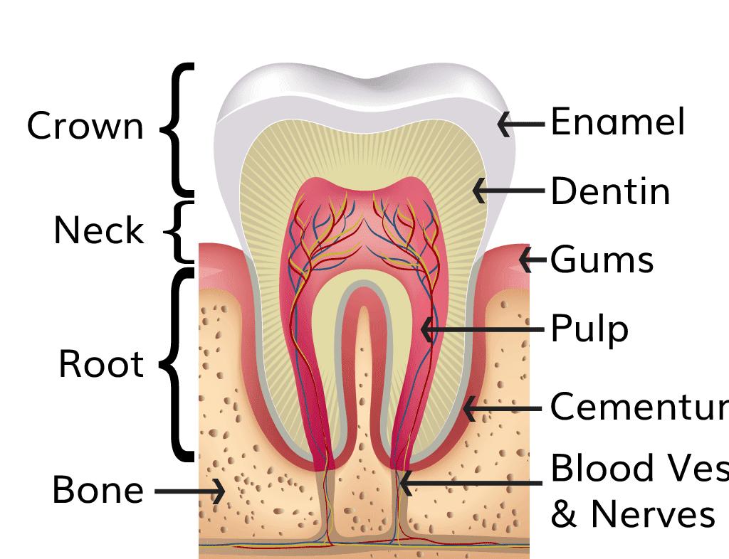
Cross-section diagram of a tooth showing its main structures for oral health education.
Oral and Dental Health

Dental History Taking and Oral Examination: Step-by-Step Guide

Dental Health · Clinical Procedures Dental History Taking and Oral Examination: A Step-by-Step Guide Dental history taking and a systematic oral examination help the clinician understand a patient’s concern, identify relevant health risks, and decide what further assessment may be needed. Neither should be rushed: the patient’s own account, a careful extraoral and intraoral examination, appropriate investigations, and clear documentation all contribute to safe care. This guide outlines a practical approach for dental and health-science learners. It is an educational framework; examinations, tests and treatment decisions must be performed by appropriately trained professionals within local scope and protocols. Learning objectives By the end of this lesson, you should be able to: organise a patient-centred dental history; describe a systematic general, extraoral and intraoral examination; record relevant positive and negative findings; select investigations only when they answer a clinical question; and recognise situations that require urgent referral. 1. Why the history and examination matter A good history clarifies what brought the patient to care, how the problem has changed, and which personal or medical factors may affect examination, diagnosis, treatment or referral. The examination then checks the areas the patient identifies and looks for other relevant signs in the mouth, face and neck. Investigations provide additional information when the clinical question cannot be answered adequately from history and examination alone. The process is also a conversation. Introduce yourself, confirm the patient’s identity, explain what you would like to ask or examine, invite questions, and obtain consent. Protect confidentiality and use an interpreter or other communication support when needed. For a child or a person who needs decision-making support, follow applicable consent, assent and safeguarding procedures. Do not promise a diagnosis before the findings are assessed. 2. Taking a structured dental history Begin with an open invitation such as “What has been troubling you?” Let the patient explain before using focused questions. Record important symptoms in the patient’s own words where possible, then clarify details without leading or suggesting an answer. History area What to ask or record Why it matters Patient details and context Confirm name, age or date of birth, contact details as required, preferred language, caregiver or interpreter, and the source of the history. Supports accurate records, communication and appropriate consent. Chief complaint Ask the main reason for the visit. Record the patient’s description and the site of concern. Keeps assessment focused on the patient’s priority. History of the present problem Explore onset, duration, site, character, severity, timing, progression, triggers, relieving factors, associated symptoms, previous episodes and care already tried. Builds a clear timeline and helps distinguish urgent from less urgent concerns. Past dental history Previous visits, procedures, extractions, restorations, periodontal treatment, trauma, dentures or appliances, complications, and prior experiences or anxiety. Reveals treatment history, patient preferences, possible complications and barriers to care. Medical history Current and past illnesses, hospital admissions, relevant surgery, pregnancy where applicable, and the names of treating clinicians if coordination is needed. Systemic health can influence oral findings, treatment choices, medication safety and referral. Medicines and allergies Prescribed and non-prescribed medicines, supplements, recent changes, allergies or adverse reactions, and what happened during the reaction. May affect bleeding, healing, saliva, infection risk, interactions or emergency planning. Social and oral-health history Tobacco or other substance use, alcohol, diet and sugary drinks, brushing and interdental cleaning, fluoride use, oral habits, work or school circumstances, and access to services. Identifies relevant risks and helps shape practical, respectful advice. Family history and review of systems Ask about relevant inherited conditions or patterns and symptoms outside the mouth when they may relate to the complaint. May point to systemic disease or a need for medical assessment. Explore the main complaint carefully For toothache, clarify which tooth or area hurts, whether pain is spontaneous or triggered, how long it lasts, and whether it disturbs sleep, chewing or daily activity. Ask about swelling, fever, difficulty opening the mouth, swallowing or breathing. For bleeding gums, ask when bleeding occurs, its duration and amount, and whether there is pain, looseness or a medication that could affect bleeding. For an ulcer, lump or patch, record when it began, whether it is changing, whether it has recurred, and any associated pain, numbness, swallowing difficulty or neck lump. After dental trauma, document the time and mechanism, the teeth or soft tissues involved, bleeding, pain, previous treatment, and any associated head or facial injury. Ask about loss of consciousness, vomiting, confusion or neck pain when relevant; these may need urgent medical evaluation. Do not let a detailed dental history delay emergency care when airway, breathing, circulation or major trauma is a concern. Medical conditions, medicines and allergies Ask specifically about conditions that may change the care plan, such as diabetes, heart or respiratory disease, bleeding disorders, immune suppression, kidney or liver disease, seizure disorders, pregnancy, or treatment with anticoagulants or medicines affecting bone metabolism. The relevance depends on the patient and the procedure being considered. Check the actual medicine name, dose and use where possible instead of relying on “blood thinner” or “allergy” alone. When a history is unclear or a condition appears unstable, obtain appropriate medical advice or referral before elective care. Do not tell a patient to stop a prescribed medicine without consultation with its prescriber. Record what was discussed, what information was verified, and any agreed follow-up. Update the health history when the patient reports a change; a form does not replace discussion. 3. Prepare for the physical examination Use standard precautions and follow the facility’s infection-prevention procedures. Perform hand hygiene, use appropriate personal protective equipment, prepare clean examination instruments, and ensure a suitable light source. Explain the sequence, check that the patient is comfortable, and ask permission before touching the face, neck or mouth. Use a mirror, gauze and other instruments appropriate to the task and the clinician’s training. Provide breaks if the patient is anxious, in pain or unable to tolerate a prolonged examination. Record general observations relevant to the presenting problem. Vital signs such as temperature, pulse, blood pressure or respiratory rate

Cross-section diagram of a tooth showing its main structures for oral health education.
Oral and Dental Health

Oral Health Education: Principles, Methods and Planning

Dental Health · Health Promotion Oral Health Education: Principles, Methods and Planning Oral health education helps people gain the knowledge, confidence and practical skills to protect their teeth and mouths, recognise problems early, and use dental services appropriately. It works best when it is planned around the needs of learners, uses respectful two-way communication, and is supported by services and environments that make healthy choices possible. This lesson outlines the meaning, principles, methods and planning of oral health education for health workers, students, teachers and community programmes. Learning objectives By the end of this lesson, you should be able to: define oral health education and distinguish it from broader oral health promotion; identify the principles of effective education; select appropriate methods and materials for different groups; plan a short education session; and describe ways to assess whether learning and programme goals have been achieved. 1. Meaning and purpose of oral health education Oral health education is a planned learning process that helps individuals and groups understand oral health, develop useful skills, and make informed choices about prevention and care. It may take place during a dental consultation, in a classroom, at a community meeting, through radio or digital messages, or in another setting where people can learn and ask questions. Its purpose is broader than transferring facts. A learner may know that brushing is important but still lack a suitable brush, fluoride toothpaste, time, confidence, clean water, or access to a dental service. Good education identifies such barriers and helps people find realistic steps and support. It can improve knowledge and skills, encourage preventive behaviours, support early help-seeking, and strengthen community participation in oral health. Oral health education and oral health promotion These terms are related but not identical. Education focuses on learning, communication, skills and informed decisions. Oral health promotion includes education, but also works to create supportive policies, services and environments. Examples include ensuring that schools have safe water and a workable toothbrushing programme, making preventive services accessible, involving communities in planning, and supporting healthy food choices. Education cannot by itself remove barriers created by poverty, distance, cost or limited services. 2. Principles of effective oral health education Start with the learner: ask what people already know, what they want to learn, and what matters in their daily lives. Avoid assuming that every group has the same needs. Use accurate, consistent information: base messages on current evidence and national guidance. Correct misinformation tactfully and acknowledge when an individual needs professional assessment. Make it practical: connect each message with an achievable action, such as demonstrating toothbrushing or agreeing on one change to a daily routine. Communicate respectfully: use plain language, listen without blame, protect privacy and respect cultural practices while explaining health risks clearly. Adapt to language and access needs: use a language learners understand, clear pictures or demonstrations where useful, and accessible formats for people with visual, hearing, learning or mobility needs. Invite participation: discussion, demonstration and questions help reveal misunderstandings and let educators adapt the session. Reinforce over time: one talk rarely changes a long-standing habit. Repeat key messages through homes, schools, clinics and community channels. Link education to services: give accurate information about where and when care is available and how to obtain referral or follow-up. 3. Core oral health messages Choose messages that fit the audience, age, health needs and available services. Keep the number of messages small enough for learners to remember and act on. Daily oral hygiene: explain regular toothbrushing with fluoride toothpaste and cleaning between teeth, in line with age-appropriate national recommendations. Demonstrate a simple technique and let learners practise or explain it back. Food and drinks: discuss limiting the amount and frequency of free-sugar foods and drinks, choosing water when available and appropriate, and planning realistic alternatives. Avoid presenting one food as the sole cause of disease. Fluoride: explain how fluoride helps prevent tooth decay and how to use products safely. Guidance should account for age, ability to spit, local recommendations and total exposure. Early care: encourage people to seek dental advice for persistent toothache, swelling, bleeding gums, ulcers that do not heal, trauma or other concerning changes. Facial swelling with fever, difficulty swallowing or difficulty breathing needs urgent medical attention. Prevention across life: include the needs of infants and caregivers, children, adolescents, pregnant people, adults, older people and people with disabilities or chronic conditions. Tobacco and harmful alcohol use: explain that these are risk factors for oral disease and that people who use them can ask a health professional for support to reduce or stop. Keep the educator’s role clear Education supports prevention and timely care; it does not replace a clinical examination. Do not diagnose a lesion or promise that a home remedy will cure it. Refer symptoms and concerns to a qualified dental or medical professional according to local pathways. 4. Identify the audience and learning need Before preparing a talk, find out who will attend, what they already know, what decisions or skills they need, and what could prevent them from acting. Consider age, language, literacy, disability, culture, family roles, previous dental experiences, cost, transport, availability of products and access to services. Use a brief conversation, a few questions, observation, routine service data or community feedback to identify a need. For example, if caregivers report that children swallow toothpaste, a demonstration about dispensing a small amount and supervising brushing may be more useful than a general lecture on tooth anatomy. If people know about prevention but cannot reach a clinic, education should be paired with service information, outreach or referral planning. 5. Plan an oral health education session Step What the educator does Example 1. Assess need Identify the group, priority concern, existing knowledge and barriers. Ask caregivers what makes twice-daily brushing difficult at home. 2. Set an objective Describe what learners should know, say or demonstrate by the end. “Participants will demonstrate a safe, systematic brushing routine.” 3. Select key messages Choose a few accurate points linked to the objective. Brush regularly with fluoride toothpaste;

Oral and Dental Health

Diet and Fluoride for Dental Health: Prevention Guide

Dental Health · Prevention and Health Promotion Diet and Fluoride for Dental Health: Practical Prevention Guide Diet and fluoride for dental health are two important parts of preventing dental caries. Food and drink choices influence how often teeth are exposed to fermentable sugars and acids, while fluoride helps teeth resist mineral loss and supports repair of early enamel damage. Neither replaces toothbrushing, daily cleaning between teeth, or professional dental care. This guide explains the key concepts, practical dietary measures, common fluoride products, and safety points for families and health workers. Learning objectives By the end of this lesson, you should be able to: explain the relationship between diet and dental caries; describe dietary measures that support oral health; identify common fluoride products and how they are used; and counsel patients on age-appropriate, safe fluoride use. 1. How diet affects teeth and oral health Dental caries is a biofilm-mediated, sugar-driven, multifactorial disease. Bacteria in dental plaque metabolise fermentable carbohydrates, especially free sugars, and produce acids. Repeated acid attacks lower the pH at the tooth surface and cause enamel minerals to dissolve (demineralisation). Saliva and fluoride help restore minerals between attacks (remineralisation). A cavity develops when mineral loss repeatedly exceeds repair. Risk depends on several interacting factors: plaque biofilm, the amount and frequency of sugar or other fermentable carbohydrate exposure, fluoride availability, saliva flow, tooth surfaces, oral hygiene, and a person’s health and circumstances. A single food does not cause or prevent caries on its own. The pattern of eating and drinking across the day matters, alongside regular preventive care. Free sugars and common sources The World Health Organization defines free sugars as sugars added by a manufacturer, cook or consumer, plus sugars naturally present in honey, syrups, fruit juices and fruit-juice concentrates. These differ from sugars naturally contained within intact fresh fruit and vegetables. Common sources include sugar-sweetened soft drinks, energy drinks, sweetened tea or coffee, confectionery, biscuits, cakes, sweetened breakfast foods, syrups, honey and fruit juice. WHO recommends reducing free sugars to less than 10% of total daily energy intake; a further reduction to below 5% can provide additional health benefits. These are population-level dietary targets, not a reason to calculate calories during every dental visit. In practice, patients can begin by identifying sugary drinks and snacks, reducing their frequency and portion size, and choosing water or unsweetened alternatives. Frequency, timing and form of intake Frequent sipping or snacking exposes teeth to repeated acid challenges and leaves less time for saliva to help neutralise acids and restore minerals. Sticky foods that remain on tooth surfaces and drinks sipped over long periods can extend exposure. Encourage patients to keep sweet foods and drinks occasional and, where possible, have them with a meal rather than repeatedly between meals. Avoid putting a child to bed with a bottle containing a sugary drink or juice. Acidic drinks and foods can also contribute to dental erosion, which is chemical wear of tooth tissue not caused by plaque bacteria. Frequent intake of fizzy drinks, some fruit juices and sour products may increase acid exposure. Water is a suitable thirst drink. If acidic drinks are consumed, avoid holding or swishing them around the mouth; a dental professional can advise people with erosion, sensitivity or high caries risk. 2. Dietary measures that support dental health Reduce free sugars: Check ingredient lists and nutrition labels, and pay attention to sugars in drinks, sauces, snacks and packaged foods. Choose unsweetened drinks and less-sweet snacks more often. Reduce the number of sugar exposures: Replace repeated grazing and sipping with regular meals and planned snacks. Keep confectionery and sweet drinks occasional. Choose water between meals: Plain safe drinking water does not bathe the teeth in sugar. Where community water fluoride levels are relevant, follow local public-health guidance. Prefer a balanced, varied diet: Encourage vegetables, whole fruit, pulses, whole grains and other nutrient-dense foods as part of a healthy diet. Whole fruit is generally preferable to juice because it retains its fibre and is eaten differently; fruit juice still contains free sugars under the WHO definition. Protect children’s teeth: Avoid frequent sweet drinks, sweetened bottles and sugary snacks between meals. Caregivers should help young children brush and should attend preventive dental visits. Consider medicines and dry mouth: Some medicines reduce saliva or contain sugar. Review the patient’s medication and symptoms when appropriate; do not stop prescribed medicine without consulting the prescriber. Ask a dentist or clinician about sugar-free formulations and preventive support. Dietary counselling should be respectful and practical. Ask what a person usually eats and drinks, when they snack, what changes are affordable and culturally acceptable, and whether food access or health conditions affect their choices. Agree on one or two realistic steps and review them at a later visit. Avoid blaming people: food environments, income, work schedules and access to clean water can shape daily choices. 3. What fluoride does Fluoride is a naturally occurring mineral used in evidence-based caries prevention. At the tooth surface, fluoride supports remineralisation and makes enamel more resistant to acid dissolution. Regular topical exposure from fluoride toothpaste is a foundation of prevention for most people. Some patients benefit from additional products or professional application after an assessment of caries risk and total fluoride exposure. Fluoride reduces risk; it does not instantly remove plaque, repair a large cavity, or replace a filling when restorative treatment is needed. Early lesions may be managed non-operatively in some cases, but diagnosis and follow-up require a dental professional. 4. Common fluoride products and measures Product or measure Main use Key safety or selection point Fluoride toothpaste Routine home brushing and topical fluoride exposure Use an age-appropriate amount; supervise young children and encourage spitting. Fluoride mouthrinse Additional topical fluoride for selected patients Use only when the patient can rinse and spit; follow label and professional advice. Fluoride varnish Professional preventive application Applied by a trained professional according to age, risk and local protocol. Fluoride gel or foam Topical prevention for selected patients Concentration and use require professional selection, especially in children.

Oral and Dental Health

Improving Access to Dental Services and Oral Health Information

Introduction to Dental Health · Promotion of dental health Improving Access to Dental Services and Oral Health Information Access to dental services means that people can obtain the right oral health care and trustworthy information when they need them. Access is more than having a dental clinic somewhere in a district: care must be reachable, affordable, acceptable, timely and appropriate to the person’s needs. Effective access also includes prevention, early assessment, treatment, referral and follow-up. These notes explain common barriers and practical ways health systems and communities can reduce them. The World Health Organization identifies uneven distribution of oral health professionals and facilities, together with out-of-pocket costs, as major access challenges. Solutions therefore need to combine prevention, primary health care, fair financing, well-distributed teams and clear information. Local service names, fees and referral routes change; confirm them with current district and Ministry of Health sources. Learning objectives Define access to oral health care and distinguish its key dimensions. Recognise personal, community and health-system barriers to dental care. Describe ways to improve access through primary care, outreach, inclusive services and referral. Explain how clear, reliable oral health information supports informed decisions. Suggest indicators for checking whether access is improving fairly. 1. What Does Access to Dental Care Mean? Access describes how well a person’s need for care connects with services that can meet it. A service may exist but remain inaccessible if the journey is too long, the cost is unaffordable, the clinic cannot accommodate a wheelchair, the opening hours conflict with work, or the patient cannot understand how to obtain help. Access is also incomplete when people receive an examination but cannot obtain treatment, referral or follow-up. Dimension Meaning Example question Availability Enough services, trained staff, equipment, medicines and supplies are present. Is an appropriate provider and service available? Geographic accessibility The service can be reached using realistic travel time, transport and cost. Can a person travel there safely and in time? Affordability Direct fees and indirect costs do not prevent people from obtaining needed care. Can a household meet the costs without harmful financial strain? Acceptability Care is respectful, confidential, culturally responsive and trusted. Does the patient feel safe, heard and treated with dignity? Accommodation and information Opening hours, communication and physical arrangements work for different users. Can the patient understand the next step and use the service? Quality and continuity Care is safe and appropriate, with referral and follow-up when required. Can the patient complete the needed pathway? 2. Why Improving Access Matters Many oral diseases are preventable, but untreated problems can cause pain, infection, difficulty eating or speaking, disturbed sleep and missed school or work. A person may delay care until symptoms become severe, when available options can be more complex and costly. Oral health is part of general health: common risk factors include high free-sugar intake, tobacco use and harmful alcohol use, and oral conditions can be associated with other noncommunicable diseases. Connecting prevention and oral assessment with everyday health services helps identify needs earlier. Fair access also supports equity. Rural communities, low-income households, people with disabilities, older adults, children, people displaced from their homes and people facing language or social barriers may have greater difficulty reaching care. A universal service can still produce unequal outcomes if it is designed only around the needs of people who live close by, can pay immediately and can navigate the system easily. 3. Common Barriers to Dental Services Barrier How it affects access Examples of a response Distance and transport Long journeys, transport fares, poor roads or difficult travel can delay routine and urgent visits. Plan services across districts; use suitable outreach; publish accurate locations and hours. Cost Consultation, tests, treatment, medicines, transport and missed work can add up. Include priority oral health services in fair health-financing arrangements; explain costs before care. Limited workforce or equipment Few providers, long waits, supply interruptions or limited service range reduce capacity. Plan and support a competent oral health workforce; maintain essential equipment and supplies. Low awareness or unclear information People may not know where to go, what services are offered or when assessment is needed. Use plain-language education and clear facility signposting; explain referral steps. Fear, stigma or poor past experiences Anxiety, shame or disrespectful care can discourage a return visit. Use non-judgmental communication, privacy, pain-aware care and shared decisions. Disability and communication needs Inaccessible buildings, forms or communication exclude some patients. Provide reasonable physical, sensory, communication and scheduling accommodations. Service organisation Limited hours, complex booking, unclear fees or weak referral links create extra steps. Simplify booking, coordinate referrals and provide follow-up information. Social and geographic inequity Some groups face several barriers at once, including poverty, isolation and discrimination. Map unmet need and involve affected communities in service design. 4. Ways to Improve Access 4.1 Integrate essential oral health care into primary health care Primary health care offers a practical place to connect oral health promotion with people’s routine contact with the health system. Trained health workers can reinforce prevention messages, recognise when a person needs an oral assessment, identify basic warning signs within their scope and support referral to an appropriate dental professional. Integration should be supported by clear protocols, training, supervision, supplies and feedback from referral services. It does not mean that untrained staff should diagnose or perform dental procedures outside their authorised role. 4.2 Put prevention and early care closer to communities Community health activities, schools, antenatal services, workplaces and other routine settings can share accurate oral health information and help people navigate services. School programmes can support age-appropriate education, prevention and referral while respecting consent, privacy and follow-up needs. Outreach or mobile clinics may help underserved communities when they are properly staffed, equipped, supplied and linked to continuing care. A one-off visit is less useful if people cannot obtain subsequent treatment. 4.3 Make services financially accessible High direct payments can deter care and put household finances at risk. Health financing can reduce this barrier when essential, cost-effective oral health services are included in broad coverage arrangements, preventive

Oral and Dental Health

Dental Instruments and Equipment: Types, Uses and Safety

Introduction to Dental Health · Concepts, tools and equipment Dental Instruments and Equipment: Types, Uses and Safety Dental instruments and equipment help a dental team examine the mouth, prevent disease, provide treatment and communicate findings. The range extends from a simple mouth mirror and cotton pliers to a dental chair, suction system, steriliser and imaging equipment. Knowing each item’s name is only the beginning: students should also understand its purpose, limitations, basic care and infection-prevention requirements. This overview introduces common equipment used in general dental care. It is for health-sciences learning. Clinical use, maintenance and reprocessing must follow local Ministry of Health requirements, facility procedures, staff training and the manufacturer’s instructions. Instruments used for invasive treatment should only be handled by appropriately trained practitioners. Learning objectives Distinguish dental equipment, instruments, handpieces and consumables. Name common examination, periodontal, restorative and surgical instruments. Explain the main parts of a dental unit and the purpose of its accessories. Describe core infection-prevention principles for reusable and single-use items. Recognise limits of equipment and basic safety considerations. 1. Key Terms: Instrument, Equipment and Consumable Dental equipment is the broader group of devices and systems used to deliver or support care, such as the dental chair, operating light, compressor, suction pump, steriliser and radiographic unit. A dental instrument is a hand-held or powered tool used for examination or treatment. A hand instrument is operated manually, while a powered instrument uses air, electricity or ultrasonic vibration. A consumable is used up during care, such as gauze, cotton rolls, local anaesthetic cartridges or disposable saliva ejectors. Reusable devices are designed for cleaning and reprocessing between patients according to validated instructions. Single-use devices are intended for one patient and one use, then discarded safely; they must not be reprocessed unless specifically authorised by the relevant regulator and manufacturer. Direct vision means looking at an area directly, whereas indirect vision uses a mirror to view surfaces that are difficult to see directly. 2. The Dental Unit and Treatment Area Equipment Main parts or function Learning point Dental chair Supports and positions the patient; controls may adjust height and backrest. Position for safe access, comfort, communication and transfer; respect the patient’s mobility needs. Operating light Illuminates the oral cavity and treatment field. Adjust without glare and avoid touching the patient or sterile instruments with a contaminated handle. Instrument tray and delivery arm Holds handpieces, syringes and selected instruments within the operator’s reach. Arrange only the items needed for the planned care; keep clean and contaminated areas distinct. Foot control Activates or regulates a connected handpiece or other unit functions. Know which device is connected before activation; protect the control from contamination. Three-way air-water syringe Delivers air, water or a combined spray through a nozzle. Useful for rinsing or drying when clinically appropriate; follow device instructions and unit waterline procedures. Suction A saliva ejector provides lower-volume fluid removal; high-volume evacuation removes larger volumes and spray near the field. Choose the appropriate tip and maintain safe positioning under supervision; tubing and traps need routine care. Compressor and vacuum system Supply compressed air and suction to compatible dental units. Require installation, maintenance and monitoring by trained personnel. Steriliser Processes compatible reusable instruments, commonly using steam under pressure. Correct cleaning, loading, cycle selection, monitoring and storage are all essential. Radiographic equipment May include an intraoral X-ray unit, sensor or film holder, or extraoral panoramic system. Imaging should be clinically justified and performed by trained personnel under local radiation-safety rules. Work surfaces and storage Provide clean preparation areas and protected storage for processed packs and supplies. Keep clean stock away from splash, dust, moisture and used instruments. The exact arrangement differs between facilities. A small primary-care clinic may have a simpler dental unit and fewer powered devices than a referral centre, but every setting still needs dependable lighting, clean water, appropriate suction or safe alternatives, hand hygiene supplies, protective equipment, safe instrument processing and a way to arrange referral when required. 3. Basic Dental Examination Instruments Mouth mirror A small mirror on a handle provides indirect vision, helps retract soft tissues gently and reflects light. Mirrors may be plane or magnifying and may be single-use or reusable. A mirror improves visibility; it does not establish a diagnosis on its own. Dental probe or explorer A fine-ended instrument can assist trained clinicians in examining surfaces and restorations. Use is guided by the examination question and current practice; forceful probing can damage tissue or a weakened surface. Visual assessment and appropriate diagnostic methods remain important. Periodontal probe A calibrated, usually blunt-ended probe measures the gingival sulcus or periodontal pocket when used by a trained practitioner with an appropriate technique. Readings must be interpreted with other findings; one measurement does not define periodontal health. Cotton pliers (college tweezers) Angled pliers transfer cotton pellets, gauze or small items into and out of the mouth. They also help keep fingers away from the working area. They are not extraction forceps. Cheek and tongue retractors Retractors improve access and visibility by moving soft tissues gently. Avoid unnecessary pressure and check patient comfort, especially when tissues are inflamed or injured. Gauze, cotton rolls and suction tips These support moisture control and absorb fluids. Their design may be disposable or reusable; follow the single-use label, infection-control policy and waste-segregation rules. 4. Instruments for Periodontal and Preventive Care Scalers are shaped to remove deposits from tooth surfaces. Hand scalers commonly have a pointed working end; curettes have rounded toes and are designed for use in periodontal instrumentation by trained practitioners. Their shape, working end and intended surface differ, so instruments should not be interchanged casually. Ultrasonic scalers use a powered tip that vibrates to disrupt deposits while irrigation helps cool and flush the working area. Tip selection, water flow, patient factors and technique require training. Prophylaxis angles and polishing cups or brushes may be used for selected preventive or polishing procedures under professional guidance. They do not replace effective daily plaque removal or a full assessment. The air-water syringe and suction support visibility and

Scroll to Top