Spread of Malignant Tumors & Epithelial Tumors
Mechanisms of Malignant Tumor Spread
Malignant neoplasms exhibit the autonomous capacity to spread via two principal modalities: local spread into contiguous surrounding structures and distant spread to non-contiguous anatomical sites (metastasis).
Genetic Initiation of Metastatic Potential
Gene Mutation & Antigenic Alteration: Malignant transformation is initiated by somatic mutations that lead to the expression of novel Tumor Associated Antigens (TAA) on the neoplastic cell surface.
Phenotypic Consequences: Alterations in surface receptor architecture result in the destabilization of intercellular junctions, loss of contact inhibition, and down-regulation of homophilic adhesion molecules. Specifically, loss of E-cadherin function dissociates cohesive sheets of cells, promoting autonomous proliferation and active tissue invasion.
Invasion of the Extracellular Matrix (ECM)
Breaching the extracellular matrix represents the initial active phase of the metastatic cascade, encompassing four discrete, coordinated steps:
| Step | Biological Event | Molecular Mechanism & Histological Impact |
|---|---|---|
| 1. Loss of Cellular Cohesion | Intercellular detachment ("loosening up") | Normal epithelial cells are held together by homophilic transmembrane cadherins. Carcinoma cells selectively downregulate or mutate E-cadherin and catenin complexes, permitting detachment from the primary tumor mass. |
| 2. Attachment to Matrix Components | Receptor-mediated anchoring to stroma | Detached cells express cell-surface integrins and laminin receptors to bind laminin within the basement membrane and fibronectin within interstitial connective tissue. This binding activates downstream intracellular invasive signaling. |
| 3. Degradation of the ECM | Proteolytic enzymatic dissolution | Secretion of matrix metalloproteinases and proteases by neoplastic cells or recruited host fibroblasts/macrophages. Type IV collagenase dissolves epithelial and vascular basement membranes; cathepsin D digests interstitial stroma to create migratory conduits. |
| 4. Active Migration (Locomotion) | Pseudopodial directed propulsion | Directed movement through degraded matrix conduits guided by Autocrine Motility Factor (AMF) and extracellular matrix degradation products. Neoplastic cells often gain negative surface charges, causing mutual electrostatic repulsion and abolishing contact inhibition. |
ECM Invasion Cascade ("L-A-D-M")
Remember the 4-step sequence of tissue invasion: Loosening (E-cadherin loss) → Attachment (laminin and fibronectin receptors) → Degradation (Type IV collagenase, Cathepsin D) → Migration (autocrine motility factor, pseudopodia).
Vascular Dissemination and Homing of Tumor Cells
Following matrix traversal, active tumor cell locomotion facilitates direct contact with vascular conduits:
- Vascular Penetration: Malignant cells readily intravasate into thin-walled lymphatic capillaries, blood capillaries, and venules; however, penetration into thick-walled, muscular arterioles is exceptionally rare due to substantial arterial basement membranes and elastic laminae.
- Intravascular Embolization: Upon entering the circulation, malignant cells form tumor emboli. The vast majority of circulating tumor cells are eliminated by hemodynamic shear stress and host immune surveillance (natural killer cells and cytotoxic T-lymphocytes).
- Platelet Aggregation & Impaction: Surviving cells bind circulating platelets, forming a protective hetero-aggregate ("cloaking") that prevents immune lysis and facilitates mechanical arrest within capillary beds.
- Extravasation & Homing: Adherent tumor emboli attach to vascular endothelial cells, exit the vessel by degrading the vascular basement membrane (recapitulating ECM invasion mechanisms), and colonize the parenchymal niche (homing).
- Metastatic Colonization: Tumor cells proliferate within the permissive microenvironment, establishing vascularized secondary tumors (metastatic deposits).
Routes of Tumor Spread
1. Local (Direct) Spread
- Direct Tissue Continuity: Carcinoma cells invade and infiltrate adjacent tissues across anatomical boundaries.
- Clinical Example: Malignant carcinoma of the tongue directly invading deep into the underlying musculature and floor of the mouth.
2. Distant Spread (Metastases)
A. Lymphatic Spread
The predominant initial pathway of metastasis for carcinomas (epithelial malignancies), whereas sarcomas typically favor hematogenous dissemination.
- Lymphatic Embolism: Malignant cells penetrate permeable lymphatic vessels, detach as emboli, and travel via afferent lymphatics to lodge in the subcapsular sinus of regional lymph nodes.
- Secondary Nodal Progression: Tumor spreads sequentially to downstream lymph node chains via efferent lymphatic channels.
- Gross Pathology: Involved lymph nodes are markedly enlarged, firm to stony-hard, and frequently matted, fused, and fixed to adjacent anatomical structures.
- Microscopic Pathology: The architectural features of the nodal metastatic deposit faithfully mirror the histological differentiation of the primary neoplasm.
- Thoracic Duct Convergence: Extensive lymphatic progression culminates in tumor emboli accessing the thoracic duct or right lymphatic duct, draining directly into the systemic venous circulation (internal jugular/subclavian veins) and initiating widespread hematogenous metastasis.
- Lymphatic Permeation: Continuous cord-like intravascular tumor growth occludes lymphatic lumina, causing complete obstruction.
- Clinical Complication: Lymphatic obstruction results in profound lymphedema. For example, breast carcinoma invading and permeating the axillary lymphatics produces severe, non-pitting edema of the entire upper limb.
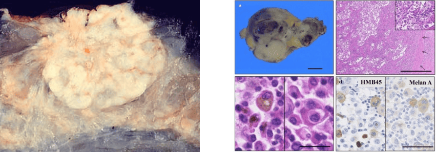
Metastasis: Breast carcinoma and lymph node metastasis
B. Hematogenous (Blood-Borne) Spread
Characteristic of sarcomas, but also frequently utilized in advanced stages of carcinomas. The anatomical distribution of secondary hematogenous deposits is largely governed by venous drainage patterns:
| Venous System | Primary Neoplastic Source | Primary Anatomical Site of Metastasis |
|---|---|---|
| Systemic Venous Circulation (Caval System) | Organs draining into the superior or inferior vena cava (e.g., breast, kidney, osteosarcoma) | Tumor emboli pass through the right cardiac chambers into pulmonary arteries → Lungs (pulmonary metastases). |
| Pulmonary Venous Circulation | Primary pulmonary carcinoma or pulmonary metastatic deposits | Emboli drain into pulmonary veins → left cardiac chambers → systemic arterial circulation → widespread deposits in liver, bone, brain, adrenal glands. |
| Portal Venous System | Gastrointestinal tract viscera (e.g., stomach, colon, rectum, pancreas) | Direct portal venous transport into hepatic sinusoids → Liver (hepatic metastases). Further spread via hepatic veins → IVC → Lungs. |
| Vertebral Venous Plexus (Batson's Plexus) | Pelvic, abdominal, or thoracic organs (e.g., prostate, urinary bladder, breast) | Emboli bypass the pulmonary circulation through valveless vertebral-portal/caval anastomoses → direct metastasis to vertebral bodies, spinal cord, and brain without preliminary lung lesions. |
Pathology of Organ Metastases
- High-Frequency Sites: Liver, lungs, skeletal bones, and brain receive high cardiac output and possess permissive vascular endothelium for arrest and homing.
- Low-Frequency (Rare) Sites: Spleen, skeletal muscle, pancreas, and small intestine rarely host metastases despite abundant perfusion (hypothesized local protease inhibitors, lactic acid, and mechanical contractile disruption).
- Gross Morphology: Metastatic visceral deposits classically appear as multiple, scattered, spherical, well-circumscribed nodules of variable dimensions, frequently showing central necrosis and umbilication.
- Microscopic Morphology: Secondary colonies preserve architectural features reminiscent of the primary site (e.g., mucin-producing adenocarcinoma glands within the pulmonary parenchyma).
- Skeletal Metastases Variants:
- Osteolytic Metastases: Predominant form (e.g., lung, breast, thyroid, kidney carcinomas); osteoclast activation produces bone resorption and punched-out lytic lesions prone to pathologic fractures.
- Osteosclerotic (Osteoblastic) Metastases: Classically seen in prostatic adenocarcinoma; neoplastic prostatic epithelial cells secrete alkaline phosphatase and osteoblast-stimulating factors, inducing dense woven bone formation surrounding metastatic deposits.
Spread Through Body Cavities (Transcoelomic Spread)
Occurs when an invasive malignant tumor penetrates through the natural serosal covering (visceral peritoneum, pleura, pericardium) of an organ, exfoliating malignant cells directly into fluid-filled serous cavities:
- Pathological Consequences: Exfoliated cells seed peritoneal or pleural surfaces, inducing inflammatory angiogenesis and exudation termed hemorrhagic effusion (hemorrhagic ascites, hemorrhagic pleural effusion).
- Transperitoneal Spread: Carcinomas of the stomach, colon, and pancreas seed the peritoneal cavity and omentum, resulting in a diffuse, nodular, fibrous omental infiltration ("omental caking") and malignant ascites.
- Krukenberg Tumors: Bilateral ovarian enlargement characterized by metastatic mucin-rich signet ring cell adenocarcinoma.
- Historical Concept: Classically attributed solely to direct transperitoneal "drop" seeding from a primary gastric adenocarcinoma.
- Current Consensus: Predominantly mediated by retrograde lymphatic transport or selective hematogenous spread, confirmed by primary origins in breast, gallbladder, biliary tract, and urinary bladder cancers.
- Transpleural and Transpericardial Spread: Direct extension of primary lung or breast carcinomas across visceral pleura/pericardium into pleural/pericardial spaces, forming extensive diaphragmatic tumor nodules accompanied by hemorrhagic effusion.
- Cerebrospinal Fluid (CSF) Seeding: Primary intracranial tumors (e.g., medulloblastoma, ependymoma) shed cells into ventricular CSF, producing seeding ("drop metastases") along the ependymal lining of cerebral ventricles, base of the skull, and spinal thecal sac.
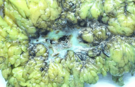
Endometrial carcinoma: transcoelomic spread
Other Methods of Spread
- 1. Transluminal Spread: Detached neoplastic cells transit within natural anatomical lumina to implant into downstream mucosa. Classically demonstrated when malignant cells from a transitional cell carcinoma (TCC) of the renal pelvis travel through the ureteral lumen to implant directly into the mucosal lining of the urinary bladder.
- 2. Surgical Implantation / Inoculation:
- Iatrogenic Mechanical Transfer: Surgical scalpels, trocars, needles, or contaminated instruments transfer viable malignant cells into the surgical incision or wound site during operative resection.
- Contact (Kissing) Implantation: Direct continuous apposition of epithelial surfaces enables local mechanical seeding, such as squamous cell carcinoma of the lower lip implanting onto the opposing surface of the upper lip.
Systemic Manifestations and Causes of Death in Malignant Disease
Malignant neoplasia causes patient mortality through a combination of systemic metabolic decompensation and critical organ failure:
| Systemic Complication | Etiological Factors | Pathophysiological Impact |
|---|---|---|
| 1. Malnutrition & Cachexia | Macrophage-derived Tumor Necrosis Factor-alpha (TNF-α / Cachectin), interleukins, toxic tumor metabolites, anorexia, mechanical luminal obstruction | Severe protein-calorie malnutrition characterized by progressive profound wasting of skeletal muscle mass and adipose tissue, accompanied by weakness, fatigue, and severe secondary immunodeficiency. |
| 2. Progressive Anemia | Erosion/invasion of blood vessels, bone marrow replacement (myelophthisis), autoimmune hemolysis, folate deficiency | Decreased tissue oxygenation causing fatigue, lethargy, pallor, and progressive high-output cardiac strain. |
| 3. Secondary Infections | Severe neutropenia, immunodepression from cachexia, mucosal barrier disruption, urinary/bronchial obstruction | Bronchopneumonia, septicemia, and opportunistic systemic infections representing direct immediate terminal events. |
| 4. Vital Organ Destruction | Mass effect, parenchymal destruction, metabolic compromise |
|
Epithelial Tumors
Neoplasms that originate from epithelial cells lining external body surfaces, internal mucosal tracts, and parenchyma of exocrine/endocrine glands. These tumors are stratified into benign (localized, non-invasive) and malignant (infiltrative, metastatic) classifications.
A. Benign Tumors of Epithelial Origin
1. Papilloma
Definition: A benign, exophytic, non-encapsulated neoplasm derived from surface epithelium.
- Classical Exophytic Architecture: Proliferating epithelial cells forming projecting, finger-like (papillary) fronds that rest upon an intact basement membrane, supported internally by a central fibrovascular connective tissue core containing nutritive blood vessels.
- Inverted (Endophytic) Architecture: Rare variant in which proliferating epithelial nests push the basement membrane and invaginate into the subepithelial stroma without crossing or degrading the basement membrane (e.g., inverted papilloma of the sinonasal tract and urinary bladder).
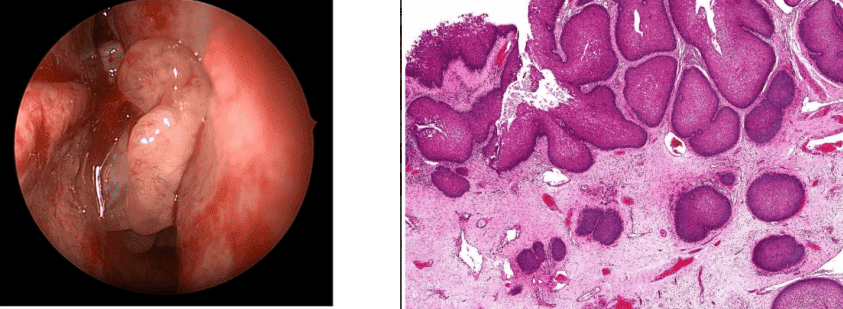
Inverted papillomas
Subtypes of Classical Papilloma
| Papilloma Type | Epithelial Origin & Anatomic Sites | Morphology & Clinical Complications |
|---|---|---|
| Squamous Cell Papilloma | Stratified squamous epithelium: Skin, lip, tongue, oral mucosa, pharynx, larynx, esophagus, uterine cervix, vagina, anal canal. | Gross: Small, sessile or pedunculated projections resembling a cauliflower. Micro: Thick, branched, hyperplastic stratified squamous epithelium covering vascular fibrous cores. |
| Transitional Cell Papilloma (Villous Papilloma) | Urothelium: Urinary bladder, ureter, renal pelvis. | Gross: Delicate, non-capsulated, velvety, friable mass of papillary fronds. Micro: Vascular cores lined by normal-appearing transitional epithelium ≤ 6 layers thick. Complications: Painless hematuria, luminal obstruction, high risk of malignant transformation into TCC. |
| Duct Papilloma | Columnar/cuboidal epithelium of major ducts: Lactiferous ducts of the breast (most common), pancreatic ducts. | Gross: Small, complex, solitary papillary projection inside a dilated duct lumen. Micro: Delicate vascular fronds lined by regular ductal epithelial cells. Complications: Serosanguinous/bloody nipple discharge, potential malignant transformation into ductal carcinoma. |
| Mucous Cell Papilloma (Adenomatous Polyp) | Mucosal columnar epithelium: Gastrointestinal tract (stomach, colon, rectum). | Gross: Sessile, pedunculated, or complex velvety mucosal projection. Micro: Crowded, proliferated mucosal glands lined by columnar cells on fibrovascular stroma. Complications: Occult/gross gastrointestinal bleeding, mechanical intussusception/obstruction (e.g., pyloric), malignant transformation into adenocarcinoma. |
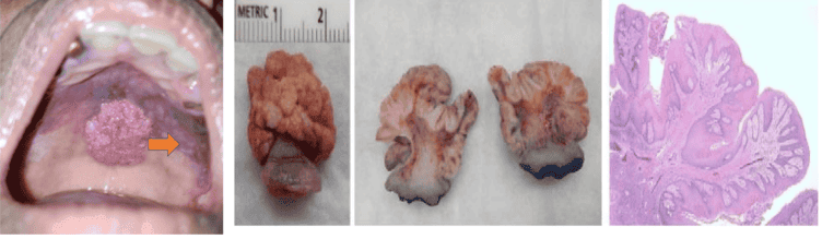
Squamous cell papillomas
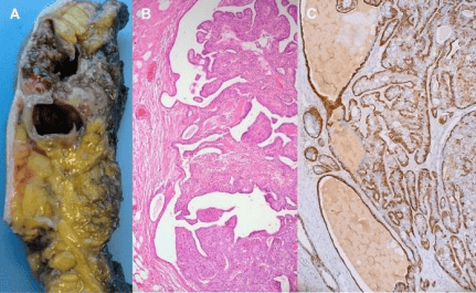
Intraductal papilloma: Gross, H&E, and Cytokeratin 14 immunohistochemistry
2. Adenoma
Definition: A benign epithelial neoplasm that arises from glandular epithelium or forms glandular patterns.
- Sites of Origin:
- Solid exocrine and endocrine glands: Breast, ovary, salivary glands, pancreas, thyroid, adrenal cortex, pituitary.
- Mucosal glandular surfaces: Gastrointestinal mucosa (colon, rectum, stomach), endometrium.
- Gross Morphology: Classically presents as a well-circumscribed, encapsulated, globular or ovoid solid mass.
- Microscopic Spectrum of Adenomas:
- Simple Adenoma (Tubular Adenoma): Proliferating acini or tubules lined by uniform epithelium, separated by delicate fibrovascular stroma.
- Fibroadenoma: Mixed proliferation of glandular epithelium and dense stromal fibrous connective tissue. The breast is the classic and primary site of occurrence.
- Cystadenoma: Adenoma with abundant retained glandular secretions leading to profound cystic dilation (e.g., ovarian cystadenoma).
- Papillary Cystadenoma: A cystadenoma characterized by marked intracystic epithelial proliferation that projects into the fluid-filled cavity as complex papillae (e.g., papillary cystadenoma of the ovary).
- Pleomorphic Adenoma: Mixed salivary gland adenoma exhibiting epithelial ductal elements embedded within a myxoid stroma rich in mucin and pseudocartilaginous/chondroid tissue.
- Warthin's Tumor (Papillary Cystadenoma Lymphomatosum): Salivary gland adenoma (parotid gland) characterized by cystic spaces lined by a double layer of large, eosinophilic oncocytic epithelial cells supported by a prominent reactive lymphoid stroma containing germinal centers.
- Complications of Adenomas:
- Autonomous, unregulated secretion of hormones (e.g., functional thyroid adenoma causing thyrotoxicosis; functional adrenocortical adenoma).
- Secondary malignant transformation into adenocarcinoma (notably high in colonic adenomatous polyps).
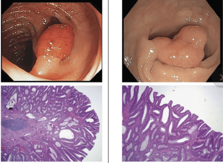
Adenoma: Small and large colonic polyps with corresponding histology
Malignant Epithelial Tumors (Carcinomas)
Carcinomas are broadly divided based on origin into carcinomas of surface epithelium and carcinomas of glandular origin.
I. Carcinomas of Surface Epithelium
1. Transitional Cell Carcinoma (TCC / Urothelial Carcinoma)
- Origin: Arises from the specialized urothelium lining the urinary tract. May arise de novo or evolve through progression of a pre-existing villous papilloma.
- Predominant Sites: Urinary bladder (most common), ureter, and renal pelvis.
- Gross Patterns:
- Papillary Type: Exophytic, villous, friable growth supported by a broad, indurated, infiltrating base.
- Non-Papillary Types: Large fungating polypoid masses, malignant excavated ulcers with everted margins, or diffuse wall-thickening infiltrative patterns.
- Microscopic Subtypes:
- Papillary TCC: Pleomorphic, hyperchromatic malignant transitional cells overlying branched fibrovascular stroma, measuring substantially more than 6 cell layers thick. Invades less deeply than non-papillary variants; associated with superior overall prognosis.
- Non-Papillary TCC: Anaplastic malignant transitional cells arranged in solid cohesive sheets and cords; early invasion of detrusor muscle layer; poorer prognosis.
- Modes of Spread: Direct local infiltration through muscularis propria into perivesical fat; distant metastasis via lymphatics (regional pelvic/iliac nodes) and late hematogenous dissemination; transluminal implantation down the urinary tract.
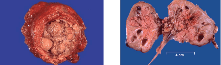
Transitional cell carcinoma (Urothelial carcinoma) of bladder and renal pelvis
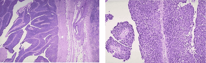
Urothelial carcinoma: Low and medium-power microscopic views
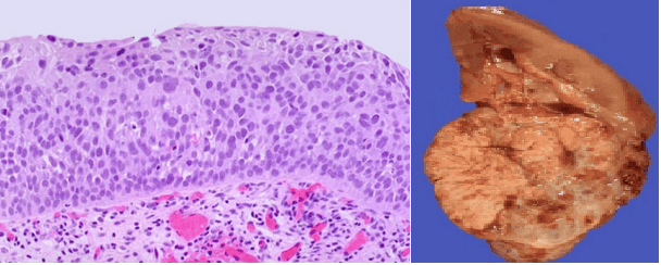
Urothelial carcinoma in situ (microscopic) and Renal cell carcinoma (gross)
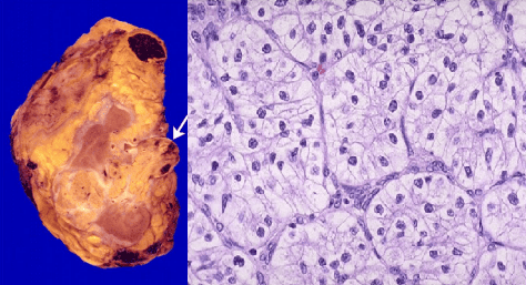
Renal cell carcinoma: Renal vein invasion and classic clear cell histology (VHL mutation)
Comparison: Squamous Cell Carcinoma vs. Basal Cell Carcinoma
| Feature | Squamous Cell Carcinoma (SCC / Epidermoid) | Basal Cell Carcinoma (BCC / Rodent Ulcer) |
|---|---|---|
| Definition & Cell of Origin | Malignant neoplasm arising from stratified squamous epithelium. | Locally aggressive malignant neoplasm arising from the basal layer of the epidermis and hair follicle adnexa. |
| Anatomical Sites |
|
|
| Predisposing Factors | Prolonged ultraviolet (sun) radiation, arsenic, industrial tars, HPV infection, chronic ulcers (Marjolin ulcer), squamous metaplasia, leukoplakia. | Prolonged, severe sun/UVB radiation exposure; light skin complexion; genetic mutations (PTCH gene / basal cell nevus syndrome). |
| Gross Morphology |
|
Initiates as a firm, smooth, pearly-white or erythematous papule with telangiectasias; breaks down centrally into an ulcer with raised, rolled-in, beaded pearly margins, an irregular rough floor, and a firm indurated base. |
| Microscopic Architecture | Infiltration of dermis by cords/islands of malignant squamous cells forming concentric lamellated "cell nests" (keratin pearls). Nest periphery reflects basal cells → prickle cells with intercellular bridges → central laminated concentric acellular keratin. Graded by Broder's Criteria based on proportion of cell nests. |
Dermis infiltrated by solid lobules/islands of small, uniform, basaloid epithelial cells. Distinctive peripheral palisading (parallel columnar cells arranged perpendicularly) surrounding central disorganized polyhedral cells. May show retraction artifact from surrounding stroma and melanin pigmentation. |
| Metastatic Potential & Spread | Aggressive potential: Infiltrates locally, invades regional lymphatics early, and disseminates via blood vessels in late stages. | Locally destructive ("rodent" action): Infiltrates and destroys deep facial cartilage and bone. Metastasis is extremely rare (< 0.1%); may rarely undergo basosquamous transformation leading to distant metastases. |
Broder's Classification of Squamous Cell Carcinoma
- Grade I: Well-differentiated; 75% to 100% of the tumor is composed of keratinizing cell nests.
- Grade II: Moderately differentiated; 50% to 75% cell nests.
- Grade III: Poorly differentiated; 25% to 50% cell nests; marked anaplasia.
- Grade IV: Anaplastic; 0% to 25% cell nests; minimal or undetectable keratinization; high mitotic activity.
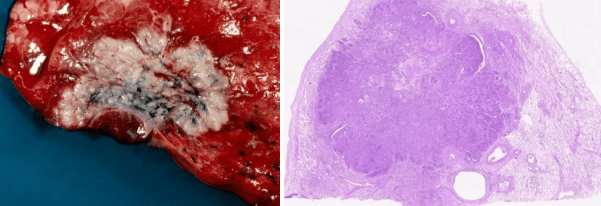
Squamous cell carcinoma: Gross appearance and histopathological section
II. Carcinomas of Glandular Origin
Malignancies arising from specialized secretory glandular epithelium located either within mucosal linings (gastrointestinal mucosa, endometrium, endocervix) or within solid parenchyma of exocrine/endocrine glandular organs (breast, prostate, pancreas, ovary, salivary glands).
- Gross Morphology:
- Mucosal Surfaces: Fungating cauliflower-like polypoid mass, malignant excavated ulcer with indurated margins, or diffuse infiltrative pattern (e.g., napkin-ring annular constriction in the left colon; diffuse leather-bottle linitis plastica in the stomach).
- Solid Glands: Hard, irregular, non-encapsulated, infiltrative masses with ill-defined borders anchoring to surrounding fat and fascia.
- Spread: Aggressive local invasion; extensive regional lymphatic drainage; late hematogenous dissemination; frequent transcoelomic seeding when reaching serosa.
Microscopic Categories of Glandular Carcinoma
| Histological Type | Distinguishing Pathological Features | Primary Anatomical Sites |
|---|---|---|
| 1. Adenocarcinoma (Classical) | Forms distinct malignant glandular acini. Differs from normal acini by showing:
|
Gastrointestinal tract (stomach, colon), gallbladder, pancreas, endometrium, endocervix, lung, breast, prostate. |
| 2. Mucin-Secreting Carcinomas | Characterized by copious mucin synthesis:
|
Stomach, colon, breast, gallbladder. |
| 3. Carcinoma Simplex (Undifferentiated / Spheroidal Cell) | High-grade, poorly differentiated malignant epithelial tumor of glandular origin. Exhibits neither glandular acinar formation nor mucin secretion. Malignant cells proliferate as solid syncytial groups, sheets, or trabeculae surrounded by fibrous stroma. | Breast (most common), gastrointestinal tract. |
| 4. Scirrhous Carcinoma | Grossly stony-hard, gritty, contracted mass with ill-defined margins. Microscopically characterized by small, compressed cords or single malignant cells embedded in a dense, prominent, desmoplastic collagenous stroma (exceedingly high stroma-to-tumor ratio). | Breast (invasive ductal carcinoma), stomach (linitis plastica). |
| 5. Medullary Carcinoma (Encephaloid) | Grossly soft, circumscribed, fleshy (brain-like: encephaloid), expansile tumor. Microscopically composed of large, cohesive sheets of vesicular malignant cells with scant fibrous stroma, prominent geographic necrosis, and a characteristic prominent lymphoplasmacytic infiltrate. | Breast, thyroid gland (medullary thyroid carcinoma derived from parafollicular C-cells). |
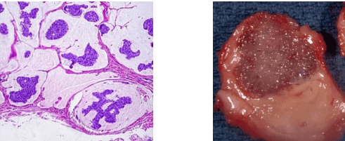
Mucin or Colloid carcinoma: Histological pools of mucin and gelatinous gross cut surface
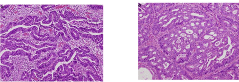
Well-differentiated (tubular) and moderately differentiated (cribriform) adenocarcinoma
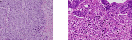
Poorly differentiated adenocarcinoma with marked tumor budding at the invasive front
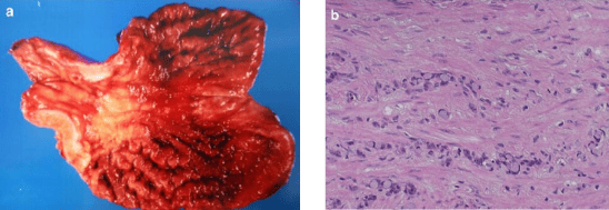
Scirrhous carcinoma (Linitis plastica): Gross gastric transmural thickening and dense fibrous stroma
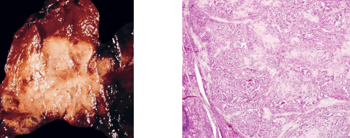
Medullary carcinoma: Solid circumscribed mass with neuroendocrine sheet-like growth
Specialized Types of Carcinoma
- Hepatocellular Carcinoma (HCC): Malignant neoplasm of the liver originating directly from mature hepatocytes. Strongly linked to chronic hepatitis B/C viral infections and hepatic cirrhosis. Grossly presents as expanding, confluent, bile-stained nodules bulging against fibrous bands; microscopically exhibits a prominent macrotrabecular and sinusoidal pattern.
- Choriocarcinoma: Highly aggressive, gestational malignant neoplasm originating from trophoblastic tissue of the placenta. Grossly soft, fleshy, friable, with extensive central ischemic necrosis and massive hemorrhage. Microscopically shows a characteristic dimorphic (biphasic) population of mononucleated cytotrophoblasts capped by multinucleated syncytiotrophoblasts, completely lacking true chorionic villi. Invades myometrium and disseminates via hematogenous routes early (lungs, brain).
- Salivary Gland Carcinomas: Primary malignant tumors of major and minor salivary glands, including:
- Adenoid Cystic Carcinoma: Characterized by an infiltrative cribriform ("Swiss cheese") pattern of basaloid cells with a high propensity for perineural invasion.
- Mucoepidermoid Carcinoma: Composed of an intimate mixture of squamoid, intermediate, and mucin-secreting cells.
- Acinic Cell Carcinoma: Composed of serous acinar cells displaying cytoplasmic zymogen-like granules.
- Renal Cell Carcinoma (RCC): Arises from renal tubular epithelium; predominantly clear cell carcinoma linked to von Hippel-Lindau (VHL) gene inactivation. Exhibits marked tendency for invasion into the renal vein and inferior vena cava.
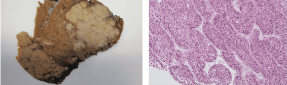
Hepatocellular carcinoma: Bulging nodular gross liver mass and macrotrabecular histology
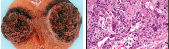
Choriocarcinoma: Gross hemorrhagic mass and biphasic trophoblastic histology
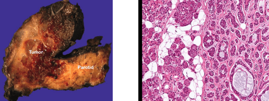
Adenoid cystic carcinoma: Infiltrative parotid mass with classic cribriform microscopic architecture
Summary of Neoplastic Spread and Epithelial Tumors
Key Takeaways
- Definition of Metastasis: The biological cascade wherein neoplastic cells detach from a primary tumor, invade extracellular stroma, intravasate into vascular or lymphatic channels, circulate, survive host defenses, extravasate, and establish viable secondary colonies at distant anatomical sites.
- Phenotypic Fidelity: Metastatic colonies consistently reproduce the biological, histological, and immunohistochemical profile of the primary tumor from which they originate.
- Epithelial Tumors Spectrum: Epithelial neoplasms arise from mucosal coverings, skin, or glandular parenchyma throughout the body. Benign epithelial tumors (papillomas, adenomas) remain localized and non-invasive, whereas malignant epithelial tumors (squamous cell carcinomas, adenocarcinomas, urothelial carcinomas) demonstrate progressive local invasion and widespread metastatic potential.