Introduction to General Pathology
Conceptual Foundations of Pathology
Key Definitions
- Pathology (Etymology & Definition): Rooted in the classical Greek terms "pathos" (signifying suffering or ailment) and "logos" (denoting systematic study or discourse). It represents the scientific exploration of bodily structural and functional aberrations induced by disease processes.
- Integrative Scientific Bridge: Pathology functions as the fundamental nexus connecting preclinical biomedical sciences (human anatomy, medical physiology, and physiological chemistry) with clinical diagnostics, therapeutics, and bedside management. A master-level comprehension of disease mechanics is an indispensable prerequisite for all clinical practitioners to decode, evaluate, and manage human disease.
- State of Health: A harmonious physiological baseline wherein an individual experiences complete biological, biochemical, and environmental equilibrium with their surroundings.
- Disease (Loss of Ease): A disruption of standard physiological harmony, characterized by structural lesions or functional impairments that cause biological disharmony or discomfort ("dis-ease").
- The Patient: The human host or biological subject experiencing the functional disruptions and structural derangements of a disease state.
- Lesion: Any measurable, discrete structural or functional alteration developed within cells, tissues, or organs as a direct consequence of a disease process or experimental exposure.
- Pathologic Morphology: The rigorous macroscopic and microscopic diagnostic inspection of diseased biological specimens to delineate specific alterations.
Curricular Learning Outcomes
Following this instructional unit, students are expected to achieve mastery across several core domains:
- Delineate the diagnostic boundaries, clinical scope, and foundational role of pathology within modern medicine.
- Differentiate general pathology (mechanisms governing basic cellular injury, inflammation, repair, and neoplasia) from systemic pathology (organ-specific and system-restricted disease presentations).
- Appraise the central utility of pathology in clinical disease identification (diagnosis), rational therapy selection, prognostic forecasting, and population-level preventive strategies.
Historical Evolution of Pathological Sciences
Humanity’s interpretation of physical suffering has evolved from primordial mystical beliefs to modern molecular medicine. This intellectual trajectory is structured across distinct historical epochs:
| Historical Era & Pioneer | Key Milestone or Scientific Discovery | Enduring Impact on Modern Pathology |
|---|---|---|
| Primitive & Supernatural Era | Attributed illness to curses, wrath of deities, malevolent magic, and demonic possession. Interventions relied on clerical prayers, ritual sacrifice, and faith healing. | Highlights historical roots; these traditional beliefs still coexist alongside evidence-based medicine in various global communities today. |
| Hippocrates of Kos (460–370 BC) "Father of Medicine" |
Departed from supernatural dogmas; introduced rational clinical inquiry, systematic symptom observation, bedside diagnostics, and professional ethics via the Hippocratic Oath. | Established clinical observation and medical ethics as the foundation of empirical practice. |
| Aulus Cornelius Celsus (c. 25 BC – 50 AD / 53–7 AD) |
Formulated the four cardinal physical signs of acute inflammation:
• Rubor (erythema/redness) • Tumor (edema/swelling) • Calor (localized warmth/heat) • Dolor (hyperalgesia/pain) |
Remains the cornerstone bedside clinical framework for recognizing acute vascular and exudative tissue inflammation. |
| Claudius Galen (130–200 AD) |
Integrated classical humoral doctrines; performed extensive mammalian dissections; described loss of function (functio laesa) as an outcome of severe inflammation. | Dominated medical dogma for over a millennium until the Renaissance anatomical revolution. |
| Andreas Vesalius (1514–1564) |
Pioneered direct human cadaveric dissection using executed criminals, correcting historical anatomical inaccuracies in his landmark work De Humani Corporis Fabrica. | Sparked the rise of European anatomical amphitheaters and made dissection central to medical training. |
| Gabriel Fallopius (1523–1562) |
Identified and comprehensively described the uterine oviducts (Fallopian tubes). | Expanded macroscopic reproductive anatomical mapping. |
| Hieronymus Fabricius (1537–1619) |
Discovered the specialized gut-associated lymphoid diverticulum in birds, termed the Bursa of Fabricius. | Laid the embryological and immunological basis for identifying B-lymphocyte maturation pathways. |
| Antonie van Leeuwenhoek (1632–1723) |
A Dutch draper who ground high-magnification optical lenses, creating the first functional single-lens microscope to visualize unicellular microorganisms and spermatozoa. | Introduced the sub-macroscopic world to biological science, making modern microscopy possible. |
| Giovanni Battista Morgagni (1682–1771) "Father of Anatomic Pathology" |
Italian anatomist who systematically matched ante-mortem clinical records with post-mortem structural autopsy discoveries in De Sedibus et Causis Morborum. | Established the principle of clinicopathologic correlation, showing that symptoms reflect specific organ lesions. |
| Sir Percival Pott (1714–1788) |
Documented an unusually high incidence of scrotal squamous cell carcinoma among young chimney sweeps in London (1775), identifying chimney soot as the culprit agent. | Pioneered occupational epidemiology and proved that chemical exposures can act as environmental carcinogens. |
| John & William Hunter (1718–1793) |
Scottish surgeons and anatomists who curated extensive museum collections of pathological human organ specimens. | Founded pathological anatomy museums, providing permanent visual references for medical education. |
| Carl Freiherr von Rokitansky (1804–1878) |
Austrian pathologist who personally conducted over 30,000 post-mortem examinations, documenting gross morbid anatomy in detail. | Standardized necropsy protocols and expanded gross pathological catalogs. |
The 19th & 20th Century: Cellular Pathology and Technological Advances
During the latter half of the 19th century, diagnostic pathology transitioned from macroscopic morbid anatomy to microscopic analysis, driven by microtomy, optical refinement, and the emergence of synthetic histological dyes:
| Investigator | Technological & Scientific Discovery | Diagnostic Significance |
|---|---|---|
| Louis Pasteur (1822–1895) |
Disproved spontaneous generation; proved the germ theory of disease; developed sterilization techniques and microbial vaccines. | Established that specific micro-organisms serve as exogenous etiologic agents of infectious diseases. |
| Gerhard H. Armauer Hansen (1841–1912) |
Identified Mycobacterium leprae (Hansen's bacillus) as the chronic pathogen causing leprosy. | Marked the first direct identification of a pathogenic bacterium responsible for a chronic human granulomatous disease. |
| Hans Christian Gram (1853–1938) |
Formulated differential bacteriological staining using crystal violet, iodine mordant, and counterstain. | Segregated the bacterial kingdom into Gram-positive and Gram-negative categories based on cell wall composition. |
| Dmitri L. Romanowsky (1861–1921) |
Developed polychromatic staining using balanced combinations of methylene blue and eosin derivatives. | Enabled fine morphological examination of peripheral blood elements, bone marrow, and intracellular parasites. |
| Robert Koch (1843–1910) |
Developed bacterial heat-fixation and aniline dye staining; isolated Mycobacterium tuberculosis (1882/1883) and Vibrio cholerae. | Established Koch's postulates, linking specific microorganisms to defined infectious pathologies. |
| May-Grünwald, Giemsa, & Leishman | Standardized modifications of Romanowsky-type blood and tissue stains. | Enabled routine staining for hematopathology, bone marrow aspiration cytology, and hemoparasite detection (such as Plasmodium). |
| Robert Feulgen (1884–1955) |
Developed the Feulgen acid-hydrolysis reaction, which selectively stains nuclear deoxyribonucleic acid. | Early biochemical proof that DNA is concentrated in nuclear chromatin. |
| Rudolf Virchow (1821–1905) "Father of Cellular Pathology" |
Formulated the dictum "Omnis cellula e cellula" (all cells arise from preexisting cells). Stated that disease represents structural and functional disruptions at the cellular level. Widely known across Europe as the "Pope of Pathology." | Shifted focus from whole-organ pathology to cellular-level analysis, establishing microscopic histopathology as an investigative standard. |
| Karl Landsteiner (1868–1943) "Father of Blood Transfusion" |
Discovered the major human ABO blood group antigens (awarded the Nobel Prize in Physiology or Medicine in 1930). | Made safe blood transfusion, immunohematology, and pre-transfusion cross-matching clinically viable. |
| Ernst Ruska & Bodo von Borries (1933) |
Constructed the first transmission electron microscope, bypassing the resolution limits of light physics. | Allowed visualization of viral particles, intracellular organelles, and submicroscopic features (e.g., glomerular podocyte foot processes). |
| George N. Papanicolaou (1883–1962) |
Developed diagnostic exfoliative cytology and the specialized polychrome stain known as the Pap smear. | Helped establish population-level screening for pre-invasive cervical dysplasia and early carcinoma. |
| William Boyd (1885–1979) |
Eminent pathologist and educator whose narrative textbooks guided pathology instruction worldwide for more than 50 years. | Shaped modern medical school curricula by emphasizing clinicopathologic reasoning. |
Modern Molecular Pathology & Genomic Medicine (1950s–Present)
From the mid-20th century into the 21st century, pathology has expanded from cellular morphologic inspection to the molecular and genetic characterization of disease:
Milestones in Molecular Pathology
- Structural Elucidation of DNA (1953): James Watson and Francis Crick described the antiparallel double-helix structure of deoxyribonucleic acid, providing the molecular model for genetic replication and translation.
- Confirmation of the Human Diploid Karyotype (1956): Joe Hin Tjio and Albert Levan determined that the normal human somatic cell nucleus contains exactly 46 chromosomes (23 pairs), rather than the previously accepted 48, establishing the foundation of human cytogenetics.
- Chromosomal Transposition & Genome Dynamism (1983 Nobel Prize): Barbara McClintock demonstrated mobile genetic elements ("jumping genes" or transposons), proving that genomes possess structural dynamism rather than static sequences.
- In Vitro Isolation of Human Stem Cells (1998): Researchers isolated and cultivated pluripotential embryonic stem cells, inaugurating the fields of regenerative medicine, tissue repair, and experimental modeling for disorders such as Alzheimer's, stroke, diabetes, and various malignancies.
- Completion of the Human Genome Project (April 2003): Successfully sequenced the complete human genome, revealing that human somatic nuclei house approximately 3 billion chemical base pairs encoding roughly 30,000 distinct genes distributed across 23 chromosome pairs. This catalog serves as the foundation for modern personalized and precision oncology.
Primary Subdivisions of Pathology
To systematically organize the study of disease, pathology is structured into two main academic domains:
| Pathological Division | Operational Scope & Focus | Representative Subject Domains |
|---|---|---|
| General Pathology | Investigates the universal, non-specific biological and biochemical reactions of cells, tissues, and systems to diverse injurious stimuli. |
• Reversible cell injury, cellular adaptations (hypertrophy, hyperplasia, atrophy, metaplasia), and cell death (necrosis vs. apoptosis) • Acute and chronic inflammation, wound healing, tissue repair, and fibrosis • Hemodynamic disruptions: edema, passive congestion, thrombosis, embolism, infarction, and circulatory shock • Immunopathology: hypersensitivity disorders, autoimmune diseases, primary and secondary immunodeficiency states • Neoplasia: benign and malignant carcinogenesis, tumor biology, grading, and staging • Medical genetics, environmental and nutritional deficiencies or toxicities • Systemic metabolic derangements (such as Diabetes Mellitus) • Fundamental functional organ evaluation (such as renal and liver function panels) |
| Systemic Pathology | Focuses on how general pathological processes manifest in specific anatomical organ systems and tissue beds. |
• Cardiovascular pathology (e.g., atherosclerosis, ischemic heart disease, vasculitis) • Respiratory pathology (e.g., lobar pneumonia, chronic obstructive pulmonary disease, pulmonary tuberculosis) • Gastrointestinal, hepatic, renal, endocrine, reproductive, musculoskeletal, and central nervous system diseases |
The Four Core Aspects of Disease
In diagnostic and academic pathology, every disease process is analyzed through four essential pillars:
Pillar 1: Etiology (The "Why")
Refers to the underlying cause or initiating trigger of a disease. If the exact causative mechanism is identified, it is designated as a primary etiology. When a disease arises without a known cause, it is termed idiopathic (essential or cryptogenic). Identifying the primary etiology remains the cornerstone for diagnostic formulation, pathophysiology analysis, and rational therapeutics.
Pillar 2: Pathogenesis (The "How")
The stepwise biological, cellular, and molecular progression through which the primary etiology generates characteristic structural and functional lesions. Pathogenetic cascades often initiate during a subclinical, latent, or incubation phase before symptoms appear. This molecular progression directly drives the morphologic changes observed in affected tissues.
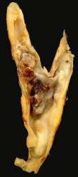
Vascular Atheroma and Thrombotic Occlusion
Pillar 3: Morphologic Changes
The observable structural, architectural, and cellular alterations that occur in tissues following pathogenetic damage. Morphology is divided into:
- Macroscopic / Gross Pathology: Structural organ derangements visible to the unaided eye during anatomical dissection, surgical inspection, or gross examination (e.g., tissue enlargement, induration, pallor, cavitation, necrosis).
- Microscopic Changes (Histopathology / Cytopathology): Cellular and sub-cellular alterations visualized using light or electron microscopy (e.g., loss of nuclear polarity, hyperchromasia, coagulative necrosis, granuloma formation).
Certain diseases display characteristic, pathognomonic morphologic changes that allow for definitive diagnosis. Examples include identifying acid-fast bacilli in sputum for tuberculosis, or detecting classic Reed-Sternberg cells to confirm Hodgkin lymphoma.
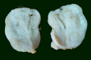
Gross Macroscopic Tissue Specimen
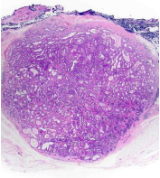
Microscopic Section (H&E Staining)
Pillar 4: Functional Derangements & Clinical Significance
Morphological changes lead to physiological and biochemical derangements that disrupt normal organ function. This functional impairment dictates the clinical presentation:
- Symptoms: Subjective functional complaints reported by the patient (e.g., pain, malaise, nausea, dyspnea).
- Signs: Objective, measurable anatomical or physiological abnormalities detected by the clinician (e.g., fever, tachycardia, jaundice, organomegaly, hypertension).
Together, these findings shape the clinical trajectory, potential complications, response to therapy, and overall prognosis of the disease.
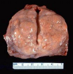
Gross Surgical Pathology Specimen
Diagnostic Modalities in Laboratory Medicine
Pathologists utilize a range of anatomical, cytological, biochemical, and molecular techniques to arrive at a definitive diagnosis:
| Diagnostic Modality | Methodology & Biological Specimen | Primary Clinical Applications |
|---|---|---|
| Histopathology | Microscopic evaluation of intact, chemically fixed, paraffin-embedded, microtome-sectioned tissue samples obtained via surgical biopsy or resection. | Considered the gold standard for diagnosing neoplasms, evaluating surgical resection margins, grading tumors, and assessing inflammatory diseases. |
| Cytopathology | Evaluation of detached cells or cellular clusters obtained via fine-needle aspiration, spontaneous exfoliation into body fluids, or manual scraping of epithelial surfaces. | Screening for pre-invasive dysplasia (e.g., Pap smear), diagnosing palpable masses (e.g., thyroid or breast nodules), evaluating cavity effusions, and monitoring cancer recurrence. |
| Hematopathology | Analysis of peripheral blood smears, bone marrow aspirates, and trephine core biopsies using Romanowsky-type stains and flow cytometry. | Diagnosing anemias, leukemias, myeloproliferative disorders, myelodysplastic syndromes, and coagulation abnormalities. |
| Immunohistochemistry (IHC) | Application of labeled antibodies to identify specific cellular antigens in tissue sections, visualized using enzymatic chromogens (e.g., DAB brown stain). | Identifying tumor lineage, classifying undifferentiated cancers, and detecting therapeutic predictive markers (e.g., ER, PR, HER2-neu in breast carcinoma). |
| Microbiological Examination | Analysis of body fluids, exudates, and tissue samples using microscopic staining (Gram, Ziehl-Neelsen), microbiological cultures, and serological assays. | Isolating and identifying bacterial, mycobacterial, fungal, and viral pathogens to guide targeted antimicrobial therapy. |
| Biochemical Examination | Automated quantitative measurement of electrolytes, enzymes, hormones, and metabolites in blood, serum, plasma, and urine. | Essential for detecting and monitoring metabolic diseases (e.g., blood glucose/HbA1c in diabetes) and evaluating organ function (renal and liver panels). |
| Cytogenetics | Evaluation of chromosome structure and number using karyotyping and molecular methods on germline or somatic cells. | Detecting congenital chromosomal anomalies (e.g., Down syndrome, Turner syndrome) and acquired translocations in hematologic cancers (e.g., Philadelphia chromosome). |
| Molecular Pathology | Targeted analysis of DNA and RNA using Polymerase Chain Reaction (PCR), Fluorescence In Situ Hybridization (FISH), and Next-Generation Sequencing (NGS). | Identifying oncogenic driver mutations (e.g., EGFR, BRAF, KRAS), detecting minimal residual disease, and guiding targeted therapies. |
| Autopsy Pathology | Systematic post-mortem dissection and macroscopic/microscopic examination of a deceased individual. Divided into forensic (medicolegal) autopsies and clinical (hospital) autopsies. | Determining the cause and manner of death, verifying diagnostic accuracy, supporting legal investigations, and providing clinical education. |
Histopathological Techniques & Biopsy Processing
Histopathology involves the microscopic study of diseased tissue sections. Tissue samples are acquired from living patients via surgical biopsy, categorized as:
- Incisional Biopsy: Surgical excision of a representative portion of a suspicious lesion for diagnostic assessment.
- Excisional Biopsy: Complete surgical excision of an entire lesion, often serving both diagnostic and therapeutic purposes.
Principles and Objectives of Tissue Fixation
Immediately following removal from the body, tissue must be placed in a sufficient volume of fixative, routinely 10% neutral buffered formalin (equivalent to a 4% aqueous solution of formaldehyde gas), unless intended for intraoperative frozen section diagnosis. The primary goals of fixation include:
- Arresting Autolysis and Putrefaction: Inactivates endogenous hydrolytic lysosomal enzymes that cause self-digestion and halts bacterial decomposition.
- Cross-Linking and Coagulation: Chemically cross-links and stabilizes cellular proteins, preventing the loss of soluble, diffusible components during subsequent processing.
- Structural Fortification: Hardens tissue architecture, preparing it to withstand the physical forces of dehydration, clearing, paraffin embedding, and microtome sectioning.
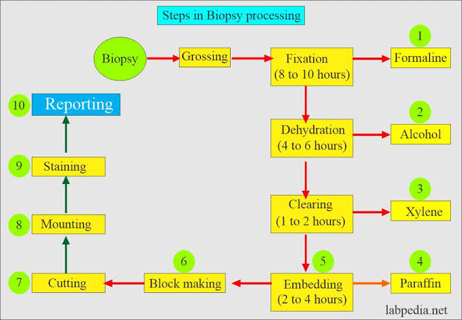
Steps in Biopsy Processing Protocol
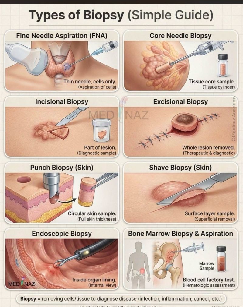
Common Clinical Biopsy Procedures
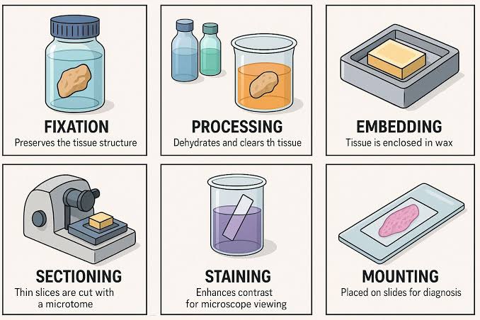
Histology Laboratory: Tissue Processing Sequence
Histological Staining Principles
Because processed tissue sections are translucent, selective chemical dyes are required to reveal cellular and tissue architecture:
Hematoxylin and Eosin (H&E) Staining
- Hematoxylin: A basic dye that binds to acidic, negatively charged cellular components (basophilic structures), particularly nuclear chromatin and ribonucleic acids, imparting a characteristic deep blue-purple coloration.
- Eosin: An acidic counterstain that binds to basic, positively charged proteins (eosinophilic structures), staining cytoplasm, intracellular proteins, collagen fibers, and the extracellular matrix a light to bright pink.
- Special & Ancillary Stains: Complementary methods utilized when H&E is insufficient, such as Periodic Acid–Schiff (PAS) for glycogen, mucin, and basement membranes; reticulin silver stains for tissue architecture; and targeted immunohistochemical markers.
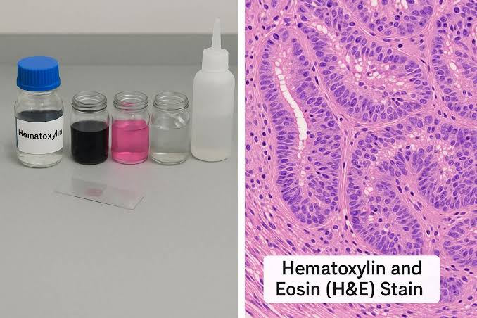
Representative Histopathology (H&E Stain)
Cytopathology: Principles & Clinical Applications
Cytopathology focuses on diagnosing disease through the evaluation of individual cells and small cell clusters detached from tissue surfaces:
| Cytopathologic Method | Mechanism of Specimen Collection | Target Tissues & Clinical Applications |
|---|---|---|
| Fine-Needle Aspiration Cytology (FNAC) | Cells are aspirated from solid or cystic lesions using a narrow-gauge needle (22–25 gauge) attached to a syringe under negative pressure. |
• Superficial palpable masses: Thyroid gland, breast, superficial lymph nodes, skin, and soft tissue nodules. • Deep non-palpable lesions: Lung, mediastinum, liver, pancreas, kidney, adrenal gland, and retroperitoneum, sampled under real-time imaging guidance (ultrasound, CT scan, or fluoroscopy). |
| Exfoliative Cytology | Examination of cells shed spontaneously into physiological secretions, natural conduits, or pathological fluid collections. |
• Sputum, bronchial washings/brushings • Cerebrospinal fluid (CSF) • Voided or catheterized urine • Body cavity effusions: pleural, peritoneal (ascites), and pericardial spaces • Nipple discharge and spontaneous vaginal discharge |
| Abrasive (Brush/Scrape) Cytology | Cells are manually dislodged from mucous membranes, epithelial surfaces, or serosal linings using a specialized spatula, brush, or swab. |
• Cervical scrapings (Pap smear) to detect pre-malignant dysplasia and carcinoma of the uterine cervix. • Gastrointestinal endoscopy brushings (esophagus, stomach, biliary tract). |
Clinical Applications & Strategic Advantages of Cytology
- Cancer Screening in Asymptomatic Populations: Routine Pap smear screening allows early detection and management of cervical intraepithelial neoplasia, helping reduce cervical cancer mortality. This is particularly relevant given that cervical carcinoma remains the most common cancer among women in Uganda.
- Primary Diagnosis in Symptomatic Disease: Works alongside physical examination and radiology (the clinical "triple assessment") to distinguish cystic, inflammatory, and neoplastic masses.
- Longitudinal Recurrence Surveillance: Serves as a non-invasive follow-up method to monitor cancer recurrence, such as using periodic voided urine cytology to detect recurrent urothelial carcinoma of the urinary bladder.
- Key Resource Advantages: Relative to formal surgical histopathology, cytology is cost-effective, rapid, and does not require general anesthesia or surgical suites. These features make it well-suited for resource-limited healthcare settings in developing regions like Uganda, while remaining a valuable complement to definitive histopathology.

Cervical Cytology Sampling Device (Pap Test)
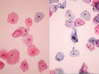
Cervical Cytology (Papanicolaou Stain)
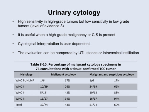
Urinary Cytology for Bladder Cancer Surveillance
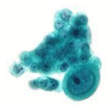
Fine-Needle Aspiration Cytologic Cluster
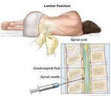
Lumbar Puncture for Exfoliative CSF Cytology
Specialized Diagnostic Laboratory Modalities
Hematological Examination
A specialized discipline focused on investigating morphological and quantitative abnormalities in peripheral blood cells (erythrocytes, leukocytes, and platelets) alongside their precursors within the bone marrow. It is key for identifying and differentiating:
- Various forms of anemia (e.g., iron deficiency, megaloblastic, hemolytic, aplastic).
- Hematologic malignancies, including acute and chronic leukemias, myelodysplastic syndromes, and plasma cell disorders.
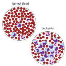
Peripheral Blood Smear: Normal vs. Leukemia
Immunohistochemistry (IHC)
Uses tagged antibodies to identify specific antigens within tissue sections. It is widely used to determine tumor origins, differentiate morphologically similar neoplasms (e.g., carcinomas, sarcomas, melanomas, lymphomas), and identify prognostic or predictive biomarkers.
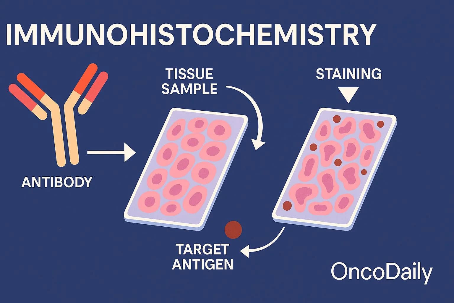
Immunohistochemistry (IHC) Mechanism
Ancillary Diagnostic Modalities
- Microbiological Examination: Evaluates bodily fluids, exudates, and tissue biopsies using direct microscopy, microbiological culture, and serology to identify causative bacterial, fungal, parasitic, or viral pathogens.
- Biochemical Examination: Assays normal and abnormal metabolites, electrolytes, enzymes, and proteins in blood and urine to detect and monitor metabolic disturbances (e.g., blood glucose/HbA1c for diabetes; urea/creatinine for renal function).
- Clinical Genetics (Cytogenetics): Investigates inherited chromosomal abnormalities in germ cells as well as acquired defects in somatic tissues using karyotyping and molecular methods.
- Molecular Diagnostic Techniques: Uses methods such as Fluorescence In Situ Hybridization (FISH), Polymerase Chain Reaction (PCR), and Southern blotting to identify specific gene mutations, translocations, and amplifications in genetic and neoplastic diseases.
- Autopsy (Post-Mortem Examination): Comprehensive anatomical dissection of a deceased body to determine the primary cause of death, assess disease progression, and serve forensic or clinical quality assurance purposes.
Microbiological Pathogen Visualization
Biochemical Examination Assays
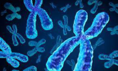
Cytogenetics: Human Chromosomal Architecture
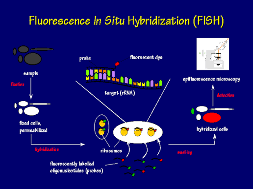
Fluorescence In Situ Hybridization (FISH)
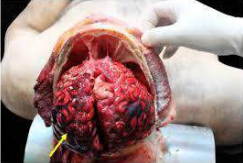
Post-Mortem Autopsy Examination
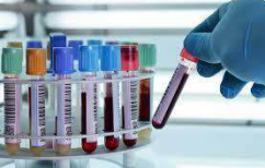
Multimodal Laboratory Diagnostics
Etiological Classification of Human Diseases
Human disease etiologies are broadly categorized into congenital (genetic) factors, acquired (environmental) factors, or a combination of both:
| Environmental Factor Category | Primary Injurious Mechanisms | Clinical Manifestations & Examples |
|---|---|---|
| Physical Agents | Excessive physical energy transferred to bodily tissues. |
• Mechanical trauma (lacerations, contusions, skeletal fractures) • Thermal extremes: hyperthermia (burns, heat stroke) and hypothermia (frostbite) • Ionizing and ultraviolet radiation (DNA damage) • Electrical energy (thermal burns, cardiac arrhythmias) |
| Chemical Agents & Drugs | Direct toxic disruption of cellular processes. Effects may be:
• Generalized toxicity: e.g., cyanide inhibiting cytochrome c oxidase. • Local damage: caustic acids or alkalis causing liquefactive or coagulative necrosis. • Organ-specific toxicity: selectively affecting sites of metabolism (liver) or excretion (kidneys). |
• Acetaminophen (paracetamol) hepatotoxicity causing acute centrilobular liver necrosis. • Chronic ethanol-induced hepatic steatosis, alcoholic hepatitis, and cirrhosis. • Heavy metal nephrotoxicity (lead, mercury). |
| Nutritional Deficiencies | Inadequate nutrient intake, malabsorption, transport defects, or metabolic utilization failure. Often presents as multiple, concurrent deficiencies. |
• Protein-energy malnutrition: Kwashiorkor (severe protein deficiency with edema) and Marasmus (combined protein and caloric wasting). • Micronutrient deficiencies: iron deficiency anemia; vitamin C deficiency (scurvy). |
| Nutritional Excesses | Caloric surplus, high intake of refined carbohydrates, and saturated fats causing metabolic dysfunction and chronic low-grade inflammation. | Increasingly prevalent globally and in Africa: drives obesity, Type 2 diabetes mellitus, systemic hypertension, metabolic dysfunction-associated steatotic liver disease, and atherosclerotic cardiovascular disease. |
| Infections & Infestations | Damage caused by pathogenic organisms via:
• Direct cytopathic effect: microbial invasion and lysis of host cells. • Exotoxin/endotoxin production: bacterial toxins disrupting cellular signaling or structural integrity. • Immunologically mediated injury: host immune response causing collateral tissue damage. |
• Direct lysis: Poliovirus destroying anterior horn motor neurons; Plasmodium species lysing red blood cells in malaria. • Toxin-mediated: Corynebacterium diphtheriae causing mucosal pseudomembrane formation; Clostridium tetani inducing spastic paralysis. |
| Immunological Factors | Immune system dysregulation categorized into:
• Hypersensitivity: Exaggerated immune response to benign antigens. • Immunodeficiency: Defective immune components leading to vulnerability to opportunistic infections. • Autoimmunity: Loss of self-tolerance resulting in immune attacks against host antigens. |
• Hypersensitivity: IgE-mediated mast cell degranulation in bronchial asthma triggered by pollen. • Immunodeficiency: CD4+ T-cell depletion by HIV resulting in AIDS. • Autoimmunity: Autoimmune destruction of pancreatic beta cells in Type 1 diabetes mellitus; systemic lupus erythematosus (SLE). |
| Psychogenic Factors | Chronic neuropsychological and environmental stress influencing physiology through neuroendocrine and autonomic pathways. | Particularly notable in urbanized and technologically demanding settings; contributes to peptic ulcer disease, stress cardiomyopathy, chronic tension syndromes, and essential hypertension. |
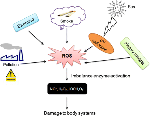
Environmental Stressors and Cellular Disease Mechanisms
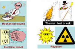
Physical Agents of Cellular Injury
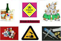
Chemical Agents of Disease
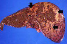
Chemical Injury: Hepatic Cirrhosis
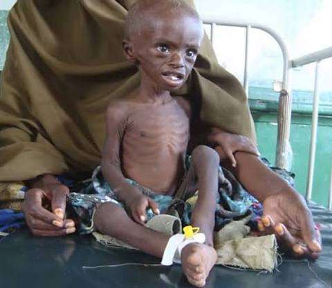
Nutritional Deficiency: Severe Infantile Marasmus
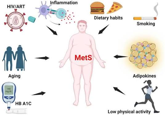
Nutritional Excess: Obesity and Cardiometabolic Risk
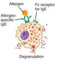
Immediate Type I Hypersensitivity Mechanism
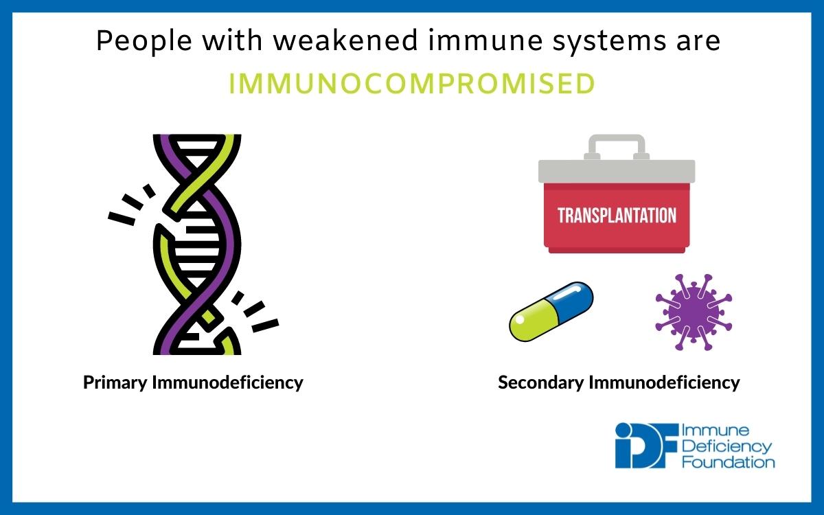
Classification of Immunodeficiency States
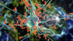
Autoimmunity: Loss of Self-Tolerance

Psychogenic Factors in Disease
Natural History of Disease & Death Pathways
The progression of disease over time in the absence of therapeutic intervention defines its natural history:
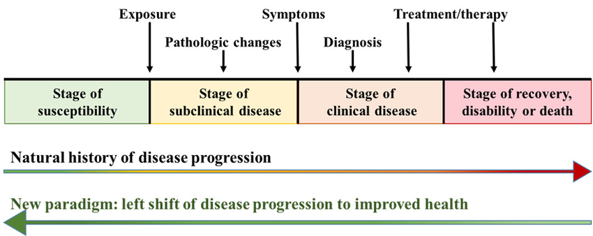
Chronological Timeline: Natural History of Disease
| Stage in Natural History | Biological & Pathological Characteristics | Clinical Manifestation & Outcomes |
|---|---|---|
| 1. Stage of Susceptibility | The host encounters predisposing environmental risk factors or harbors genetic vulnerabilities that initiate disease risk. | Completely asymptomatic; primary prevention measures (e.g., vaccination, smoking cessation, lifestyle modifications) are most effective at this stage. |
| 2. Stage of Subclinical Disease (Latency / Incubation) | Marks the biological onset of the disease process. Cellular and structural changes begin without triggering conscious symptoms or overt signs. Often termed the incubation period for infectious agents and the induction/latency period for chronic conditions. | Asymptomatic; secondary prevention strategies (screening modalities such as Pap smears, screening mammography, or fasting lipid panels) can detect early disease during this window. |
| 3. Stage of Clinical Disease | Structural and functional derangements cross a clinical threshold, producing noticeable symptoms and detectable physical signs. | Usual time point of diagnosis; disease manifestations vary widely in severity, from mild self-limiting forms to severe or life-threatening presentations. |
| 4. Stage of Recovery, Disability, or Demise | The disease process reaches its resolution, stabilization, or end point. Can follow several trajectories:
• Full resolution: complete anatomical and functional recovery without residual sequelae. • Chronic sequelae: disease activity settles, but leaves permanent tissue damage or functional disability. • Demise: progressive organ failure leading to death. |
Tertiary prevention and rehabilitation aim to minimize long-term functional impairment when permanent sequelae occur. Natural recovery can occur at various points along this progression. |
Pathological Framework of Death: Clinical vs. Biological Death
Clinical Death (Potentially Reversible Phase)
- Definition: The cessation of respiratory effort, blood circulation, and overt brainstem activity, during which prompt cardiopulmonary resuscitation may restore life. It begins with either the final agonal breath or the last spontaneous cardiac contraction, serving as a reversible transition zone between life and permanent biological demise.
- Clinical Signs:
- Absent central and peripheral pulses; blood pressure is completely unmeasurable.
- Loss of consciousness and unresponsiveness to deep, painful stimuli.
- Bilateral, widely dilated and fixed pupils.
- Certain deep brainstem or spinal reflexes may transiently persist in response to stimulation.
- Resuscitation Mechanism: Endotracheal intubation can stimulate sensory afferents of the superior laryngeal nerve. Because its sensory nucleus resides in the medulla oblongata adjacent to the respiratory center, this input can occasionally stimulate the central respiratory network and help restore spontaneous breathing during active resuscitation.
Biological Death (Irreversible Somatic Demise)
- Definition: Follows unreversed clinical death and represents permanent, irreversible cellular necrosis, tissue breakdown, and somatic death. It is defined by the irreversible cessation of circulatory and respiratory function, or the irreversible loss of all brain functions, including the brainstem.
- Diagnostic Criteria: Declared according to standardized, internationally accepted neurological and cardiopulmonary criteria, confirming the permanent cessation of cerebral, brainstem, and circulatory activity.
Clinical Vignettes: Pathological Principles in Practice
Clinical Case 1: Acute Myocardial Infarction
Clinical Vignette: A 55-year-old male presents with sudden, severe substernal chest pressure that radiates down his left arm. He appears anxious and profusely diaphoretic. An electrocardiogram (ECG) demonstrates marked ST-segment elevations across the anterior leads. Serum cardiac troponin levels are significantly elevated.
- Pathogenesis: Atherosclerotic plaque rupture within a coronary artery → exposure of subendothelial collagen → acute luminal thrombosis → sudden cessation of myocardial blood flow → ischemia → coagulative necrosis of cardiac myocytes.
- Morphology:
- Macroscopic: Early pale, ischemic myocardium, progressing to yellow-tan softening with a hyperemic border, and ultimately forming a dense, grayish-white fibrous collagen scar over subsequent weeks to months.
- Microscopic: Early wave-like myofibers, contraction band necrosis, hypereosinophilic myocytes, loss of striations, nuclear karyolysis, dense neutrophilic infiltration, followed by macrophage clearance and replacement by vascularized granulation tissue and collagen.
- Clinical Correlation: Myocardial ischemia and lactic acidosis stimulate local sensory afferents, causing crushing substernal chest pain with radiation along the T1–T4 dermatomes. Transmural injury currents produce ST-segment elevation on the ECG, while myocyte membrane rupture allows diagnostic cardiac enzymes (troponins I and T, CK-MB) to leak into the bloodstream.
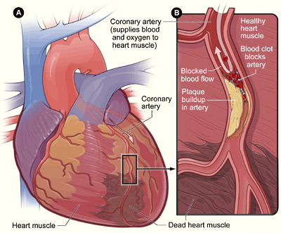
Pathogenesis of Coronary Artery Thrombosis
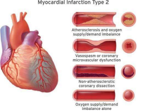
Ischemic Mechanisms in Type 2 Myocardial Infarction
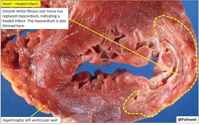
Healed Myocardial Infarction (Gross Pathology)
Clinical Case 2: Cancer Biopsy – Invasive Ductal Carcinoma
Clinical Vignette: A 48-year-old female presents with a painless, progressively enlarging mass in her left breast noted over the preceding 6 months. Physical examination reveals an irregular, firm, immobile nodule. A targeted core needle biopsy is performed, leading to a diagnosis of invasive (infiltrating) ductal carcinoma.
- Pathogenesis: Stepwise accumulation of genetic and epigenetic alterations: Normal ductal epithelium → ductal epithelial hyperplasia → atypical ductal hyperplasia → ductal carcinoma in situ (DCIS) → degradation of the epithelial basement membrane → invasive carcinoma infiltrating surrounding stroma.
- Morphology:
- Macroscopic: A firm to hard, gritty, gray-white irregular mass with infiltrative borders that invade adjacent yellow adipose tissue.
- Microscopic: Atypical malignant epithelial cells forming irregular cords, nests, and poorly formed glands, showing marked nuclear pleomorphism, hyperchromasia, frequent mitoses, and induction of a dense, reactive fibrous stroma (desmoplasia).
- Clinical Correlation: Tissue biopsy is essential to confirm malignancy, determine histologic grade, and assess therapeutic receptor status (ER, PR, and HER2-neu) by immunohistochemistry, which guides surgical planning, chemotherapy, and endocrine therapies.
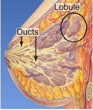
Anatomy of the Human Breast
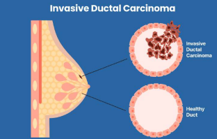
Invasive Ductal Carcinoma vs. Normal Ductal Architecture
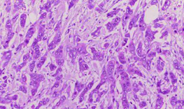
Invasive Ductal Carcinoma (Histopathology, H&E)
Clinical Case 3: Infectious Disease – Pulmonary Tuberculosis
Clinical Vignette: A 23-year-old student presents with a 6-week history of low-grade fever, productive cough, drenching night sweats, and unexplained weight loss. A chest radiograph demonstrates cavitary lesions in the right pulmonary upper lobe. A sputum smear stained with the Ziehl-Neelsen technique reveals acid-fast bacilli.
- Pathogenesis: Inhalation of aerosolized droplets containing Mycobacterium tuberculosis → phagocytosis by unactivated alveolar macrophages → intracellular bacterial replication → presentation of antigens to CD4+ T-lymphocytes → generation of a Type IV cell-mediated immune response → release of interferon-gamma → macrophage activation into epithelioid histiocytes → caseating granulomatous inflammation.
- Morphology:
- Macroscopic: Well-defined, yellowish-white, friable, cheese-like central necrosis (caseous necrosis), often accompanied by parenchymal cavitations and apical scarring.
- Microscopic: Distinct granulomas characterized by central, structureless granular eosinophilic necrosis, bordered by transformed epithelioid macrophages, multinucleated Langhans giant cells with horseshoe-shaped nuclear arrangements, and an outer mantle of mature T-lymphocytes and fibroblasts.
- Clinical Correlation: Tissue necrosis and caseous drainage into adjacent bronchi leave visible radiolucent cavitary lesions on chest X-rays. Systemic release of macrophage cytokines (TNF-alpha and IL-1) accounts for chronic constitutional symptoms, including fever, night sweats, and marked weight loss.
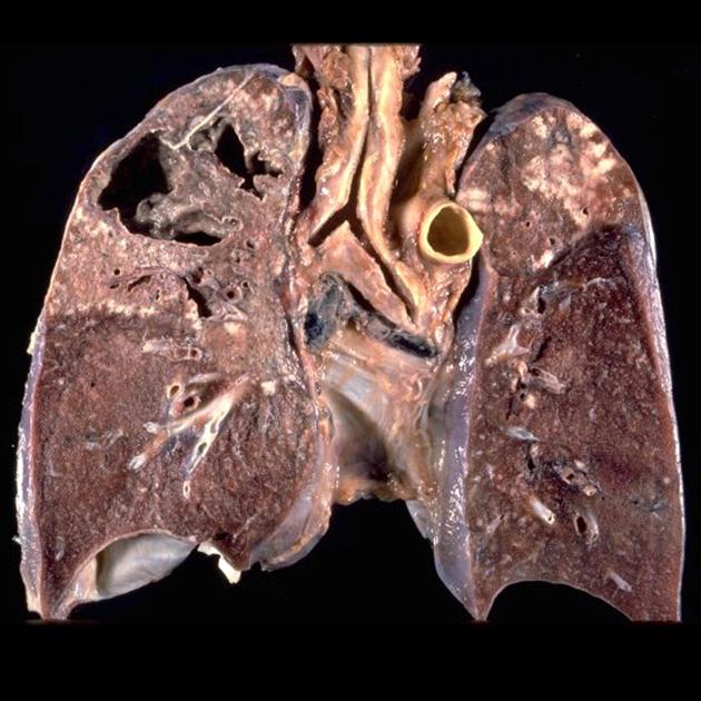
Secondary Pulmonary Tuberculosis with Cavitation (Gross Pathology)
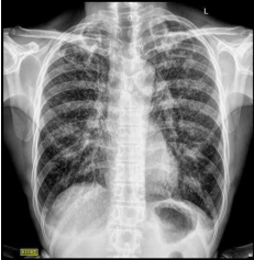
Chest Radiograph: Cavitary Pulmonary Tuberculosis
Core Competency Synthesis
The foundational concepts of this unit are summarized through seven core competency questions:
Summary of Foundational Concepts
- What is pathology? The scientific discipline that investigates the underlying structural, biochemical, and functional abnormalities in cells, tissues, and organs that lead to disease.
- What core aspects of disease does it study? It focuses on the four pillars of disease: Etiology (causes), Pathogenesis (mechanisms), Morphologic changes (structural alterations), and Functional derangements (clinical significance, symptoms, signs, and prognosis).
- Why should medical students study pathology? Pathology serves as the bridge between basic sciences and clinical medicine. It provides the mechanistic framework needed to understand clinical presentations, interpret diagnostic findings, select rational therapies, and forecast prognosis.
- What is pathogenesis? The progressive sequence of molecular, cellular, and tissue events that unfold from the initial etiologic trigger to the full development of a disease's structural and clinical features.
- What diagnostic modalities are utilized by the pathologist? Histopathology, cytopathology (including FNAC, exfoliative, and abrasive cytology), hematopathology, immunohistochemistry, clinical chemistry/biochemistry, medical microbiology, cytogenetics, molecular pathology, and post-mortem autopsy.
- What are the primary purposes of tissue fixation? Arrests enzymatic autolysis and microbial putrefaction, chemically coagulates and cross-links proteins to prevent loss of cellular constituents, and fortifies tissue architecture to withstand subsequent histotechnical processing and microtome cutting.
- What constitutes the natural history and course of disease? The chronological evolution of a disease in the absence of treatment, transitioning through the stage of susceptibility, the stage of subclinical disease (biological onset, latency, incubation), the stage of clinical disease (symptoms, signs, diagnostic confirmation), and concluding in the stage of recovery, residual disability/sequelae, or demise (clinical vs. biological death).