DIENCEPHALON
Inner Part of Cerebral Hemispheres
The inner part of Cerebral Hemispheres comprises of:
- Lateral Ventricles.
- 3rd ventricle.
- White matter.
- Subcortical Nuclei (Basal Ganglia).
- Limbic System.
- “Diencephalon”.
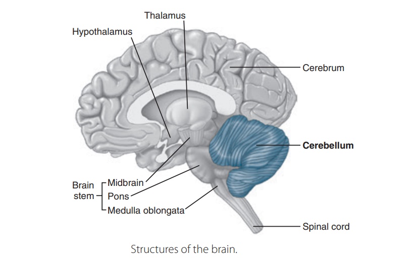
Inner components of the cerebral hemispheres
Diencephalon Overview
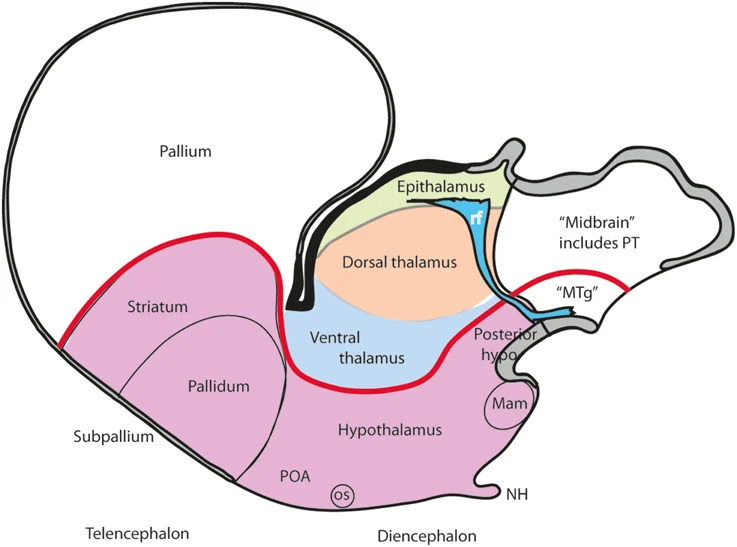
Diencephalon is composed of the structures that surround the 3rd ventricle.
Position within the forebrain
- Deep to the cerebral hemispheres
- Rostral to the midbrain
- Paired structures surround the third ventricle
- The hypothalamus reaches the ventral brain surface
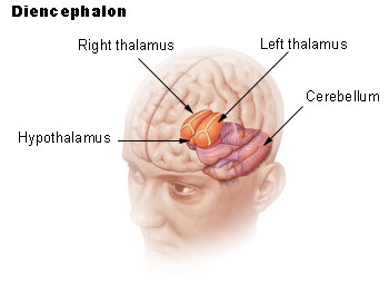
Position of the diencephalon within the forebrain
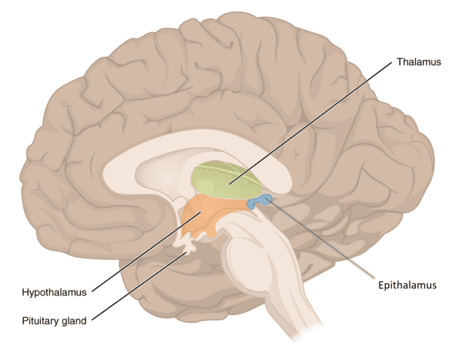
Diencephalic anatomical boundaries and brainstem transitions
Subdivisions of the Diencephalon
- Epithalamus
- Thalamus
- Metathalamus
- Hypothalamus
- 3rd Ventricle
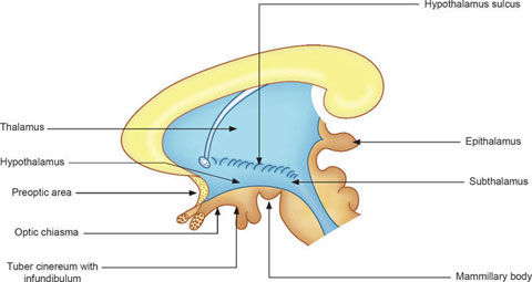
Subdivisions of the Diencephalon
THIRD VENTRICLE
- Narrow midline cavity of the diencephalon
- Interventricular foramina connect lateral ventricles
- Cerebral aqueduct leads to the fourth ventricle
- Ependyma lines its walls
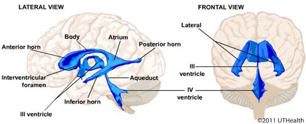
Third ventricle and ventricular communications
WALLS OF THE THIRD VENTRICLE
- Lateral walls: thalamus and hypothalamus
- Roof: ependyma with tela choroidea and choroid plexus
- Anterior wall: lamina terminalis and adjacent landmarks
- Floor: optic and hypothalamic structures
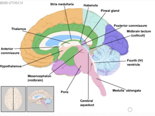
Walls of the Third Ventricle
RECESSES AND POSTERIOR LANDMARKS
- Optic recess near the optic chiasm
- Infundibular recess enters the stalk
- Pineal and suprapineal recesses posteriorly
- Posterior commissure near the aqueduct inlet
| Third Ventricle Boundary / Recess | Anatomical Composition | Clinical & Functional Landmark |
|---|---|---|
| Anterior Wall | Lamina terminalis, anterior commissure | Site of third ventriculostomy; embryonic rostral neuropore closure site |
| Roof | Ependyma, tela choroidea, choroid plexus | Suspended from stria medullaris thalami; synthesizes cerebrospinal fluid |
| Floor | Optic chiasm, infundibulum, tuber cinereum, mammillary bodies | Ventral hypothalamic surface; direct contact with subarachnoid cisterns |
| Lateral Wall | Medial surfaces of thalamus and hypothalamus | Divided horizontally by the hypothalamic sulcus |
| Diverticular Recesses | Optic, infundibular, pineal, and suprapineal recesses | Outpouchings projecting into adjacent neural and glandular stalks |
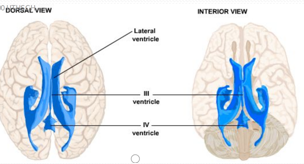
Recesses of the Third Ventricle
THALAMUS
THE GATEWAY TO THE CERBRAL CORTEX
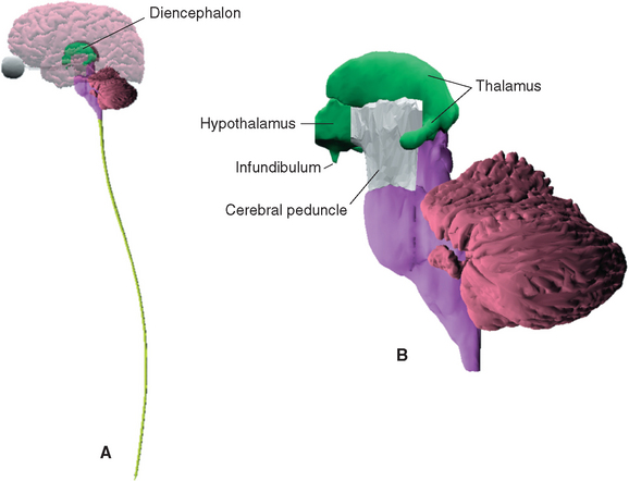
Thalamus: The Gateway to the Cerebral Cortex
Thalamic Anatomy and Dimensions
- Large, paired, structure of grey matter
- Dimensions: 3 x 1.5 x 1.5 cm
- Lies immediately lateral to third ventricle
- Between cerebral cortex and brain stem (Both anatomically and functionally)
- Sends fibres out to cerebral cortex in all directions
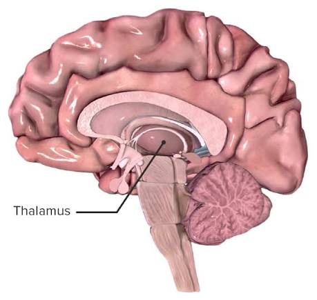
THALAMUS
THALAMUS AND ITS RELATIONS
- Paired ovoid gray matter masses
- Medial surface borders the third ventricle
- Lateral surface faces the internal capsule
- Posterior enlargement forms the pulvinar
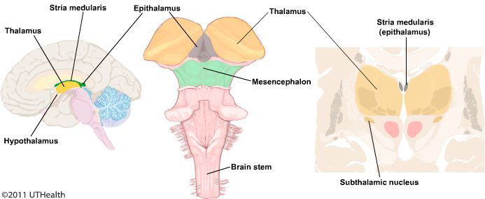
Thalamus and its anatomical relations
THALAMUS FUNCTIONS
- Integration of sensory information
- Regulation of motor activity
- Regulation of consciousness
INTERNAL ORGANIZATION OF THE THALAMUS
- Internal medullary lamina divides nuclear groups
- Anterior, medial and lateral groups
- Intralaminar nuclei lie within the lamina
- Reticular nucleus lies along the lateral surface
FUNCTIONAL GROUPS OF THALAMIC NUCLEI
- Specific relays connect defined inputs to cortex
- Association nuclei interact with association cortex
- Intralaminar nuclei support arousal and broad integration
- Reticular neurons regulate thalamic activity
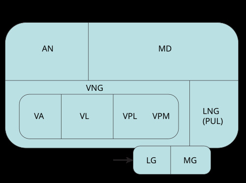
Thalamic Nuclei (lateral view; MNG on other side)
VPL AND VPM SENSORY RELAYS
- VPL receives somatosensory input from the body
- VPM receives somatosensory input from the face
- Both project to primary somatosensory cortex
- The gustatory relay includes VPM parvocellular regions
VPL vs. VPM Somatosensory Relay Distinction
VPL = Ventral Posterolateral: Think "L for Limbs/Legs" → receives spinothalamic and medial lemniscus fibers from the body and neck.
VPM = Ventral Posteromedial: Think "M for Mandible/Mouth/Makeup" → receives trigeminothalamic fibers from the face, and gustatory taste fibers via the solitary tract to the parvocellular division.
VISUAL AND AUDITORY RELAYS
- Lateral geniculate nucleus receives retinal input
- Optic radiations reach primary visual cortex
- Medial geniculate nucleus receives inferior colliculus input
- Auditory radiations reach primary auditory cortex
Metathalamus Geniculate Relay Mnemonic
LGN (Lateral Geniculate Nucleus): "L for Light / Look" → receives optic tract axons → sends optic radiations (including Meyer's loop) to the primary visual cortex (calcarine sulcus, area 17).
MGN (Medial Geniculate Nucleus): "M for Music / Media" → receives brachium of the inferior colliculus → projects auditory radiations to the primary auditory cortex (transverse temporal gyri of Heschl, areas 41 & 42).
MOTOR THALAMIC NUCLEI
- VA receives prominent basal ganglia input
- VL receives cerebellar and basal ganglia input
- Outputs reach motor and premotor cortex
- Thalamic relays help regulate movement
ANTERIOR AND MEDIODORSAL NUCLEI
- Anterior nuclei participate in memory circuits
- Mammillothalamic tract reaches anterior nuclei
- Mediodorsal nucleus connects with prefrontal cortex
- Lesions may affect memory and behavior
PULVINAR AND RETICULAR NUCLEUS
- Pulvinar links visual and association networks
- It contributes to attention and integration
- Reticular nucleus provides inhibitory regulation
- Thalamocortical rhythms contribute to sleep
| Thalamic Nucleus | Major Afferent Input | Primary Cortical Projection | Functional Domain |
|---|---|---|---|
| Ventral Posterolateral (VPL) | Medial lemniscus, Spinothalamic tract | Primary somatosensory cortex (Brodmann 3, 1, 2) | Somatosensory relay for body and limbs |
| Ventral Posteromedial (VPM) | Trigeminal lemniscus, Solitary tract (taste) | Primary somatosensory cortex, Gustatory cortex | Somatosensory relay for head/face; gustation |
| Lateral Geniculate (LGN) | Optic tract (retinal ganglion cells) | Primary visual cortex (Brodmann 17) via optic radiation | Visual sensory relay |
| Medial Geniculate (MGN) | Inferior colliculus (brachium of inferior colliculus) | Primary auditory cortex (Brodmann 41, 42) | Auditory sensory relay |
| Ventral Anterior (VA) | Basal ganglia (Globus pallidus internus, SNr) | Premotor and supplementary motor cortex | Motor planning and execution initiation |
| Ventral Lateral (VL) | Dentate nucleus of cerebellum, Basal ganglia | Primary motor cortex (Brodmann 4), Premotor cortex | Motor coordination, feedback, and smoothing |
| Anterior Nucleus (AN) | Mammillary body (via mammillothalamic tract) | Cingulate gyrus (Papez circuit) | Limbic memory consolidation and emotional tone |
| Mediodorsal (MD) | Amygdala, olfactory cortex, temporal neocortex | Prefrontal cortex | Executive cognition, affect, personality, and mood |
| Pulvinar (PUL) | Superior colliculus, parietal/occipital/temporal areas | Visual and parieto-occipito-temporal association cortex | Visual attention, feature binding, and sensory salience |
| Reticular Nucleus | Collaterals of thalamocortical and corticothalamic fibers | Back to other thalamic nuclei (GABAergic inhibition) | Thalamic gating; regulates sleep spindle rhythms |
THALAMIC BLOOD SUPPLY AND LESIONS
- Perforators arise mainly from PCA and PCom arteries
- Territories include anterior, paramedian and inferolateral regions
- Sensory lesions cause contralateral deficits
- Bilateral paramedian injury can impair arousal
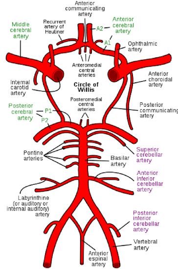
Thalamic arterial territories and perforating vessels
Functions of the thalamus
- Sensory integration/relay station for all sensory pathways (Except olfaction)
- Capable of recognition of pain, thermal & tactile sensations
- Influences voluntary movements through basal ganglia & cerebellum – cerebral cortex – cortico-nuclear / cortico-spinal pathways
- Maintains state of wakefulness and alertness
- Impulses received from hypothalamus projected to prefrontal & cingulate gyrus – Determination of mood
- Recent memory and emotions
Lesions of the thalamus
- Sensory loss (light touch, discrimination, muscle joint sense)
- Thalamic Pain (thalamic overreaction)
- Motor Dysfunctions
- Changes in consciousness and alertness
The lesions of the thalamus usually affect the neighboring structures producing symptoms from these structures……
Thalamic Pain Syndrome (Dejerine-Roussy Syndrome)
Etiology: Ischemia or lacunar infarction of the inferolateral territory (thalamogeniculate branches of the Posterior Cerebral Artery), damaging the VPL / VPM nuclei.
Clinical Features: Initial complete contralateral hemianesthesia (loss of tactile discrimination, pain, temperature, and conscious proprioception), followed weeks to months later by severe, intractable central neuropathic pain and hyperpathia/allodynia (severe pain triggered by light touch).
HYPOTHALAMUS
HYPOTHALAMUS AND SURFACE LANDMARKS
- Below the hypothalamic sulcus
- Medial to the subthalamic and capsular regions
- Optic chiasm marks the anterior inferior region
- Mammillary bodies mark the posterior inferior region
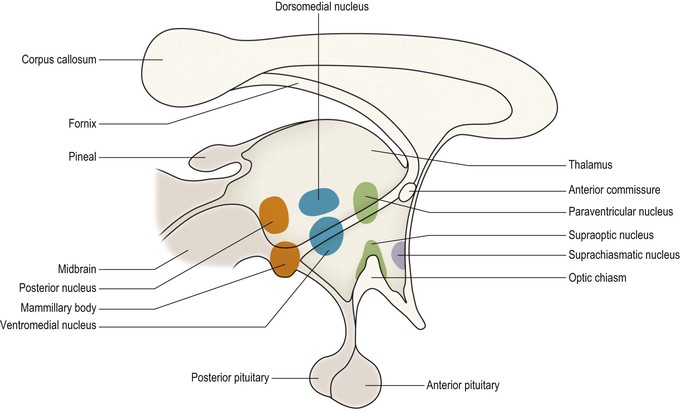
Hypothalamus and surface landmarks
HYPOTHALAMIC REGIONS AND ZONES
- Rostrocaudal regions: preoptic, anterior, tuberal, posterior
- Mediolateral zones: periventricular, medial, lateral
- Nuclei vary by region and zone
- Location helps predict major connections
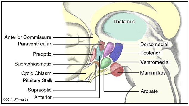
Hypothalamic regions, zones, and nuclear groups
SUPRAOPTIC AND PARAVENTRICULAR NUCLEI
- Magnocellular neurons synthesize vasopressin and oxytocin
- Axons descend to the posterior pituitary
- The posterior pituitary stores and releases these hormones
- PVN also contains endocrine and autonomic cell populations
POSTERIOR PITUITARY PATHWAY
- Hypothalamic cell bodies synthesize the hormones
- The hypothalamo-neurohypophyseal tract carries axons
- Action potentials trigger hormone release from terminals
- Stalk injury can disrupt vasopressin delivery
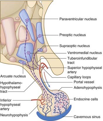
Circuitry of Neurohypophyseal tract
ANTERIOR PITUITARY PATHWAY
- Hypothalamic factors enter the primary capillary plexus
- Portal veins carry them to the anterior pituitary
- Releasing and inhibitory signals regulate secretion
- Dopamine provides tonic inhibition of prolactin
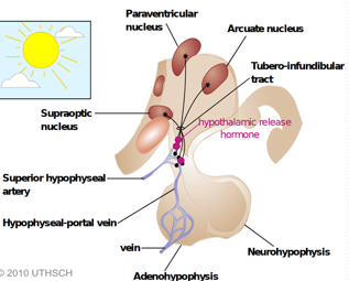
Anterior pituitary tubero-infundibular portal pathway
Pituitary Stalk Transection Effect
Stalk Disruption: Interruption of the hypophyseal portal system prevents hypothalamic releasing hormones from reaching the adenohypophysis, causing a decrease in GH, TSH, ACTH, LH, and FSH.
Prolactin Paradox: In contrast, serum prolactin levels RISE because dopamine (prolactin-inhibiting factor) tonic suppression is severed.
SUPRACHIASMATIC NUCLEUS: CIRCADIAN TIMING
- SCN lies just above the optic chiasm
- Retinohypothalamic input conveys environmental light
- SCN coordinates daily physiological rhythms
- Pineal melatonin reflects a downstream timing pathway
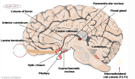
Suprachiasmatic nucleus circadian pathway
TEMPERATURE AND AUTONOMIC REGULATION
- Preoptic networks detect and integrate thermal signals
- Heat loss includes sweating and skin vasodilation
- Posterior hypothalamic circuits support cold defense
- Descending pathways reach autonomic centers
Thermoregulation Mnemonic ("A/C & Post-Warm")
Anterior / Preoptic = "A/C" (Air Conditioning): Mediates cooling. Activated by warmth → parasympathetic firing → diaphoresis (sweating) and peripheral vasodilation. Lesion causes hyperthermia.
Posterior Hypothalamus = Preserves Heat: Activated by cold → sympathetic firing → shivering and peripheral vasoconstriction. Lesion causes hypothermia / poikilothermia.
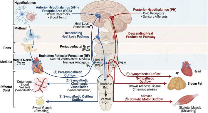
Hypothalamic temperature and autonomic connectivity
FEEDING AND ENERGY BALANCE
- Arcuate neurons integrate peripheral metabolic signals
- POMC and AgRP circuits have opposing effects
- Lateral hypothalamic networks support feeding and arousal
- Ventromedial regions contribute to energy regulation
Hypothalamic Feeding Centers
Lateral Nucleus = Hunger Center: "Late for dinner" → stimulates appetite via orexin. Destruction/lesion leads to anorexia and severe starvation.
Ventromedial Nucleus = Satiety Center: "Very Much full" → signals satiety (leptin receptor site). Destruction/lesion leads to hyperphagia, massive obesity, and rage.
Feeding and energy balance circuit
THIRST AND WATER BALANCE
- Circumventricular structures detect circulating signals
- Hypothalamic networks coordinate thirst and vasopressin
- Vasopressin increases renal water conservation
- Deficiency produces polyuria and dilute urine
Central Diabetes Insipidus
Etiology: Loss of vasopressin (ADH)-secreting magnocellular neurons in supraoptic/paraventricular nuclei or high stalk damage.
Manifestations: Inability to concentrate urine → severe polyuria (>3 L/day), polydipsia, hypernatremia, and inappropriately dilute urine with low urine specific gravity (<1.006) and low osmolality (<300 mOsm/kg).
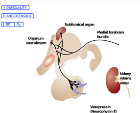
Thirst and water balance pathways
MAMMILLARY BODIES AND MEMORY
- Mammillary bodies form part of the hypothalamus
- Fornix conveys hippocampal connections
- Mammillothalamic tract reaches anterior thalamus
- Damage can contribute to memory impairment
Wernicke-Korsakoff Syndrome
Pathology: Thiamine (Vitamin B1) deficiency, classically in chronic alcoholism, causing petechial hemorrhages and atrophy of the mammillary bodies and anterior thalamic nuclei.
Features: Acute Wernicke triad (confusion, ataxia, ophthalmoplegia), followed by Korsakoff psychosis: irreversible anterograde amnesia, retrograde amnesia, and confabulation.
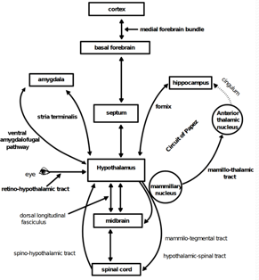
Mammillary bodies in memory circuitry (Circuit of Papez)
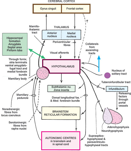
HYPOTHALAMIC CONNECTIONS
- Fornix links hippocampal and hypothalamic regions
- Medial forebrain bundle links limbic and brainstem systems
- Dorsal longitudinal fasciculus carries descending connections
- Pituitary pathways provide endocrine outputs
| Pathway / Tract | Originating Structure | Termination Target | Primary Functional Role |
|---|---|---|---|
| Fornix | Hippocampal subiculum & CA fields | Mammillary bodies, septal nuclei | Major limbic conduit for episodic memory processing |
| Mammillothalamic Tract | Mammillary bodies | Anterior nucleus of thalamus | Relay step within Papez memory consolidation loop |
| Medial Forebrain Bundle (MFB) | Septal area, basal forebrain | Lateral hypothalamus, brainstem reticular formation | Bidirectional communication linking limbic structures to brainstem; reward circuit |
| Dorsal Longitudinal Fasciculus (DLF) | Periventricular hypothalamus | Periaqueductal gray, autonomic brainstem nuclei | Primary descending pathway for hypothalamic autonomic control |
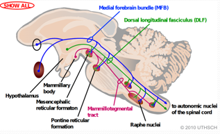
Major hypothalamic fiber connections
EPITHALAMUS
- Pineal gland near the posterior third ventricle
- Habenular nuclei and their connections
- Stria medullaris thalami carries habenular afferents
- Habenular commissure crosses the midline
Epithalamus: Pineal gland and habenular complex
PINEAL GLAND AND CLINICAL RELATIONS
- Pinealocytes secrete melatonin
- Sympathetic innervation links secretion to light-dark timing
- Pineal masses may obstruct the aqueduct
- Adjacent dorsal midbrain compression may impair upgaze
Parinaud Syndrome (Dorsal Midbrain Syndrome)
Mechanism: Germinoma or pinealoma of the pineal gland compressing the pretectal area and superior colliculi of the dorsal midbrain (tectum), frequently accompanied by non-communicating hydrocephalus due to cerebral aqueduct obstruction.
Classic Signs:
- Upward gaze paralysis (conjugate supranuclear vertical gaze palsy)
- Pseudo-Argyll Robertson pupils (light-near dissociation; pupils constrict to accommodation but fail to react to light)
- Convergence-retraction nystagmus on attempted upward gaze
- Eyelid retraction (Collier's sign)
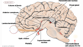
Pineal gland and clinical relations
SUBTHALAMUS
- Below the thalamus and lateral to the hypothalamus
- Includes subthalamic nucleus and zona incerta
- Related fiber fields contain pallidal and cerebellar pathways
- STN neurons provide excitatory motor input
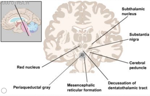
Subthalamic nucleus and surrounding neural structures
STN IN MOTOR CONTROL
- Indirect pathway recruits the subthalamic nucleus
- STN excites GPi and SNr
- These output nuclei inhibit the thalamus
- The hyperdirect pathway reaches STN from cortex
| Basal Ganglia Pathway | Role of Subthalamic Nucleus (STN) | Net Effect on Thalamus & Motor Cortex |
|---|---|---|
| Indirect Pathway | Striatum inhibits GPe → disinhibits STN → STN uses glutamate to excite GPi / SNr | Inhibits thalamus (GPi fires GABA → dampens unwanted movements) |
| Hyperdirect Pathway | Cortex directly fires glutamatergic projections to STN, bypassing striatum | Rapid global motor braking; fast cancellation of ongoing movement programs |
Hemiballismus
Etiology: Unilateral destructive lesion (most commonly a lacunar stroke from posterior cerebral or anterior choroidal artery branches) of the contralateral subthalamic nucleus.
Mechanism: Loss of STN excitatory drive to the GPi → loss of normal GABAergic thalamic inhibition → hyperkinetic motor output.
Clinical Manifestation: Involuntary, violent, flinging, wide-amplitude movements of the contralateral limbs (arm and leg).
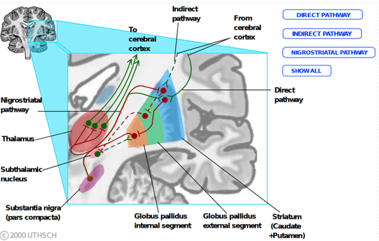
Subthalamic nucleus (STN) in motor control circuitry