Neural Tube
Development and Histogenesis
Neurulation
The events of neurulation occur as follows:
- A. The notochord
- induces the overlying ectoderm to differentiate into neuroectoderm and form the neural plate.
- The notochord forms the nucleus pulposus of the intervertebral disk in the adult.
- B. The neural plate
- folds to give rise to the neural tube,
- which is open at both ends at the anterior and posterior neuropores.
- The anterior and posterior neuropores
- connect the lumen of the neural tube with the amniotic cavity.
- 1. The anterior neuropore
- closes during week 4 (day 25).
- becomes the lamina terminalis.
- Failure of the anterior neuropore to close results in upper neural tube defects (NTDs; e.g., anencephaly).
- 2. The posterior neuropore
- closes during week 4 (day 27).
- Failure of the posterior neuropore to close results in lower NTDs (e.g., spina bifida with myeloschisis).
- C. As the neural plate folds
- some cells differentiate into neural crest cells.
Neuropore Closure Timeline ("25 Anterior, 27 Posterior")
The Anterior (rostral) neuropore closes first on Day 25 (leads to brain and lamina terminalis; failure causes anencephaly). The Posterior (caudal) neuropore closes 2 days later on Day 27 (leads to lower spinal cord; failure causes spina bifida). Both close during Week 4.
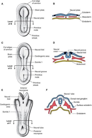
Diagrams illustrating the dorsal aspect of the human embryo. (A) Late presomite and early neural plate stage. (B) Early somite stage and neural groove stage. (C) Eight-somite stage and early neural tube stage. The anterior and posterior neuropores
- D. The rostral part of the neural tube becomes the adult brain.
- E. The caudal part of the neural tube becomes the adult spinal cord.
- F. The lumen of the neural tube gives rise to the ventricular system of the brain and central canal.
Molecular consideration
The following factors appear to play a role in neurulation:
- BMP-4 (bone morphogenetic protein)
- noggin (an inductor protein)
- chordin (an inductor protein)
- FGF-8 (fibroblast growth factor)
- and N-CAM (neural cell adhesion molecule).
Protein factors and their roles
| Molecular Factor | Signaling Mechanism | Primary Embryological Role in Neurulation |
|---|---|---|
| Bone Morphogenetic Proteins (BMP) | Interacts with the apicobasal polarity pathway | BMPs regulate neural tube closure; influence formation of dorsolateral hinge points (DLHPs) through antagonistic interaction with Noggin |
| Sonic Hedgehog (Shh) | Ventral morphogen gradient from notochord & floor plate | Essential for ventralizing the neural tube, inducing the floor plate and motor neurons; regulates gene expression in neural development |
| Noggin | BMP antagonist (inductor protein) | Interacts with BMPs to regulate DLHP formation and neural tube closure |
| Pax3 | Dorsal transcription factor | Crucial role in neural tube closure, particularly the dorsal neural fold |
| Cdx2 | Caudal transcription factor | Regulates neural tube closure, particularly in the caudal region |
| Zic2 | Transcriptional regulator / co-factor | Regulates Pax3 expression and acts as a co-factor for Cdx proteins; controls dorsal restriction of Pax3 expression in caudal neural tube |
| Fibronectin | Extracellular matrix glycoprotein | Essential for neural tube closure; its deletion or blockade can lead to neural tube defects |
| Protease-Activated Receptors (PARs) | G-protein-coupled transmembrane receptors | PARs, such as Par1 and Par2, are involved in regulating neural tube closure |
Dorsal-Ventral Neural Tube Axis
Dorsalizing Signals (Sensory/Alar): Mediated by BMP-4, BMP-7, and Pax3/Pax7 from overlying surface ectoderm and roof plate.
Ventralizing Signals (Motor/Basal): Mediated by Sonic Hedgehog (Shh) secreted by the notochord and floor plate. High Shh concentration induces motor neuroblasts; low Shh allows alar plate sensory specialization.
NEURAL CREST CELLS
- The neural crest cells:
- differentiate from cells located along the lateral border of the neural plate.
- It is mediated by BMP-4 and BMP-7.
- Neural crest cells undergo a prolific migration throughout the embryo (both the cranial region and trunk region) and ultimately differentiate into a wide array of adult cells and structures.
A. Cranial neural crest cells
- 1. There is a remarkable relationship between the origin of cranial neural crest cells from the rhombencephalon (hindbrain) and their final migration into pharyngeal arches.
- 2. The rhombencephalon is divided into eight segments called rhombomeres (R1–R8).
- 3. Cranial neural crest cells from R1 and R2 migrate into pharyngeal arch 1 (which also receives neural crest cells from the midbrain area).
- 4. Cranial neural crest cells from R4 migrate into pharyngeal arch 2.
- 5. Cranial neural crest cells from R6 and R7 migrate into pharyngeal arch 3.
Cranial neural crest cells differentiate into the following adult cells and structures:
- a. Pharyngeal arch skeletal and connective tissue components
- b. Bones of neurocranium
- c. Pia and arachnoid
- d. Parafollicular (C) cells of thyroid
- e. Aorticopulmonary septum
- f. Odontoblasts (dentin of teeth)
- g. Sensory ganglia of cranial nerve (CN) V, CN VII, CN IX, and CN X
- h. Ciliary (CN III), pterygopalatine (CN VII), submandibular (CN VII), and otic (CN IX) parasympathetic ganglia
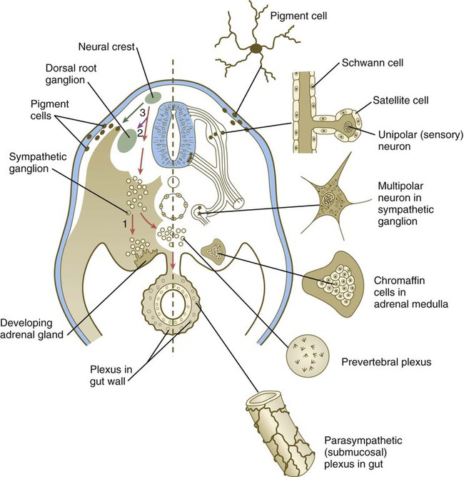
Major neural crest migratory pathways and derivatives in the trunk. Left, Pathways in the early embryo. The first emigrating cells (pathway 1) follow the ventral (sympathoadrenal) pathway (red arrows). The second wave of emigrating cells (pathway 2) follows the ventrolateral pathway indicated by the purple arrow. The last cells to leave the neural tube (pathway 3) follow the dorsolateral pathway (green arrow) as they go on to differentiate into pigment cells.
Trunk neural crest cells
- 1. Trunk neural crest cells:
- extend from somite 6 to the most caudal somites
- and migrate in a dorsolateral, ventral, and ventrolateral direction throughout the embryo.
- 2. Trunk neural crest cells differentiate into the following adult cells and structures:
- a. Melanocytes
- b. Schwann cells
- c. Chromaffin cells of adrenal medulla
- d. Dorsal root ganglia
- e. Sympathetic chain ganglia
- f. Prevertebral sympathetic ganglia
- g. Enteric parasympathetic ganglia of the gut (Meissner and Auerbach; CN X)
- h. Abdominal/pelvic cavity parasympathetic ganglia
Neural Crest Derivatives Mnemonic ("SOME PATH")
Schwann cells
Odontoblasts / Ossicles
Melanocytes
Enteric ganglia (Auerbach & Meissner plexuses; absent in Hirschsprung disease)
Pia and arachnoid mater / Parafollicular C cells of thyroid
Adrenal chromaffin cells / Aorticopulmonary septum
Trigeminal and other cranial nerve sensory/autonomic ganglia
Head & pharyngeal arch skeletal components
VESICLE DEVELOPMENT OF THE NEURAL TUBE
A. The three primary brain vesicles and two associated flexures develop during week 4.
1. Primary brain vesicles
- a. Prosencephalon (forebrain)
- is associated with the appearance of the optic vesicles
- and gives rise to the telencephalon and diencephalon.
- b. Mesencephalon (midbrain)
- remains as the mesencephalon.
- c. Rhombencephalon (hindbrain)
- gives rise to the metencephalon and myelencephalon.
2. Flexures
- a. Cephalic flexure (midbrain flexure)
- is located between the prosencephalon and the rhombencephalon.
- b. Cervical flexure
- is located between the rhombencephalon and the future spinal cord.
B. The five secondary brain vesicles become visible in week 6 of development and form various adult derivatives of the brain:
- 1. Telencephalon
- gives rise to the cerebral hemispheres, caudate, and putamen.
- 2. Diencephalon
- gives rise to the epithalamus, subthalamus, thalamus, hypothalamus, mammillary bodies, neurohypophysis, pineal gland, globus pallidus, retina, iris, ciliary body, optic nerve (CN II), optic chiasm, and optic tract.
- 3. Mesencephalon
- gives rise to the midbrain.
- 4. Metencephalon
- gives rise to the pons and cerebellum.
- 5. Myelencephalon
- gives rise to the medulla.
| Primary Vesicle (Week 4) | Secondary Vesicle (Week 6) | Adult Neural Derivatives | Associated Neural Cavity |
|---|---|---|---|
| Prosencephalon (Forebrain) | Telencephalon | Cerebral hemispheres, caudate, putamen, amygdaloid, claustrum, lamina terminalis, olfactory bulbs, hippocampus | Lateral ventricles |
| Diencephalon | Epithalamus, subthalamus, thalamus, hypothalamus, mammillary bodies, neurohypophysis, pineal gland, globus pallidus, optic cup derivatives (retina, CN II) | Third ventricle | |
| Mesencephalon (Midbrain) | Mesencephalon | Midbrain (superior and inferior colliculi, cerebral peduncles, red nucleus, substantia nigra) | Cerebral aqueduct (of Sylvius) |
| Rhombencephalon (Hindbrain) | Metencephalon | Pons and cerebellum | Upper fourth ventricle |
| Myelencephalon | Medulla oblongata | Lower fourth ventricle |
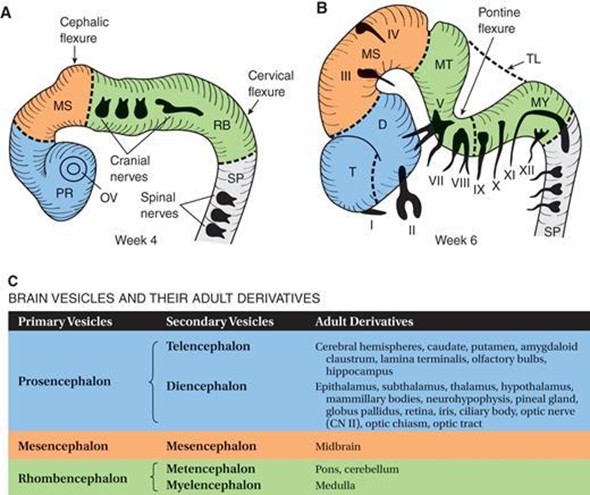
Schematic illustrations of the developing brain vesicles. (A) Three-vesicle stage of the brain in a 4-week-old embryo. Divisions are indicated by dashed lines. PR prosencephalon; MS mesencephalon; RB rhombencephalon; SP spinal cord; OV optic vesicle. (B) Five-vesicle stage of the brain in a 6-week-old embryo. Divisions are indicated by dashed lines. Cranial nerves (CN) are indicated by Roman numerals. CN VI is not shown because it exits the brain stem from the ventral surface. (C) T telencephalon; D diencephalon; MS mesencephalon; MT metencephalon; MY myelencephalon; TL tela choroidea; SP spinal cord. (D) Table indicating the brain vesicles and their adult derivatives..
HISTOGENESIS OF THE NEURAL TUBE
The cells of the neural tube are neuroectodermal (or neuroepithelial) cells that give rise to the following cell types:
- A. Neuroblasts
- form all neurons found in the CNS.
- B. Glioblasts (spongioblasts)
- are, for the most part, formed after cessation of neuroblast formation.
- Radial glial cells are an exception and develop before neurogenesis is complete.
- Glioblasts form the supporting cells of the CNS and include the following:
1. Astrocytes
Have the following characteristics and functions:
- project foot processes to capillaries; this contributes to the blood–brain barrier.
- play a role in the metabolism of neurotransmitters (e.g., glutamate, γ-aminobutyrate [GABA], serotonin).
- buffer the [K+] of the CNS extracellular space.
- form the external and internal glial-limiting membrane in the CNS.
- form glial scars in a damaged area of the CNS (i.e., astrogliosis).
- undergo hypertrophy and hyperplasia in reaction to CNS injury and contain the glial fibrillary acidic protein (GFAP) and glutamine synthetase, which are good markers for astrocytes.
2. Oligodendrocytes
- produce the myelin in the CNS.
- A single oligodendrocyte can myelinate several (up to 30) axons.
3. Ependymocytes
- line the central canal and ventricles of the brain.
- These cells are not joined by tight junctions, so that exchange between the cerebrospinal fluid (CSF) and CNS extracellular fluid occurs freely.
4. Tanycytes
- are modified ependymal cells that mediate transport between CSF in the ventricles and the neuropil.
- These cells usually project to hypothalamic nuclei that regulate the release of gonadotropic hormones from the adenohypophysis.
5. Choroid plexus cells
- are a continuation of the ependymal lining that is reflected over the choroid plexus villi and secrete CSF by selective transport of molecules from blood.
- These cells are joined by tight junctions (zonula occludens), which are the basis of the blood–CSF barrier.
- CSF is normally clear.
6. Microglia (Hortega cells)
- are the macrophages of the CNS.
- They arise from monocytes (mesoderm).
- and invade the developing nervous system in week 3 along with the developing blood vessels.
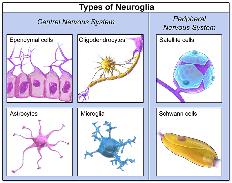
There are four major glia in the CNS. (a) Oligodendrocytes myelinate parts of several axons. (b) Astrocytes have multiple processes and form perivascular feet that completely enclose all capillaries (only a few such feet are shown here to allow their morphology to be seen). (c) Ependymal cells are epithelial-like cells that line the ventricles and central canal. (d) Microglial cells have a protective, phagocytic, immune-related function. Two glial cells occur in the PNS. (e) Schwann cells, also called neurolemmocytes, form a series ensheathing axons. (f) Satellite cells are restricted to ganglia where they cover and support the large neuronal cell bodies.
Embryological Origin of Glial Cells
Neuroectoderm (Neural Tube): Gives rise to Astrocytes, Oligodendrocytes, Ependymocytes, Tanycytes, and Choroid plexus cells.
Neural Crest: Gives rise to Schwann cells and Satellite cells of the PNS.
Mesoderm (Monocyte Lineage): Gives rise exclusively to Microglia (scavenger macrophages of the CNS).
LAYERS OF THE EARLY NEURAL TUBE
A. Ventricular Zone
- 1. The early neural tube consists of neuroectoderm arranged in a pseudostratified columnar arrangement.
- 2. A first wave of proliferation and differentiation of the neuroectoderm gives rise to neuroblasts, which migrate into the intermediate zone.
- 3. A second wave of proliferation and differentiation of the neuroectoderm gives rise to glioblasts, which migrate into the intermediate zone and marginal zone.
- 4. The neuroectoderm that remains in the ventricular zone gives rise to ependymocytes, tanycytes, and choroid plexus cells.
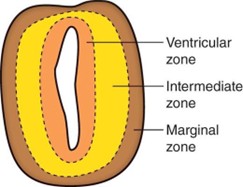
Transverse section showing the concentric layers of the early neural tube: Ventricular zone, Intermediate zone, and Marginal zone.
B. Intermediate Zone
- 1. It contains neuroblasts; these cells differentiate into neurons with dendrites and axons.
- 2. Also contains glioblasts, which differentiate into astrocytes and oligodendrocytes.
- 3. It forms the gray matter of the central nervous system.
- 4. The intermediate zone is divided into:
- the alar plate: associated with sensory (afferent) functions.
- and the basal plate: associated with motor (efferent) functions.
C. Marginal Zone
- 1. The marginal zone contains axons from neurons within the intermediate zone.
- 2. The marginal zone also contains glioblasts, which differentiate into astrocytes and oligodendrocytes.
- 3. The marginal zone forms the white matter of the central nervous system.
Neural Tube Concentric Layers
Ventricular Zone (Inner): Ependymal lining & mitotic germinal matrix source.
Intermediate / Mantle Zone (Middle): Cell bodies → forms Gray Matter.
Marginal Zone (Outer): Axonal nerve fibers → forms White Matter.
DEVELOPMENT OF THE SPINAL CORD
The spinal cord develops from the neural tube caudal to the fourth pair of somites.
A. Alar (sensory) plate
- It is a dorsolateral thickening of the intermediate zone of the neural tube.
- It gives rise to sensory neuroblasts of the dorsal horn (general somatic afferent [GSA] and general visceral afferent [GVA] cell regions).
- It receives axons from the dorsal root ganglia that become the dorsal (sensory) roots.
- It becomes the dorsal horn of the spinal cord.
B. Basal (motor) plate
- 1. The basal plate is a ventrolateral thickening of the intermediate zone of the neural tube.
- 2. It gives rise to motor neuroblasts of the ventral and lateral horns:
- general somatic efferent [GSE]
- and general visceral efferent [GVE] cell regions.
- 3. It projects axons from motor neuroblasts; these fibers exit the spinal cord and become the ventral (motor) roots.
- 4. The basal plate becomes the ventral horn of the spinal cord.
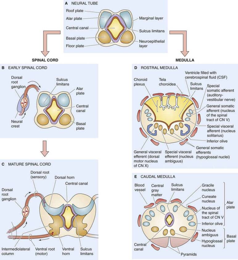
Schematic illustration of three successive stages in the development of the spinal cord. The neural crest gives rise to the dorsal root ganglion and the alar and basal plates give rise to the dorsal and ventral horns, respectively on the left and medulla on the right side.
C. Sulcus limitans (SL)
- 1. The SL is a longitudinal groove in the lateral wall of the neural tube that appears during week 4 of development and separates the alar and basal plates.
- 2. It disappears in the adult spinal cord but is retained in the rhomboid fossa of the brain stem.
- 3. It extends from the spinal cord to the rostral midbrain.
D. The roof plate
- is the nonneural roof of the central canal, which connects the two alar plates.
E. The floor plate
- is the nonneural floor of the central canal, which connects the two basal plates.
- The floor plate contains the ventral white commissure.
F. Myelination
- 1. Myelination of the spinal cord begins during month 4 in the ventral (motor) roots.
- 2. Oligodendrocytes accomplish myelination in the CNS.
- 3. Schwann cells accomplish myelination in the PNS.
- 4. Myelination of the corticospinal tracts is not completed until the end of 2 years of age (i.e., when the corticospinal tracts become myelinated and functional).
- 5. Myelination of the association neocortex extends to 30 years of age.
G. Positional changes of the spinal cord
- 1. At week 8 of development: the spinal cord extends the entire length of the vertebral canal.
- 2. At birth: the conus medullaris extends to the level of the third lumbar vertebra (L3).
- 3. In adults: the conus medullaris terminates at the L1–L2 interspace.
- 4. Disparate growth between the vertebral column and the spinal cord results in the formation of the cauda equina consisting of dorsal and ventral roots (L3–Co), which descends below the level of the conus medullaris.
- 5. Disparate growth results in the nonneural filum terminale, which anchors the spinal cord to the coccyx.
Conus Medullaris Ascension & Spinal Tap Levels
Because the vertebral column elongates faster than the spinal cord:
- Neonate/Infant: Cord ends at L3. Lumbar puncture must be performed at the L4–L5 or L5–S1 interspace to avoid conus damage.
- Adult: Cord ends at L1–L2. Lumbar puncture is safely performed at the L3–L4 or L4–L5 interspace (supracristal plane at the iliac crests).
DEVELOPMENT OF THE MYELENCEPHALON
The myelencephalon develops from the caudal rhombencephalon and gives rise to the medulla oblongata.
A. Alar plate sensory neuroblasts give rise to the following:
- 1. Cochlear and vestibular nuclei: These form the special somatic afferent (SSA) column and in the medullopontine junction.
- 2. Spinal trigeminal nucleus: This forms the general somatic afferent (GSA) column.
- 3. Solitary nucleus: This forms the special visceral afferent (SVA; taste) and general visceral afferent (GVA) columns.
- 4. Dorsal column nuclei: These consist of the gracile and cuneate nuclei.
- 5. Inferior olivary nuclei: are cerebellar relay nuclei and are derived from the alar plate by migrating to a ventral position.
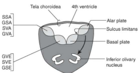
Transverse section through the open medulla demonstrating the alar (sensory) and basal (motor) columns and ventral migration of the inferior olivary nucleus.
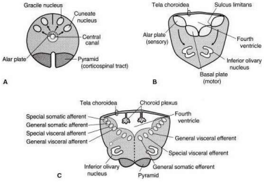
Schematic illustrations of the development of the medulla. (A) Transverse section of the caudal medulla showing the development of the gracile and cuneate nuclei from the alar plates. (B) Schematic sketch through the rostral (open) medulla showing the relationships of the alar and basal plates; the inferior olivary nucleus is derived from the alar plate. (C) A later stage of B shows the four sensory modalities of the alar plate and the three motor modalities of the basal plate; the white arrow indicates the lateral migration of the SVE column (CN IX, CN X, and CN XI); pyramids consist of motor fibers, the corticospinal tracts.
B. Basal plate motor neuroblasts give rise to the following:
- 1. Dorsal motor nucleus of the vagus nerve (CN X) and the inferior salivatory nucleus of the glossopharyngeal nerve (CN IX): which form the general visceral efferent (GVE) column.
- 2. Nucleus ambiguus: which forms the special visceral efferent (SVE) column (CN IX, CN X, and CN XI).
- 3. Hypoglossal nucleus: which forms the general somatic efferent (GSE) column.
C. The roof plate
- forms the roof of the fourth ventricle.
- The roof plate is called the tela choroidea, which is a monolayer of ependymal cells covered with pia mater.
- The tela choroidea is invaginated by pial blood vessels to form the choroid plexus of the fourth ventricle.
D. The open (rostral) medulla
- extends from the obex to the striae medullares of the rhomboid fossa.
- The lateral walls of the rostral medulla open like a book and form the rhomboid fossa (i.e., the floor of the fourth ventricle) due to the formation of the pontine flexure.
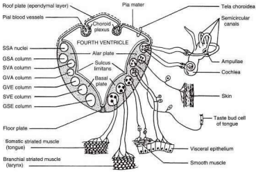
Schematic diagram of the brainstem illustrating the cell columns derived from the alar and basal plates. The seven cranial nerve modalities are shown. GSA = general somatic afferent, GVA = general visceral afferent, GVE = general visceral efferent; SSA = special somatic afferent, SSE = special somatic efferent; SVA = special visceral afferent, SVE = special visceral efferent.
DEVELOPMENT OF THE METENCEPHALON
The metencephalon develops from the rostral rhombencephalon and gives rise to the pons and cerebellum.
A. Pons
1. Alar plate sensory neuroblasts give rise to the following:
- a. Cochlear and vestibular nuclei: which form the special somatic afferent (SSA) column of CN VIII.
- b. Spinal and principal trigeminal nuclei: which form the general somatic afferent (GSA) column of CN V.
- c. Solitary nucleus: which forms the special visceral afferent (SVA; taste) and general visceral afferent (GVA) columns of CN VII.
- d. Pontine nuclei: which consist of cerebellar relay nuclei (pontine gray) and are derived from the alar plate by migrating to a ventral position.
2. Basal plate motor neuroblasts give rise to the following:
- a. Superior salivatory nucleus: which forms the general visceral efferent (GVE) column of CN VII.
- b. Facial (CN VII) and motor trigeminal (CN V) nuclei: which form the special visceral efferent (SVE) column.
- c. Abducent (CN VI) nucleus: which forms the general somatic efferent (GSE) column.
3. Base of the pons
The base of the pons contains the following:
- a. Pontine nuclei from the alar plate
- b. Corticobulbar, corticospinal, and corticopontine fibers: whose cell bodies are located in the cerebral cortex.
- c. Pontocerebellar fibers
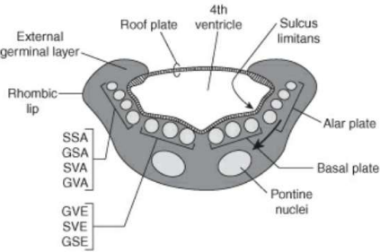
Transverse section of the developing metencephalon showing the rhombic lip, ventricular floor, and functional cranial nerve columns.
B. Cerebellum
- 1. The cerebellum is formed from the rhombic lips, which are the two dorsolateral thickened alar plates.
- 2. The rhombic lips thicken at week 6 to form the cerebellar plate, which has a dumbbell appearance.
- 3. The cerebellar plate is separated into cranial and caudal portions by a transverse groove.
- 4. The caudal portion forms the flocculonodular lobe, which is the most primitive part of the cerebellum.
- 5. The cranial portion forms the vermis and the cerebellar hemispheres, both of which undergo extensive formation of fissures and folia.
- 6. Rhombic lips consist of neuroectoderm arranged in the ventricular zone, intermediate zone, and marginal zone.
- 7. In month 3: the neuroectoderm in the ventricular zone undergoes another wave of proliferation to form the internal germinal layer. The internal germinal layer gives rise to the following:
- a. Deep cerebellar nuclei (i.e., dentate, emboliform, globose, and fastigial nuclei)
- b. Purkinje cells
- c. Golgi cells
- 8. Some neuroectodermal cells from the internal germinal layer migrate through the marginal zone to form the external germinal layer. The external germinal layer gives rise to the following:
- a. Basket cells
- b. Granule cells
- c. Stellate cells
- 9. Both the external and internal germinal layers give rise to astrocytes, Bergmann cells, and oligodendrocytes within the cerebellum.
| Cerebellar Germinal Layer | Embryological Location | Neuronal & Glial Cell Derivatives |
|---|---|---|
| Internal Germinal Layer | Ventricular neuroectoderm (Month 3 proliferation) | Deep cerebellar nuclei (Dentate, Emboliform, Globose, Fastigial), Purkinje cells, Golgi cells |
| External Germinal Layer | Migrates outward through marginal zone to cerebellar surface | Granule cells, Basket cells, Stellate cells |
| Shared Glial Progenitors | Derived from both germinal layers | Astrocytes, Bergmann glia, Oligodendrocytes |
Deep Cerebellar Nuclei Mnemonic ("Don't Eat Greasy Foods")
From Lateral to Medial: Dentate, Emboliform, Globose, Fastigial. All arise from the internal germinal layer.
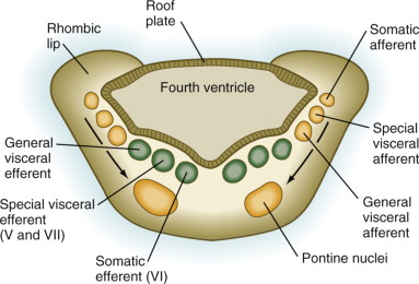
Schematic illustration (transverse section) of the development of the pons and cerebellum. The alar plate (rhombic lip) gives rise to the cerebellum, the four sensory cell columns, and the pontine nuclei. The basal plate gives rise to the three motor columns. The base of the pons contains the descending corticospinal tracts, which originate from the motor and sensory strips of the cerebral cortex. The white arrow indicates the lateral migration of the SVE column (V and CN VII).
DEVELOPMENT OF THE MESENCEPHALON
- The mesencephalon remains unchanged during primary to secondary vesicle formation and gives rise to the midbrain.
A. Alar plate sensory neuroblasts
- gives rise to the superior colliculi and the inferior colliculi.
B. Basal plate motor neuroblasts give rise to the following:
- 1. Edinger-Westphal nucleus of CN III: which forms the general visceral efferent (GVE) column.
- 2. Oculomotor (CN III) nucleus: which forms the general somatic efferent (GSE) column.
- 3. Substantia nigra: (arrows indicate direction of migration).
- 4. Red nucleus: (arrows indicate direction of migration).
C. Crus cerebri
- contain corticobulbar, corticospinal, and corticopontine fibers
- derived from the cerebral cortex of the telencephalon.
D. Migratory cranial nerve nuclei
- The trochlear (CN IV) nucleus and a portion of the sensory mesencephalic trigeminal (CN V) nucleus originate in the metencephalon and secondarily migrate into the mesencephalon.
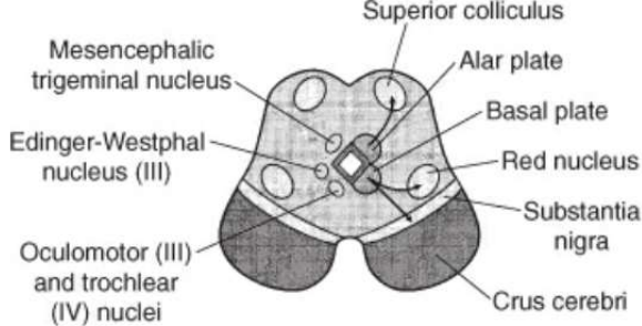
Transverse section through the midbrain illustrating the derivatives of the alar plate (colliculi) and basal plate (oculomotor complex, red nucleus, substantia nigra, crus cerebri).
DEVELOPMENT OF THE DIENCEPHALON, OPTIC STRUCTURES, AND HYPOPHYSIS
A. Diencephalon
- develops from the prosencephalon within the walls of the primitive third ventricle.
- The alar plates remain prominent in the prosencephalon, but the basal plates regress.
- The diencephalon gives rise to the epithalamus, thalamus, subthalamus, and hypothalamus.
1. Epithalamus
- develops from the embryonic roof plate and dorsal parts of alar plates.
- It gives rise to the pineal body (epiphysis), habenular nuclei, habenular commissure, posterior commissure, tela choroidea, and the choroid plexus of the third ventricle.
2. Thalamus
- is an alar plate derivative that gives rise to the thalamic nuclei, lateral geniculate body, and medial geniculate body.
3. Subthalamus
- is an alar plate derivative.
- It includes the subthalamic nucleus, zona incerta, and lenticular and thalamic fasciculi (fields of Forel).
- It gives rise to neuroblasts that migrate into the telencephalic white matter to become the globus pallidus (pallidum), which is a basal ganglion.
4. Hypothalamus
- develops from the alar plate and floor plate ventral to the hypothalamic sulcus.
- It gives rise to hypothalamic nuclei, mammillary bodies, and neurohypophysis.
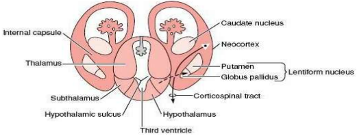
Schematic illustration (transverse section) of the development of the forebrain. The cerebral cortex and basal ganglia are shown. The internal capsule divides the corpus striatum into the caudate nucleus and the lentiform nucleus. The alar plate of the diencephalon gives rise to the thalamus and the hypothalamus. Cells from the subthalamus give rise to the globus pallidus.
B. Optic vesicles, cups, and stalks
- are derivatives of the diencephalon.
- They give rise to the retina, iris, ciliary body, optic nerve (CN II), optic chiasm, and optic tract.
C. Hypophysis (pituitary gland)
- is attached to the hypothalamus by the pituitary stalk and consists of the anterior lobe (adenohypophysis) and the posterior lobe (neurohypophysis).
| Pituitary Lobe | Embryological Precursor | Germ Layer Source |
|---|---|---|
| Anterior Lobe (Adenohypophysis) | Rathke's pouch (upward evagination from stomodeum) | Surface oral ectoderm |
| Posterior Lobe (Neurohypophysis) | Infundibulum (downward neuroectodermal evagination of floor of diencephalon) | Neuroectoderm |
Craniopharyngioma
Benign, calcified suprasellar tumor arising from epithelial remnants of Rathke's pouch (oral ectoderm). Most common suprasellar mass in children; compresses the optic chiasm causing bitemporal hemianopsia and pituitary/hypothalamic dysfunction.
DEVELOPMENT OF THE TELENCEPHALON
The telencephalon develops from the prosencephalon.
- The telencephalon gives rise to the cerebral hemispheres, caudate, putamen, amygdaloid, claustrum, lamina terminalis, olfactory bulbs, and hippocampus.
A. Cerebral hemispheres
- develop as bilateral evaginations of the lateral walls of the prosencephalic vesicle and contain the cerebral cortex, cerebral white matter, basal ganglia, and lateral ventricles.
- The cerebral hemispheres are interconnected by three commissures:
- the corpus callosum
- anterior commissure
- and hippocampal (fornical) commissure.
- Continuous hemispheric growth gives rise to frontal, parietal, occipital, and temporal lobes, which overlie the insula and dorsal brain stem.
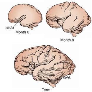
The diagram shows the development of the cerebral cortex at month 6, month 8, and term. Note the change in the cerebral cortex from a smooth surface or lissencephalic structure to a convoluted surface or gyrencephalic structure. As growth proceeds, a complex pattern of sulci (grooves) and gyri (elevations) develops.
B. Cerebral cortex (pallium)
- 1. Like the rest of the neural tube, the wall of the telencephalon consists of neuroectoderm arranged in the ventricular zone, intermediate zone, and marginal zone.
- 2. Neuroblasts from the ventricular zone and the intermediate zone migrate peripherally to form a transient layer called the cortical plate.
- 3. Neuroblasts in the ventricular zone undergo a wave of proliferation to form the subventricular zone.
- 4. Neuroblasts from the subventricular zone migrate peripherally to form the subplate zone.
- 5. The cortical plate and subplate zone together form the cerebral neocortex.
- 6. The intermediate zone becomes devoid of neuroblasts and develops into the white matter of the cerebral hemispheres.
- 7. The marginal zone develops into the molecular layer of the cerebral cortex.
- 8. The cerebral cortex is classified as the neocortex and allocortex:
- a. Neocortex (isocortex): is a six-layered cortex that represents 90% of the cortical mantle.
- b. Allocortex: is a three-layered cortex that represents 10% of the cortical mantle. The allocortex is subdivided into:
- the archicortex (which includes the hippocampal formation)
- and the paleocortex (which includes the olfactory cortex).
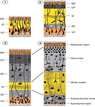
Cerebral cortex (pallium). MZ marginal zone; IZ intermediate zone; VZ ventricular zone; CP cortical plate; SP subplate zone; SZ subventricular zone.
C. Corpus striatum & Basal Ganglia
- 1. It appears in week 5 of development in the floor of the telencephalic vesicle.
- 2. It gives rise to the basal ganglia: the caudate nucleus, putamen, amygdaloid nucleus, and claustrum.
- 3. is divided into the caudate nucleus and the lentiform nucleus by corticofugal and corticopetal fibers (which make up the internal capsule).
- 4. The neurons of the globus pallidus (also a basal ganglion):
- have their origin in the subthalamus
- these neurons migrate into the telencephalic white matter
- and become the medial segments of the lentiform nucleus.
D. Commissures
- are fiber bundles that interconnect the two cerebral hemispheres; cross the midline via the embryonic lamina terminalis (commissural plate).
- 1. Anterior commissure: is the first commissure to appear and interconnects the olfactory structures and the middle and inferior temporal gyri.
- 2. Hippocampal (fornical) commissure: is the second commissure to appear and interconnects the two hippocampi.
- 3. Corpus callosum:
- is the third commissure to appear (between weeks 12 and 22).
- The corpus callosum is the largest commissure of the brain and interconnects homologous neocortical areas of the two cerebral hemispheres.
- The corpus callosum does not project commissural fibers from the visual cortex (area 17) or the hand area of the motor or sensory strips (areas 4 and 3, 1, 2).
CONGENITAL MALFORMATIONS OF THE CENTRAL NERVOUS SYSTEM
A. Variations of spina bifida
Spina bifida: occurs when the bony vertebral arches fail to form properly thereby creating a vertebral defect, usually in the lumbosacral region.
1. Spina bifida occulta
- is evidenced by a tuft of hair in the lumbosacral region.
- It is the least severe variation and occurs in 10% of the population.
- MRI of spina bifida occulta shows the presence of the bony vertebral bodies (VB) along the entire length of the vertebral column.
- However, the bony spinous processes terminate much higher because the vertebral arches fail to form properly. This creates a bony vertebral defect.
- The spinal cord is intact.
2. Spina bifida with meningocele
- occurs when the meninges protrude through a vertebral defect and form a sac filled with CSF.
- The spinal cord remains in its normal position.
3. Spina bifida with meningomyelocele
- occurs when the meninges and spinal cord protrude through a vertebral defect and form a sac filled with CSF.
4. Spina bifida with rachischisis
- occurs when the posterior neuropore of the neural tube fails to close during week 4 of development.
- This condition is the most severe type of spina bifida, causing paralysis from the level of the defect caudally.
- This variation presents clinically as an open neural tube that lies on the surface of the back.
- This condition also falls into a classification called neural tube defects (NTDs).
| Type of Spina Bifida | Dural / Meningeal Status | Neural Tissue Involvement | Clinical Manifestations & Severity |
|---|---|---|---|
| Spina Bifida Occulta | Intact; no external sac | Spinal cord normal and intact | Mildest (10% of population); marked by overlying skin dimple or tuft of hair; normal AFP |
| Spina Bifida with Meningocele | Protrudes into CSF-filled subcutaneous sac | Spinal cord remains in vertebral canal | Moderate; neurological deficits variable; repaired surgically |
| Spina Bifida with Meningomyelocele | Protrudes into external cystic sac | Spinal cord & nerve roots herniate into sac | Severe; sensory/motor deficit below lesion, neurogenic bladder, Chiari II malformation |
| Spina Bifida with Rachischisis (Myeloschisis) | Completely open; failure of posterior neuropore closure | Neural plate exposed directly on dorsal surface | Most severe; flaccid paralysis below lesion, high mortality/infection risk; elevated maternal serum AFP & AChE |
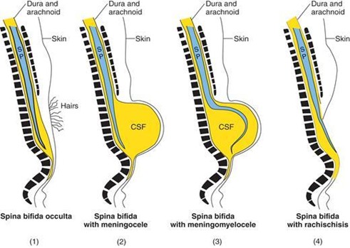
Schematic drawings illustrating the various types of spina bifida. SP spinal cord; CSF cerebrospinal fluid.
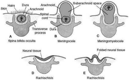
Schematic drawings illustrating a variety of neural tube defects involving the spinal cord. The term spina bifida applies to all of the defects because the bony arch of one or more vertebrae has failed to fuse dorsal to the spinal cord.
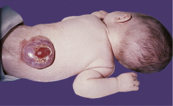
Clinical photograph demonstrating open neural tube defect (rachischisis) over the lumbosacral spine.
B. Variations of cranium bifida
Cranium bifida: occurs when the bony skull fails to form properly, thereby creating a skull defect, usually in the occipital region.
- 1. Cranium bifida with meningocele: occurs when the meninges protrude through the skull defect and form a sac filled with CSF.
- 2. Cranium bifida with meningoencephalocele:
- occurs when the meninges and brain protrude through the skull defect and form a sac filled with CSF.
- This defect usually comes to medical attention within the infant's first few days or weeks of life.
- The outcome is poor (75% of the infants die or have severe retardation).
- The MRI of a meningoencephalocele demonstrates a large protrusion extending through an occipital bone defect, which contains brain tissue.
- 3. Cranium bifida with meningohydroencephalocele: occurs when the meninges, brain, and a portion of the ventricle protrude through the skull defect.
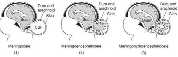
Variations of cranium bifida: (1) Meningocele, (2) Meningoencephalocele, (3) Meningohydroencephalocele.
C. Anencephaly (meroanencephaly)
- is a type of upper NTD that occurs when the anterior neuropore fails to close during week 4 of development.
- This results in:
- failure of the brain to develop (however, a rudimentary brain is present)
- failure of the lamina terminalis to form
- and failure of the bony cranial vault to form.
Biochemical Markers of Open Neural Tube Defects
Elevated Maternal Serum Alpha-Fetoprotein (MSAFP): Fetal serum leaks into amniotic fluid through open defects (anencephaly, rachischisis, meningomyelocele), leading to elevated maternal blood levels.
Amniotic Acetylcholinesterase (AChE): Elevated exclusively in open neural tube defects (confirmatory test distinguishing open NTD from ventral wall defects like gastroschisis).
Prophylaxis: Daily maternal intake of folic acid (0.4 mg standard; 4.0 mg high risk) initiated ≥1 month prior to conception reduces NTD incidence by up to 70%.
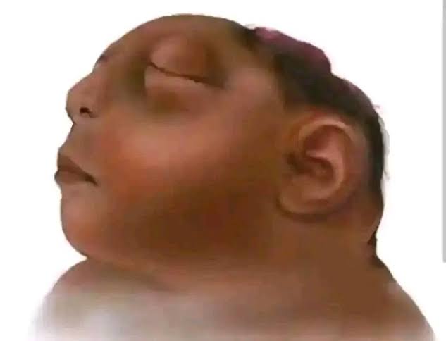
Infant with anencephaly displaying absence of the cranial vault and exposed rudimentary vascular neural tissue.
