Mechanism of Blood Clotting
Complete study notes covering the physiological phases of hemostasis, the biochemical coagulation cascade, modern cell-based models, fibrinolysis, and clinical laboratory diagnostics.
3.1 Definition and Overview
Hemostasis is the physiological process that stops bleeding from a damaged blood vessel. It is a rapid, localized, and carefully regulated process that involves three overlapping phases:
- Vascular spasm (vasoconstriction)
- Platelet plug formation (primary hemostasis)
- Coagulation cascade (secondary hemostasis)
- Clot stabilization and fibrinolysis
3.2 Vascular Spasm
This is the immediate reflex constriction of a damaged vessel to reduce blood loss. It is most effective in smaller vessels and is mediated by:
- Local myogenic spasm: Direct damage to vascular smooth muscle triggers contraction.
- Endothelin release: Released from damaged endothelial cells.
- Neural reflexes: Triggered by pain receptors.
3.3 Platelet Plug Formation (Primary Hemostasis)
This process converts a temporary leak into a mechanical plug through three distinct steps:
Step 1: Adhesion
- Vessel injury exposes subendothelial collagen.
- von Willebrand factor (vWF) binds to the exposed collagen.
- Platelet glycoprotein Ib (GPIb) receptors bind to vWF, causing platelets to adhere to the damaged wall.
Step 2: Activation
- Adhesion triggers a platelet shape change (pseudopod formation).
- Release of granule contents:
— Dense granules: Release ADP, serotonin, and Ca2+.
— Alpha granules: Release fibrinogen, factor V, vWF, and PDGF. - Synthesis and release of thromboxane A2 (TXA2), a potent vasoconstrictor and platelet activator.
- Platelet membrane expresses glycoprotein IIb/IIIa (GPIIb/IIIa) receptors.
Step 3: Aggregation
- GPIIb/IIIa receptors bind fibrinogen, linking platelets together.
- This forms a loose platelet plug (white thrombus), which is later reinforced by fibrin.
Antiplatelet Therapy
Aspirin irreversibly inhibits COX-1, blocking the synthesis of Thromboxane A2 (TXA2). This effectively reduces platelet aggregation and forms the basis for secondary prevention of myocardial infarction and stroke.
3.4 Coagulation Cascade (Secondary Hemostasis)
Secondary hemostasis involves a series of enzymatic reactions involving plasma proteins called clotting factors. Most factors are synthesized in the liver. Factors II, VII, IX, and X are Vitamin K-dependent.
3.4.1 Clotting Factors
| Factor | Name | Source | Function |
|---|---|---|---|
| I | Fibrinogen | Liver | Converted to fibrin (clot structure) |
| II | Prothrombin | Liver | Converted to Thrombin (central enzyme) |
| III | Tissue Factor | Tissue | Initiates the Extrinsic Pathway |
| IV | Calcium (Ca2+) | Diet | Cofactor for multiple reactions |
| V | Proaccelerin | Liver | Cofactor for Factor X activation |
| VII | Proconvertin | Liver | Initiates extrinsic pathway |
| VIII | Antihemophilic A | Liver | Cofactor for Factor IX (Deficient in Hemophilia A) |
| IX | Antihemophilic B | Liver | Activates Factor X (Deficient in Hemophilia B) |
| X | Stuart-Prower | Liver | Forms Prothrombinase complex |
| XI | PTA | Liver | Activates Factor IX |
| XII | Hageman Factor | Liver | Initiates Intrinsic Pathway |
| XIII | Fibrin-stabilizing | Liver | Cross-links fibrin monomers |
*Note: Factor VI was originally assigned to activated factor V but is no longer used in modern nomenclature.
3.4.2 The Three Pathways
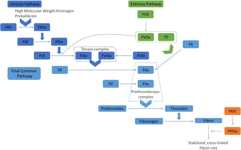
A. Extrinsic Pathway (Tissue Factor Pathway)
- Trigger: Tissue injury exposing Tissue Factor (Factor III) to blood.
- Sequence: Factor III + Factor VII activates Factor X.
- Speed: Fast-acting; generates thrombin within seconds.
- Lab Test: Prothrombin Time (PT).
B. Intrinsic Pathway (Contact Activation Pathway)
- Trigger: Blood exposure to negatively charged subendothelial surfaces.
- Sequence: Factor XII → XI → IX (with VIII as cofactor) → activates Factor X.
- Speed: Slower; requires several minutes.
- Lab Test: Activated Partial Thromboplastin Time (aPTT).
C. Common Pathway
- Both pathways converge at Factor X activation.
- Factor Xa + Va (Prothrombinase complex) + Ca2+ + phospholipids converts Prothrombin (II) to Thrombin (IIa).
- Thrombin converts Fibrinogen (I) to Fibrin monomers.
- Fibrin monomers polymerize into a mesh.
- Factor XIIIa cross-links the fibrin to create a stable, permanent clot.
3.4.3 Cell-Based Model (Modern Understanding)
Modern hematology emphasizes that coagulation occurs in three phases on cell surfaces:
- Initiation: Tissue factor-bearing cells activate small amounts of X and thrombin.
- Amplification: Thrombin activates platelets and cofactors (V, VIII, XI).
- Propagation: Large-scale thrombin generation (thrombin burst) on platelet surfaces produces a massive fibrin clot.
3.5 Clot Retraction and Repair
- Platelets contain actin and myosin filaments (contractile proteins).
- Clot contraction pulls wound edges together.
- This squeezes serum out of the clot (Serum = Plasma without clotting factors).
- Platelet-Derived Growth Factor (PDGF) stimulates fibroblast migration and tissue repair.
3.6 Fibrinolysis (Clot Removal)
Fibrinolysis prevents excessive clot propagation and removes the clot once healing has occurred.
Mechanism:
- Plasminogen (inactive) is incorporated into the clot during formation.
- Tissue plasminogen activator (t-PA) from endothelial cells converts plasminogen to Plasmin.
- Plasmin digests fibrin into fibrin degradation products (FDPs).
- D-dimer is a specific FDP from cross-linked fibrin.
D-dimer
Elevated D-dimer levels indicate active fibrinolysis and are used to screen for thrombotic conditions like DVT or Pulmonary Embolism (PE). However, it is non-specific and can also be elevated in infection, pregnancy, or malignancy.
3.7 Natural Anticoagulant Mechanisms
| Mechanism | Action | Deficiency State |
|---|---|---|
| Antithrombin III | Inhibits thrombin and factors Xa, IXa | Thrombophilia |
| Protein C | Inactivates factors Va and VIIIa | Thrombophilia; Warfarin skin necrosis |
| Protein S | Cofactor for Protein C | Thrombophilia |
| TFPI | Inhibits Tissue Factor-VIIa complex | — |
| Fibrin Clot | Adsorbs 85-90% of thrombin to prevent spread | — |
3.8 Clinical Laboratory Tests of Coagulation
| Test | Measures | Normal Range | Prolonged In... |
|---|---|---|---|
| PT | Extrinsic + Common (VII, X, V, II, I) | 11–13 sec | Warfarin use, Vit K deficiency, Liver disease |
| aPTT | Intrinsic + Common (XII, XI, IX, VIII, X, V, II, I) | 25–35 sec | Heparin use, Hemophilia, vWD |
| INR | Standardized PT ratio | 0.9–1.1 | Warfarin monitoring |
| TT | Fibrinogen → Fibrin conversion | 15–19 sec | Hypofibrinogenemia, DIC |
| D-dimer | Fibrin degradation products | <0.5 ug/mL | DVT, PE, DIC, Post-surgery |
Hemophilia A vs. B
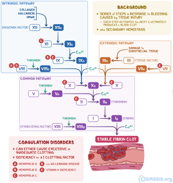
- Hemophilia A: Deficiency of Factor VIII.
- Hemophilia B: Deficiency of Factor IX.
- Both present with bleeding into joints (hemarthrosis), muscle hematomas, and a prolonged aPTT with a normal PT.
