
The orbit and its contents: osteology, eyeball, extraocular muscles & neurovasculature
On this page
Overview & orbital axes
The orbits are bilateral, symmetrical bony cavities within the craniofacial skeleton that encase, protect, and support the eyeballs (globes), their extrinsic extraocular musculature, neurovascular bundles, lacrimal apparatus, and retrobulbar adipose tissue.
Geometric Configuration
Each orbit resembles a hollow, four-sided quadrangular pyramid:
- Base (Orbital Opening): Directed anterolaterally toward the face; outlined by the thickened, protective orbital rim (orbital margin).
- Apex: Directed posteromedially toward the intracranial cavity at the optic canal.
- Medial Walls: The medial walls of both orbits are separated by the ethmoidal air cells and the superior nasal cavity; they run essentially parallel to each other.
- Lateral Walls: Form an approximate right angle (90°) to each other.
- Orbital Axis vs. Optical Axis:
- The orbital axis (line from apex through the center of the orbital opening) diverges at approximately 45° from the contralateral orbital axis (or 22.5° to 23° lateral to the sagittal midline).
- The optical axis (line of sight / axis of gaze connecting the anterior pole to the posterior pole of the globe) runs strictly parallel and anteriorly when looking straight ahead (“primary position of gaze”).
Osteology of the orbital walls
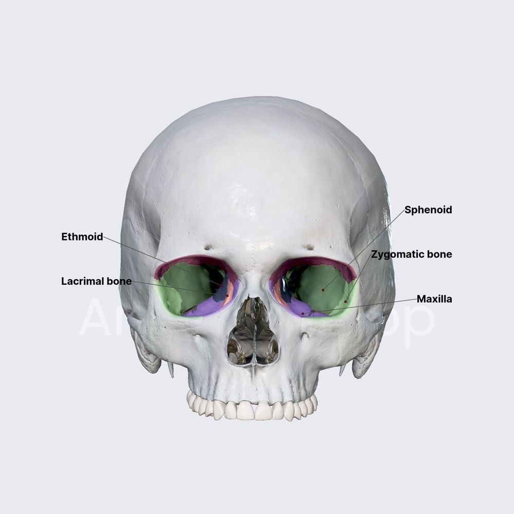
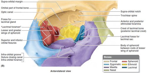
Seven individual bones articulate to construct each bony orbit: the frontal, zygomatic, maxilla, ethmoid, lacrimal, sphenoid, and palatine bones.
1. The Orbital Margin (Base)
The base of the pyramid forms the reinforced orbital margin surrounding the orbital opening:
- Superior Margin: Formed entirely by the frontal bone. Features the supra-orbital notch (or foramen) at the junction of its medial and middle thirds, transmitting the supra-orbital nerve (CN V1) and vessels.
- Medial Margin: Formed by the frontal process of the maxilla and the lacrimal process of the frontal bone; indented by the anterior lacrimal crest.
- Inferior Margin: Formed by the maxilla medially and the zygomatic bone laterally.
- Lateral Margin: Formed by the zygomatic process of the frontal bone and the frontal process of the zygomatic bone.
- Orbital Septum Attachment: The peripheral bone provides anchoring for the orbital septum, a fibrous sheet extending into the eyelids that creates a physical barrier against preseptal periorbital infections entering the postseptal orbit.
2. The Superior Wall (Roof)
- Bony Composition: Formed primarily by the horizontal orbital part of the frontal bone, with a small contribution near the apex from the lesser wing of the sphenoid.
- Anatomical Separations: Forms a thin shelf of bone separating the orbit from the anterior cranial fossa and frontal air lobes.
- Special Depressions:
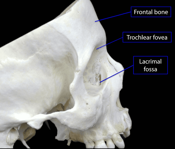
- Lacrimal Fossa (Fossa for Lacrimal Gland): Located anterolaterally in the frontal bone; lodges the orbital lobe of the lacrimal gland.
- Trochlear Fovea (or Spine): Located anteromedially; provides a fibrous or cartilaginous pulley (trochlea) for the superior oblique muscle tendon.
3. The Medial Wall
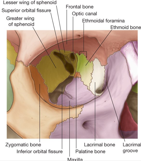
- Bony Composition: Formed mainly by the thin, paper-like orbital plate of the ethmoid (lamina papyracea), supplemented anteriorly by the lacrimal bone and frontal process of the maxilla, and posteriorly by the body of the sphenoid.
- Ethmoidal Air Cell Relations: The lamina papyracea is extremely thin (~0.2 to 0.4 mm). It directly borders the ethmoidal air sinuses, allowing ethmoid sinusitis to easily erode or spread into the orbit, causing subperiosteal abscesses or orbital cellulitis.
- Lacrimal Groove & Fossa: Anteriorly, the lacrimal bone and maxilla form the lacrimal groove (fossa for the lacrimal sac), bounded by the anterior and posterior lacrimal crests, leading down into the nasolacrimal canal.
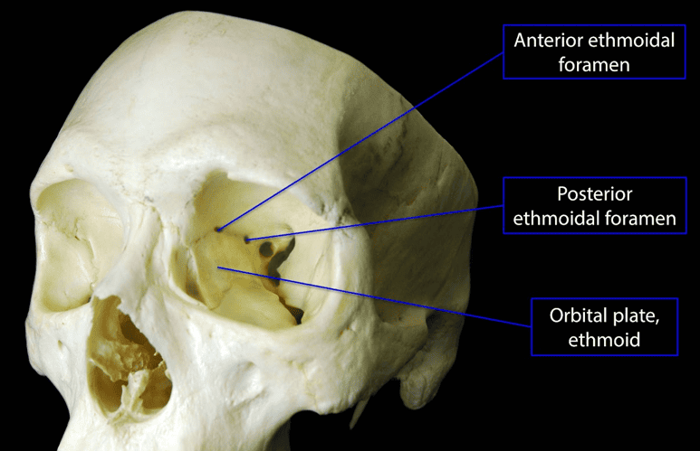
- Ethmoidal Foramina: Located along the fronto-ethmoidal suture line:
- Anterior Ethmoidal Foramen: Transmits the anterior ethmoidal nerve and vessels.
- Posterior Ethmoidal Foramen: Transmits the posterior ethmoidal nerve and vessels.

4. The Inferior Wall (Orbital Floor)
- Bony Composition: Formed mainly by the orbital surface of the maxilla, with contributions from the orbital surface of the zygomatic bone anterolaterally and the small orbital process of the palatine bone at the posterior apex.
- Maxillary Sinus Roof: The orbital floor is very thin and forms the roof of the maxillary sinus. It slants downward and forward toward the inferior orbital margin.
- Infra-Orbital Groove & Canal: Traversed by the infra-orbital groove, which continues anteriorly into the infra-orbital canal and exits via the infra-orbital foramen, transmitting the infra-orbital nerve (CN V2) and vessels.
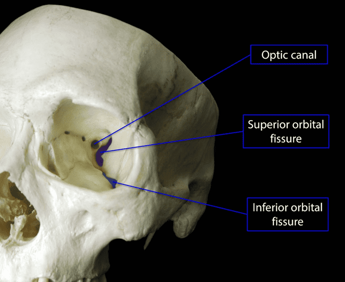
- Inferior Orbital Fissure: Separates the orbital floor from the lateral wall; represents an elongated gap between the maxilla, sphenoid, and palatine bones.
5. The Lateral Wall
- Bony Composition: Formed by the frontal process of the zygomatic bone anteriorly and the orbital surface of the greater wing of the sphenoid posteriorly.
- Strength & Thickness: This is the strongest and thickest of the four orbital walls, providing structural protection against lateral direct trauma.
- Fossa Separations: Separates the orbital cavity anteriorly from the temporal fossa, and posteriorly from the middle cranial fossa.
Foramina, fissures & apex
The orbital apex serves as the primary gateway for neurovascular structures entering and exiting the cranial cavity, pterygopalatine fossa, and infratemporal fossa.
Scroll the table sideways on a smaller screen.
| Aperture / Foramen | Bony Boundaries | Communicating Space | Transmitted Neurovascular Structures |
|---|---|---|---|
| Optic Canal | Between roots of lesser wing of sphenoid | Middle cranial fossa | • Optic nerve (CN II) encased in dural sheath • Ophthalmic artery |
| Superior Orbital Fissure (SOF) | Between greater and lesser wings of sphenoid | Middle cranial fossa |
Outside Tendinous Ring: Lacrimal nerve, Frontal nerve, Trochlear nerve (CN IV), Superior ophthalmic vein. Inside Tendinous Ring: Superior & inferior divisions of Oculomotor nerve (CN III), Nasociliary nerve, Abducent nerve (CN VI), sympathetic pupillomotor roots. |
| Inferior Orbital Fissure (IOF) | Between greater wing of sphenoid, maxilla, and palatine | Pterygopalatine & infratemporal fossae | • Infra-orbital nerve (CN V2) and vessels • Zygomatic nerve (CN V2) • Inferior ophthalmic vein tributaries to pterygoid plexus • Orbital branches of pterygopalatine ganglion |
| Anterior Ethmoidal Foramen | Fronto-ethmoidal suture | Anterior cranial fossa / nasal cavity | • Anterior ethmoidal nerve and artery |
| Posterior Ethmoidal Foramen | Fronto-ethmoidal suture | Posterior ethmoidal air cells / sphenoid sinus | • Posterior ethmoidal nerve and artery |
| Supra-orbital Notch / Foramen | Superior orbital margin (frontal bone) | Forehead and scalp | • Supra-orbital nerve (lateral branch of frontal nerve, CN V1) and vessels |
| Infra-orbital Canal / Foramen | Maxillary bone (orbital floor) | Midface and upper lip | • Infra-orbital nerve (terminal CN V2) and vessels |
| Nasolacrimal Canal | Formed by maxilla, lacrimal, and inferior nasal concha | Inferior meatus of nasal cavity | • Nasolacrimal duct |
The eyeball: coats & fascial sheath
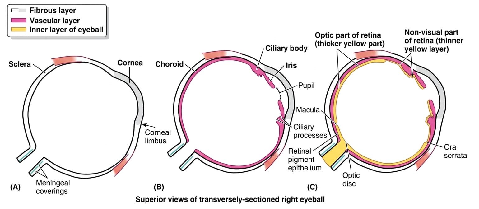
The eyeball (globe) contains the light-refracting optical apparatus of vision. It occupies the anterior half of the orbit, suspended within retrobulbar orbital fat by six extrinsic extraocular muscles and a specialized fascial suspensory apparatus. The eyeball measures approximately 25 mm in diameter and is constructed of three concentric anatomical layers (tunics).
The Fascial Sheath (Tenon’s Capsule)
- Bulbar Fascia (Tenon’s Capsule): A thin, tough fibroelastic membrane that envelops the globe from the optic nerve insertion posteriorly up to the corneoscleral junction (limbus) anteriorly. It forms the true socket in which the eyeball moves.
- Episcleral Space: A potential space filled with loose, delicate connective tissue trabeculae situated between Tenon’s capsule and the outer sclera. It acts as a low-friction bursa, facilitating unhindered multi-axial ocular rotations.
- Suspensory Ligament of Lockwood: Thickening of the lower part of Tenon’s capsule, blended with the fascial sheaths of the inferior rectus and inferior oblique muscles, forming a hammock-like sling that supports the globe. It prevents downward displacement of the eyeball even after traumatic destruction of the bony orbital floor.
- Check Ligaments: Triangular expansions from the sheaths of the medial and lateral rectus muscles attached to the lacrimal bone and zygomatic bone (Whitnall tubercle), respectively, limiting excessive ocular adduction and abduction.
Fibrous layer: sclera & cornea
The fibrous tunic forms the rigid outer skeleton of the globe, maintaining its spherical shape, protecting delicate intraocular elements, and resisting physiological intraocular pressure.
1. The Sclera (“White of the Eye”)
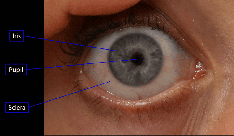
- Extent & Composition: Covers the posterior five-sixths (5/6) of the globe. Composed of dense, irregular, interwoven collagen bundles and elastic fibers that appear opaque white through the translucent bulbar conjunctiva.
- Muscle Insertions: Provides mechanical anchorage for the tendons of all extraocular rectus and oblique muscles.
- Lamina Cribrosa: A perforated sieve-like zone in the posterior sclera (~1 mm medial to the posterior pole) through which unmyelinated retinal ganglion cell axons exit to form the optic nerve. It represents the weakest area of the outer coat, prone to posterior bulging (cupping) in chronic glaucoma.
- Vascularity: The sclera itself is largely avascular; it receives its blood supply from the overlying episcleral vascular plexus and the underlying choroid.
2. The Cornea
- Extent & Curvature: Forms the transparent anterior one-sixth (1/6) of the globe. It has a steeper curvature than the sclera, protruding anteriorly as a clear dome. It provides approximately +40 to +44 diopters (over two-thirds) of the eye’s total refractive power.
- Avascularity & Optical Clarity: Completely devoid of blood and lymphatic vessels. Nourishment and oxygen are derived via:
- Diffusion from loop capillaries of the surrounding corneal limbus.
- Aqueous humor filling the anterior chamber internally.
- Oxygen absorbed directly from atmospheric air dissolved in precorneal lacrimal tear fluid externally.
- Innervation & Sensitivity: Exquisitely sensitive to pain and touch, innervated by the ophthalmic nerve (CN V1) via the long and short ciliary nerves. Touching the cornea triggers the rapid corneal blink reflex (sensory limb: CN V1; motor limb: facial nerve, CN VII, to orbicularis oculi). Desiccation of the epithelial surface leads to painful corneal ulcerations.
3. The Corneal Limbus
The corneoscleral junction (limbus) is a 1-mm-wide, gray, translucent transitional zone between the clear cornea and opaque sclera. Internally, it houses the trabecular meshwork and the circular scleral venous sinus (canal of Schlemm), which drains aqueous humor from the anterior chamber into the episcleral veins.
Vascular layer: choroid, ciliary body & iris
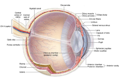
The vascular tunic (uvea / uveal tract) is the pigmented middle coat of the eyeball, comprising the choroid posteriorly, the ciliary body intermediately, and the iris anteriorly.
1. The Choroid
- Structure: A dark, reddish-brown, highly vascular and heavily pigmented layer lining the posterior 5/6 of the inner sclera. It firmly adheres internally to the retinal pigment epithelium (RPE), but separates easily from the sclera via the suprachoroidal space.
- Choriocapillaris: The innermost capillary bed of the choroid, positioned adjacent to the avascular outer third of the retina (photoreceptor rods and cones), supplying it with oxygen and nutrients.
- Perfusion Dynamics: Possesses the highest blood flow rate per gram of tissue of any organ system in the human body. This rich blood volume produces the characteristic “red eye” artifact seen in flash photography.
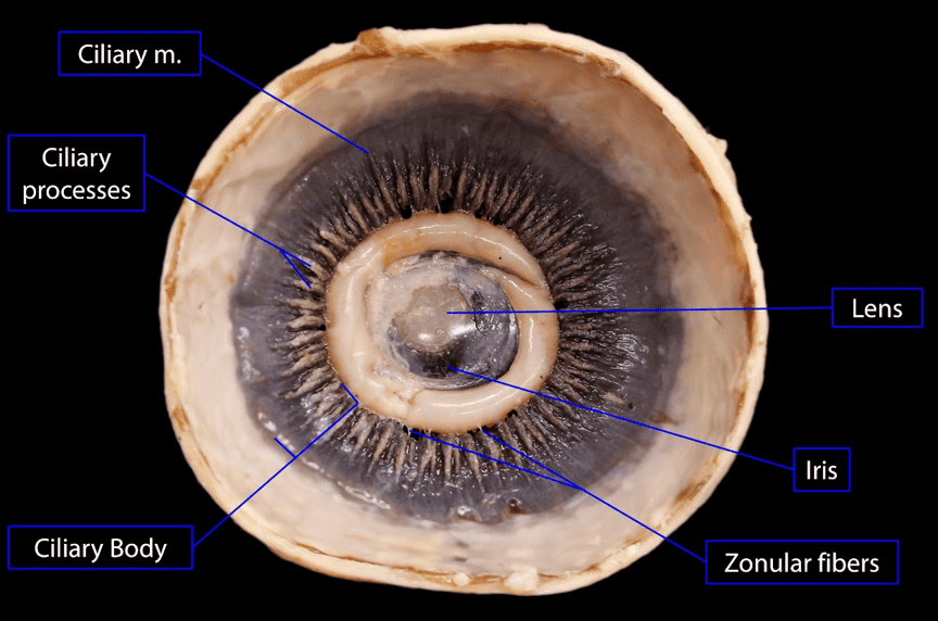
2. The Ciliary Body
A ring-shaped thickening of the uvea connecting the choroid with the iris circumference. It has dual muscular and secretory functions:
- Ciliary Muscle (Accommodation): Smooth muscle fibers arranged in circular, radial, and meridional vectors, innervated by parasympathetic fibers from CN III via the ciliary ganglion. Contraction draws the ciliary ring forward and inward toward the lens, relaxing tension on the suspensory zonular fibers. Relieved of tension, the elastic crystalline lens bulges and becomes more convex, increasing its refractive power for near vision accommodation.
- Ciliary Processes: Approximately 70 to 80 radial vascular folds on the inner surface of the ciliary body lined by a double-layered epithelium. They actively produce and secrete aqueous humor into the posterior chamber.
3. The Iris & Pupillary Motility
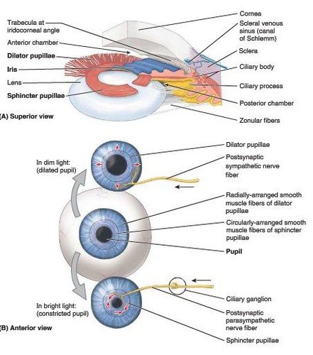
The iris is a thin, pigmented contractile diaphragm floating on the anterior surface of the crystalline lens, with a central aperture termed the pupil. It regulates the quantity of light admitted to the retina via two antagonistically arranged smooth muscles:
Dual Autonomic Control of the Pupil
- Sphincter Pupillae (Constrictor):
- Orientation: Circular smooth muscle fibers encircling the pupillary margin.
- Innervation: Parasympathetic preganglionic fibers from the Edinger-Westphal nucleus (CN III) synapsing in the ciliary ganglion; postganglionic fibers travel via the short ciliary nerves.
- Action: Constricts the pupil (miosis) in response to bright light and during near-vision accommodation. Constriction is rapid and nearly instantaneous.
- Dilator Pupillae:
- Orientation: Radially arranged myoepithelial fibers running from the ciliary margin to the sphincter.
- Innervation: Sympathetic postganglionic fibers from the superior cervical ganglion traversing the internal carotid plexus, nasociliary nerve, and long ciliary nerves.
- Action: Enlarges the pupil (mydriasis) in low-light environments and during sympathetic stress. While sympathetic responses can initiate quickly, full dark-adaptation dilation in low lighting can take up to 20 minutes.
Inner layer: retina, disc & macula
The retina forms the innermost light-sensitive sensory tunic of the eyeball, developing as an outpocketing of the embryonic diencephalon (optic vesicle).
1. Subdivisions of the Retina
- Optic Part of the Retina: The posterior light-sensitive visual retina, terminating anteriorly at the scalloped margin called the ora serrata. It comprises two primary layers:
- Neural Layer: Light-receptive, containing photoreceptors (rods for dim-light monochromatic vision; cones for bright-light color vision and acuity), bipolar interneurons, and ganglion cells whose unmyelinated axons form the nerve fiber layer.
- Pigmented Layer: Single layer of melanin-containing cuboidal cells adhering to the choroid. It absorbs scattered light, metabolizes vitamin A (retinal), forms the blood-retinal barrier, and phagocytoses shedding photoreceptor outer segments. In retinal detachment, the neural retina separates from this pigmented layer.
- Non-Visual Part of the Retina: The anterior, non-light-receptive continuation of the pigmented layer and supporting cells forward beyond the ora serrata, draping over the ciliary body (ciliary part) and posterior iris surface (iridial part) to the pupillary edge.
2. Landmarks of the Ocular Fundus
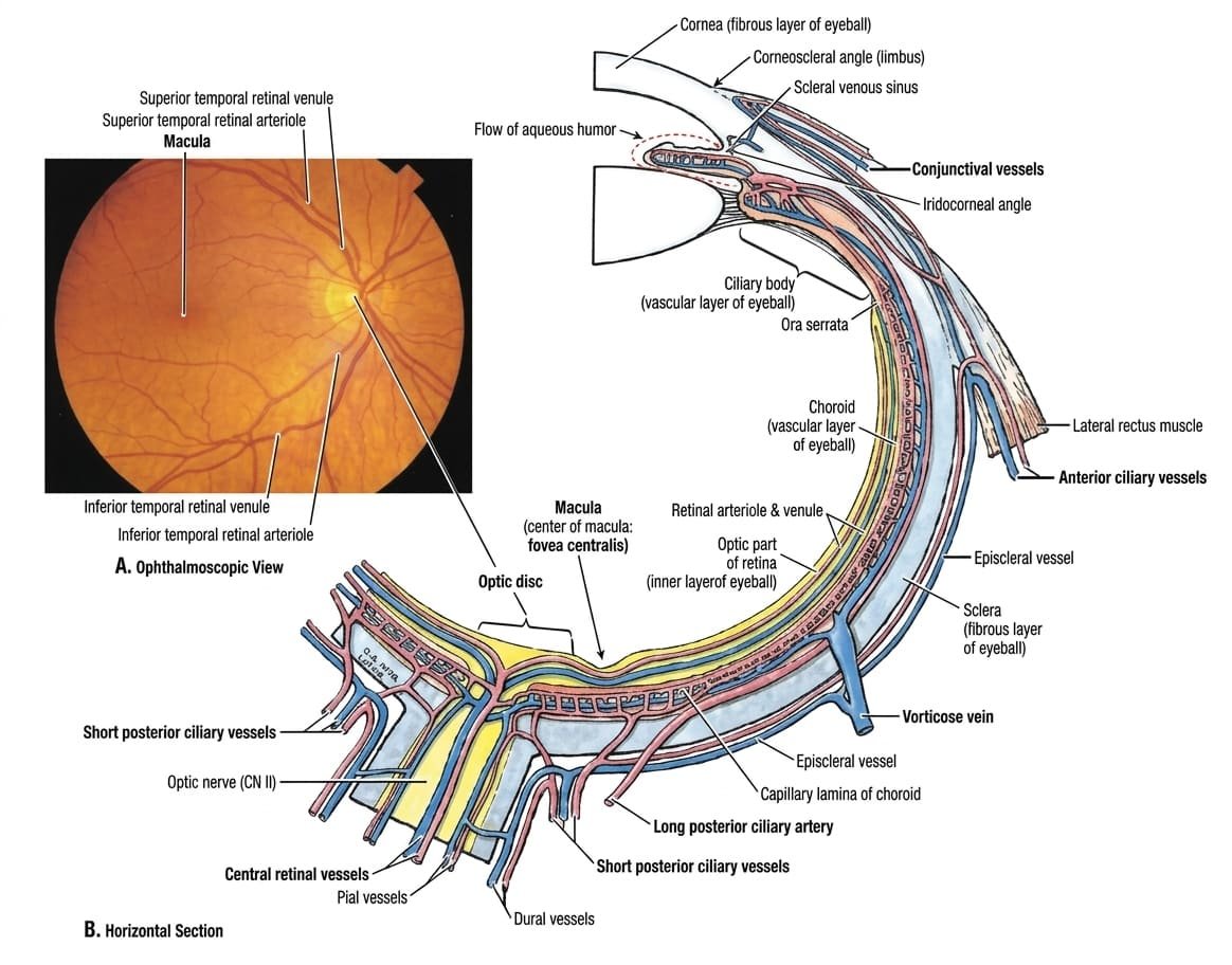
- Optic Disc (Optic Papilla): A pinkish, circular pale region (~1.5 mm diameter) located ~3 mm medial to the posterior pole. Point where ganglion cell axons gather to form the optic nerve and where central retinal vessels emerge. Because it contains no photoreceptor rods or cones, it corresponds to the physiological “blind spot” of the visual field. Has a central depression termed the physiological cup.
- Macula Lutea (“Yellow Spot”): A small oval yellowish area (~5.5 mm diameter) situated lateral to the optic disc at the visual center of the posterior retina. It is specialized for high-resolution visual acuity and contains yellow carotenoid pigments (lutein and zeaxanthin).
- Fovea Centralis & Foveola: A distinct central pit (~1.5 mm diameter) within the macula where inner retinal layers are displaced laterally, allowing light to hit photoreceptors directly without optical scattering. Composed exclusively of densely packed cones. The very center of this pit is the foveola (0.35 mm), which provides the point of maximal visual acuity. It is avascular, nourished purely by diffusion from the underlying choriocapillaris.
Chambers & aqueous humor
The interior of the eyeball is divided by the lens and its suspensory ligament into an anterior segment (filled with clear aqueous humor) and a posterior segment (filled with gelatinous vitreous humor).
Anterior and Posterior Chambers
- Anterior Chamber: The space bounded anteriorly by the posterior surface of the cornea and posteriorly by the anterior surface of the iris and central pupil.
- Posterior Chamber: The smaller space situated posterior to the iris and anterior to the crystalline lens, suspensory zonules, and ciliary body.
- Pupillary Communication: The anterior and posterior chambers are continuous with each other through the central pupillary aperture.
Aqueous Humor Flow & Glaucoma
Aqueous humor is a clear, protein-poor fluid secreted continuously at ~2 to 3 μL/min by the ciliary processes into the posterior chamber:
- Flow Pathway: Flows from the posterior chamber forward through the pupillary opening into the anterior chamber.
- Filtration & Drainage: Passes peripherally into the iridocorneal angle, filtering through the trabecular meshwork into the scleral venous sinus (canal of Schlemm), and ultimately draining into the episcleral venous system. (A minor 10% drains via the uveoscleral pathway).
- Physiological Functions: Supplies oxygen, glucose, and amino acids to the avascular cornea and lens, removes metabolic wastes, and maintains intraocular pressure (IOP), which normally ranges between 10 and 21 mmHg.
- Pathophysiology of Glaucoma: Obstruction of aqueous outflow causes IOP to rise pathologically. Elevated pressure compresses retinal nerve fibers and reduces perfusion at the lamina cribrosa, causing progressive optic disc cupping, optic neuropathy, and irreversible peripheral visual field loss.
The lens & vitreous body
The Crystalline Lens
A transparent, biconvex, elastic, avascular disc situated immediately posterior to the iris and pupil, separating the anterior one-fifth (1/5) of the globe from the posterior four-fifths (4/5):
- Lens Capsule: A highly elastic basement membrane encasing the entire lens, under continuous tension from the radially radiating zonular fibers (suspensory ligament of the lens) anchored to the ciliary processes.
- Refractive Adjustability: Contraction of the ciliary muscle relaxes zonular tension, allowing the inherent elasticity of the capsule to increase the anterior-posterior thickness and curvature of the lens, dynamically increasing its refractive power for near focus (accommodation).
- Presbyopia & Cataract: With aging, the lens nucleus loses elasticity, leading to impaired near accommodation (presbyopia). Opacification of the lens fibers or capsule produces a cataract, which impairs light transmission.
The Vitreous Body (Vitreous Humor)
Fills the large postremal (vitreous) chamber occupying the posterior 4/5 of the globe, extending from the posterior lens capsule to the sensory retina:
- Composition: A transparent, gelatinous matrix composed of 99% water bound within a network of type II collagen fibrils and hydrophilic glycosaminoglycans (primarily hyaluronic acid).
- Irreplaceability: Unlike aqueous humor, the vitreous body is not continuously replenished; loss of vitreous during ocular trauma or surgery is permanent and replaced clinically with physiological gas or silicone oil.
- Structural Roles: Maintains the spherical shape of the globe, supports the delicate sensory retina against the underlying pigmented epithelium and choroid, transmits light rays to the retina without refraction distortion, and acts as a shock-absorbing buffer during ocular movements.
Extraocular muscles & motility
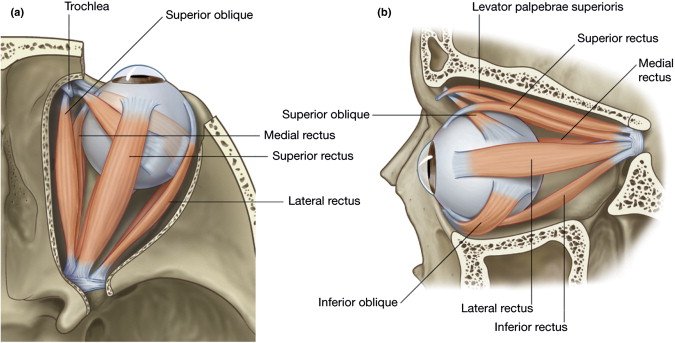
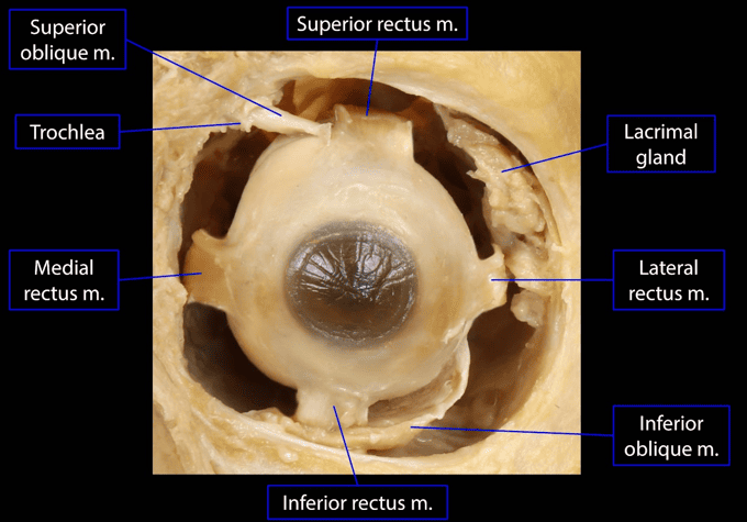
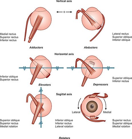
Ocular movements are coordinated by seven extrinsic striated muscles: one that raises the upper eyelid (levator palpebrae superioris) and six that act directly on the globe (four recti and two obliques).
Scroll the table sideways on a smaller screen.
| Muscle Name | Bony Origin | Scleral / Tissue Insertion | Cranial Nerve Innervation | Primary & Secondary Actions |
|---|---|---|---|---|
| Levator Palpebrae Superioris (LPS) | Lesser wing of sphenoid (superior & anterior to optic canal) | Anterior surface of superior tarsal plate and skin of upper eyelid | Oculomotor nerve (CN III, superior division); deep layer (superior tarsal / Müller’s muscle) by sympathetic postganglionics | Elevates upper eyelid (retracts lid) |
| Superior Rectus (SR) | Common tendinous ring (Annulus of Zinn) at orbital apex | Anterosuperior sclera (~7.7 mm posterior to corneal limbus) | Oculomotor nerve (CN III, superior division) | Primary: Elevation Secondary: Intorsion (medial rotation), Adduction |
| Inferior Rectus (IR) | Common tendinous ring (Annulus of Zinn) | Anteroinferior sclera (~6.5 mm posterior to corneal limbus) | Oculomotor nerve (CN III, inferior division) | Primary: Depression Secondary: Extorsion (lateral rotation), Adduction |
| Medial Rectus (MR) | Common tendinous ring (Annulus of Zinn) | Anteromedial sclera (~5.5 mm posterior to corneal limbus) | Oculomotor nerve (CN III, inferior division) | Primary: Adduction (pure adductor) |
| Lateral Rectus (LR) | Common tendinous ring (spans superior orbital fissure) | Anterolateral sclera (~6.9 mm posterior to corneal limbus) | Abducent nerve (CN VI) | Primary: Abduction (pure abductor) |
| Superior Oblique (SO) | Body of sphenoid superomedial to optic canal | Passes through fibrocartilaginous trochlea, reflects posterolaterally, inserting into posterosuperolateral sclera | Trochlear nerve (CN IV) | Primary: Intorsion (internal rotation) Secondary: Depression, Abduction |
| Inferior Oblique (IO) | Anterior orbital floor (orbital surface of maxilla lateral to lacrimal groove) | Courses posterolaterally under inferior rectus, inserting into postero-inferolateral sclera | Oculomotor nerve (CN III, inferior division) | Primary: Extorsion (external rotation) Secondary: Elevation, Abduction |
Arterial supply: ophthalmic artery
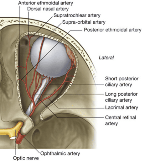
The arterial supply to the orbit, eyeball, and surrounding facial tissues arises primarily from the ophthalmic artery, a major branch of the internal carotid artery given off immediately after it exits the cavernous sinus.
Course & Spatial Relationships
The ophthalmic artery enters the orbit through the optic canal within the lesser wing of the sphenoid, enclosed in the dural sheath inferolateral to the optic nerve (CN II). In the apex, it lies lateral to the nerve, then courses medially by crossing superior to the optic nerve (in 85% of individuals) alongside the nasociliary nerve, running anteriorly along the medial wall of the orbit between the medial rectus and superior oblique muscles.
Ten Principal Branches of the Ophthalmic Artery
- Central Retinal Artery: Arises inferolateral to the optic nerve. Pierces the dural sheath ~1 cm behind the globe, passes down the center of the optic nerve, and enters the retina at the optic disc. Divides into superior and inferior nasal and temporal branches. True anatomical end-arteries; occlusion produces sudden, painless, complete blindness.
- Lacrimal Artery: Arises lateral to the optic nerve, courses forward along the superior border of the lateral rectus muscle, supplying the lacrimal gland, conjunctiva, and lateral eyelids (via lateral palpebral branches). Gives off a recurrent meningeal branch to the middle cranial fossa.
- Short Posterior Ciliary Arteries (6 to 12): Arise as two trunks that divide into multiple vessels, piercing the sclera around the optic nerve entrance to form the choriocapillaris, nourishing the choroid and the outer layers of the retina. Also form an anastomotic ring around the optic disc: the circle of Zinn-Haller.
- Long Posterior Ciliary Arteries (2): Pierce the sclera medially and laterally to the optic nerve, coursing forward in the suprachoroidal space to the ciliary body. Anastomose with anterior ciliary arteries to form the major arterial circle of the iris.
- Muscular Branches: Supply extraocular muscles. Those supplying the four rectus muscles give off anterior ciliary arteries, which pierce the sclera near the corneal limbus to join the major arterial circle of the iris.
- Supra-Orbital Artery: Arises after crossing the optic nerve, travels anteriorly along the superior wall, exiting via the supra-orbital foramen with the supra-orbital nerve to supply the forehead and scalp up to the vertex.
- Posterior Ethmoidal Artery: Exits via the posterior ethmoidal foramen to supply posterior ethmoidal air cells and the superior nasal cavity.
- Anterior Ethmoidal Artery: Exits via the anterior ethmoidal foramen, enters the anterior cranial fossa (giving off the anterior meningeal artery), passes down into the nasal cavity through the cribriform plate to supply the septum and lateral wall, and terminates externally as the external nasal artery.
- Medial Palpebral Arteries (Superior & Inferior): Emerge near the trochlea to supply the medial aspects of the upper and lower eyelids, forming palpebral arterial arches with lateral palpebral arteries.
- Terminal Branches:
- Supratrochlear Artery: Exits the superomedial orbital margin with the supratrochlear nerve to supply the medial forehead.
- Dorsal Nasal Artery: Leaves the orbit below the trochlea to supply the bridge and dorsum of the nose, anastomosing with the angular branch of the facial artery (an important internal-to-external carotid anastomosis).
Venous drainage & cavernous sinus
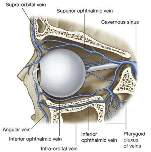
Venous blood from the eyeball and orbit drains predominantly through two large, valveless venous systems: the superior and inferior ophthalmic veins.
1. Vorticose Veins (Venae Vorticosae)
The uveal tract (choroid, ciliary body, and iris) is drained by four large vorticose veins. They emerge through the sclera in the posterior quadrants of the globe (superomedial, superolateral, inferomedial, inferolateral), draining into the superior and inferior ophthalmic veins.
2. Superior Ophthalmic Vein
- Origin & Course: Formed in the superomedial anterior orbit by the confluence of the supra-orbital vein and the angular vein (from the facial vein). Accompanies the ophthalmic artery across the superior orbit, collecting tributaries from ethmoidal, lacrimal, and central retinal veins, as well as the superior vorticose veins.
- Termination: Passes through the superior orbital fissure (outside the common tendinous ring) to drain directly into the cavernous sinus.
3. Inferior Ophthalmic Vein
- Origin & Branches: Begins as a network on the orbital floor; receives tributaries from extraocular muscles and the inferior vorticose veins.
- Dual Exit Pathways: Posteriorly, it splits:
- Joins the superior ophthalmic vein or passes independently through the superior orbital fissure into the cavernous sinus.
- Passes through the inferior orbital fissure to communicate with the pterygoid venous plexus in the infratemporal fossa.
Orbital innervation
The orbit contains diverse motor, sensory, and autonomic nerve networks, including cranial nerves II, III, IV, V1, and VI, alongside the parasympathetic ciliary ganglion and sympathetic plexuses.

1. The Common Tendinous Ring (Annulus of Zinn)
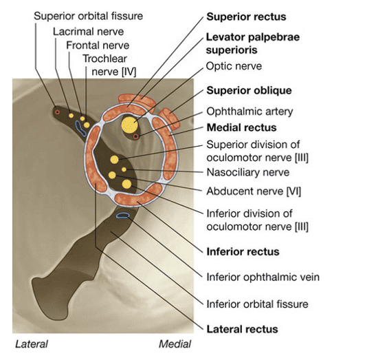
A fibrous cuff encircling the optic canal and the central narrow waist of the superior orbital fissure. It provides the origin for the four rectus muscles and organizes neurovascular entry into the orbit:
- Structures Entering OUTSIDE (Superior/Lateral to) the Ring:
- Lacrimal nerve (CN V1)
- Frontal nerve (CN V1)
- Trochlear nerve (CN IV)
- Superior ophthalmic vein
- Mnemonic: “Live Free To See” (Lacrimal, Frontal, Trochlear, Superior ophthalmic vein).
- Structures Entering INSIDE the Ring:
- Optic Canal: Optic nerve (CN II) and Ophthalmic artery.
- Oculomotor Foramen (Central SOF): Superior division of Oculomotor nerve (CN III), Nasociliary nerve (CN V1), Abducent nerve (CN VI), Inferior division of Oculomotor nerve (CN III), and sympathetic pupillomotor fibers.
2. Branches of the Ophthalmic Nerve (CN V1)
Divides into three main terminal branches within the anterior cavernous sinus before entering the orbit:
- Frontal Nerve: Largest branch; enters above the ring, coursing between the roof and levator palpebrae superioris. Divides into the larger supra-orbital nerve (exits via supra-orbital notch to supply the forehead and scalp) and the smaller supratrochlear nerve (exits above the trochlea to supply the medial forehead and upper eyelid).
- Lacrimal Nerve: Smallest branch; passes above the ring along the lateral rectus muscle. Receives a postganglionic parasympathetic secretomotor branch from the zygomaticotemporal nerve (CN V2, originating from the pterygopalatine ganglion) and delivers it to the lacrimal gland, then continues to sensory innervation of the lateral upper eyelid skin.
- Nasociliary Nerve: Enters inside the ring between the two divisions of CN III. Runs obliquely across the optic nerve toward the medial wall. Gives off:
- Sensory root to the ciliary ganglion.
- Long ciliary nerves (2 to 3): Carry sensory fibers from the cornea, iris, and ciliary body, as well as sympathetic fibers to the dilator pupillae.
- Posterior and anterior ethmoidal nerves: Supply ethmoidal sinuses, nasal cavity, and the external nose (as the external nasal nerve).
- Infratrochlear nerve: Terminal branch supplying the medial eyelids, caruncle, and lacrimal sac.
3. The Ciliary Ganglion
A small (~2 mm) parasympathetic peripheral ganglion situated in the posterior orbit between the optic nerve and the lateral rectus muscle:
- Parasympathetic Root (Motor): Carries preganglionic fibers arising from the Edinger-Westphal nucleus (CN III) via the nerve to the inferior oblique. These fibers synapse within the ciliary ganglion.
- Sympathetic Root: Postganglionic fibers from the superior cervical ganglion traversing the internal carotid plexus pass through the ganglion without synapsing to supply intraocular blood vessels.
- Sensory Root: Afferent fibers from the globe travel via the nasociliary nerve, passing through without synapsing.
- Short Ciliary Nerves (6 to 10): Emerge from the anterior border of the ganglion, pierce the sclera around the optic nerve, and convey postsynaptic parasympathetic fibers to the sphincter pupillae (miosis) and ciliary muscle (accommodation), alongside sensory and sympathetic fibers.
Clinical correlations
1. Orbital Blowout Fractures
Direct blunt trauma to the anterior orbit from a large object (such as a fist or tennis ball) causes a sudden surge in intra-orbital hydraulic pressure. Because the orbital rim is reinforced, the force decompresses through the weakest bony boundaries: the orbital floor (maxillary bone) and the medial wall (lamina papyracea).
- Clinical Features:
- Enophthalmos: Posterior displacement of the globe into the expanded orbital space.
- Diplopia on Upgaze: Herniation and mechanical entrapment of the inferior rectus muscle and inferior oblique within the fractured floor prevents upward gaze, producing vertical double vision.
- Infra-Orbital Hypoesthesia: Entrapment or shearing of the infra-orbital nerve (CN V2) causes numbness over the ipsilateral cheek, upper lip, and upper gingiva.
- Orbital Emphysema: Medial wall rupture allows air from ethmoidal sinuses to enter periorbital tissues, causing palpable crepitus upon nose-blowing.
2. Cranial Nerve Palsies Affecting the Orbit
- Oculomotor Nerve (CN III) Palsy: Complete unilateral paralysis of SR, IR, MR, IO, and LPS:
- The affected eye adopts a characteristic “down-and-out” resting posture due to unopposed tone of the lateral rectus (CN VI) and superior oblique (CN IV).
- Severe ptosis (loss of LPS innervation).
- Fixed, dilated pupil (mydriasis) and loss of near accommodation due to disrupted parasympathetics from the Edinger-Westphal nucleus.
- Trochlear Nerve (CN IV) Palsy: Paralysis of the superior oblique muscle:
- Produces vertical and torsional diplopia, exacerbated on attempted downgaze and adduction (e.g., walking downstairs or reading).
- Patients present with a compensatory head tilt toward the contralateral shoulder to relieve extorsion.
- Abducent Nerve (CN VI) Palsy: Paralysis of the lateral rectus muscle:
- The affected eye cannot abduct, presenting with convergent strabismus (esotropia) and horizontal binocular diplopia worsening on lateral gaze toward the affected side.
3. Horner’s Syndrome (Oculosympathetic Paresis)
Interruption of the three-neuron cervical sympathetic pathway (e.g., from a Pancoast tumor at the lung apex compressing the stellate ganglion or a carotid dissection):
- Mild Ptosis: Denervation of the superior tarsal muscle (Müller’s muscle).
- Miosis: Unopposed parasympathetic tone on the sphincter pupillae causes pupil constriction due to dilator pupillae paralysis.
- Anhidrosis: Loss of sympathetic innervation to facial sweat glands.
- Apparent Enophthalmos: Due to narrowing of the palpebral fissure.
4. Central Retinal Artery Occlusion (CRAO)
Embolic occlusion (typically from carotid atheroma or cardiac vegetations) of the central retinal artery produces sudden, profound, painless loss of vision in the affected eye.
- Funduscopic Hallmark: Diffuse pale, ischemic retina with a distinct “cherry-red spot” at the fovea centralis. Because the foveola contains no inner retinal layers and is nourished purely by the underlying choriocapillaris, its vascular choroidal background shines through the surrounding edematous retina.
Quick recap
References
The following authoritative biomedical reference works were verified in compiling this comprehensive study note:
- Moore’s Clinically Oriented Anatomy: Dalley, A.F. and Agur, A.M.R. (2022) Moore’s Clinically Oriented Anatomy. 9th edn. Philadelphia: Wolters Kluwer. [Chapter 8: Head – Orbit, Eyeball, and Orbital Contents].
- Gray’s Anatomy: Standring, S. (2021) Gray’s Anatomy: The Anatomical Basis of Clinical Practice. 42nd edn. London: Elsevier. [Section 4: Head and Neck – Chapter 40: The Orbit and its Contents].
- Snell’s Clinical Anatomy by Regions: Waugh, A. and Grant, A. (2019) Snell’s Clinical Anatomy by Regions. 10th edn. Philadelphia: Wolters Kluwer. [Chapter 11: The Head and Neck – The Orbit].
- Kanski’s Clinical Ophthalmology: Salmon, J.F. (2020) Kanski’s Clinical Ophthalmology: A Systematic Approach. 9th edn. Edinburgh: Elsevier. [Chapter 1: Examination Techniques; Chapter 13: Glaucoma; Chapter 17: Strabismus].
- Uganda Clinical Guidelines: Ministry of Health Uganda (2023) Uganda Clinical Guidelines: National Guidelines for the Management of Common Conditions. Kampala: Ministry of Health. [Section 18: Eye Conditions – Ocular Trauma, Cellulitis, and Red Eye Protocols].