
The scalp and face: surgical layers, neurovascular architecture & clinical anatomy
On this page
Overview & boundaries
The scalp is the multilayered soft-tissue envelope that covers the cranial vault (calvaria). Functionally and surgically, it provides physical protection to the neurocranium, supports hair follicles, and enables facial and emotional expression through the anterior epicranial muscles.
Composition & Anatomical Extent
- Tissue Components: The scalp consists of skin (normally hair-bearing and rich in sebaceous glands) and specialized subcutaneous tissues that overlie the cranial bones.
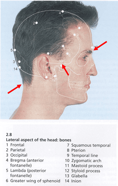
- Anterior Boundary: Demarcated by the supraorbital margins of the frontal bones.
- Posterior Boundary: Formed by the superior nuchal lines of the occipital bone, extending from the external occipital protuberance (inion) laterally toward the mastoid process.
- Lateral Boundary: Extends inferiorly over the temporal fascia to attach along the zygomatic arches.
Five layers of the scalp (SCALP)
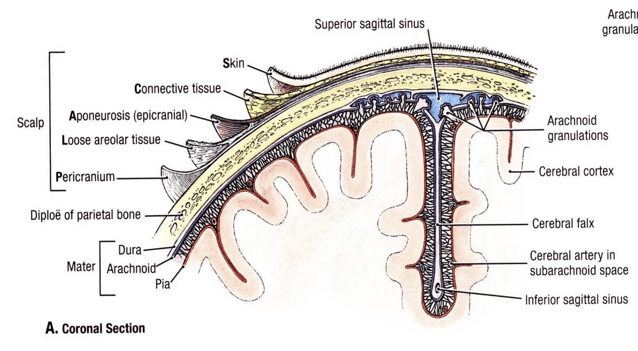
The scalp comprises five distinct tissue layers. The first letter of each layer forms the classic anatomical mnemonic SCALP.
Layer 1: S – Skin
The outer surface is thick, hair-bearing (except in baldness), and contains the highest density of sweat (eccrine) and sebaceous glands in the human body. Because of the abundance of sebaceous glands, the scalp is the most common site for epidermoid and pilar cysts (often referred to clinically as sebaceous cysts).
Layer 2: C – Connective Tissue (Superficial Fascia)
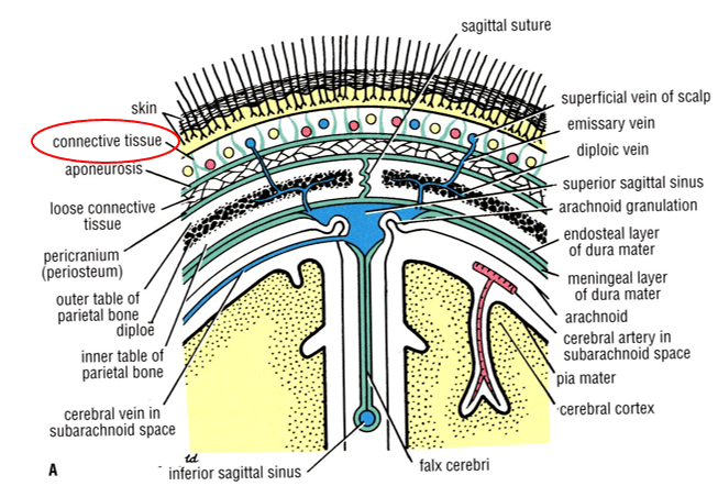
A dense, fibro-fatty layer composed of tough, inelastic fibrous septa that run vertically to anchor the overlying skin firmly to the underlying epicranial aponeurosis. It houses the main arterial, venous, and nerve trunks of the scalp.
- Surgical Hemostasis Pearl: The fibrous septa bind the adventitia of scalp blood vessels tightly to surrounding connective tissue. When lacerated, transected arteries cannot retract or contract their muscular walls, resulting in profuse, continuous arterial hemorrhage from even minor wounds. Hemostasis requires direct mechanical compression, surgical clips, or deep interlocking galeal sutures.
Layer 3: A – Aponeurosis (Galea Aponeurotica)
A broad, tough, thin tendinous sheet (also called the epicranial aponeurosis) that unites the anterior frontal and posterior occipital bellies of the occipitofrontalis muscle. Laterally, it thins and continues over the temporalis muscle to attach to the zygomatic arch. Together, Layers 1, 2, and 3 are bound tightly by fibrous septa and slide together as a single surgical unit (the “scalp proper”).
Layer 4: L – Loose Areolar Tissue (Subaponeurotic Space)
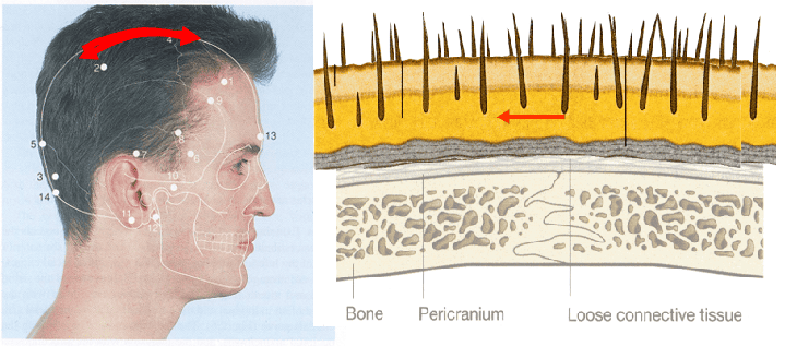
A potential cleft filled with loose areolar meshwork beneath the galea aponeurotica. It loosely connects the aponeurosis to the underlying pericranium, allowing the outer three layers to slide freely over the calvaria.
- The “Danger Area of the Scalp”: This layer houses valveless emissary veins that traverse skull foramina to connect extracranial veins directly to the intracranial diploic veins and dural venous sinuses. Infections within this space easily spread through emissary channels to cause osteomyelitis, meningitis, or septic dural venous sinus thrombosis.
Layer 5: P – Pericranium
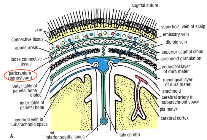
The specialized periosteum covering the outer cortical surface of the cranial bones. Although loosely attached to the bone surface over the convexities, it is firmly adherent at cranial suture lines, where it becomes continuous with the sutural ligaments and the endosteal layer of the dura mater inside the skull.
Scalp muscles & mechanics
The primary muscular driver of the scalp is the occipitofrontalis (epicranius) muscle, a digastric muscle comprising two pairs of symmetrical bellies united by an intermediate aponeurosis.
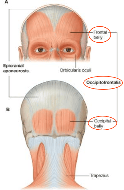
Bellies of the Occipitofrontalis
- Occipital Bellies (Occipitalis):
- Origin: Arise from the lateral two-thirds of the highest nuchal line of the occipital bone and the mastoid part of the temporal bone.
- Insertion: Project forward to insert into the posterior margin of the galea aponeurotica.
- Characteristics: Smaller, separated by a distinct midline aponeurotic interval.
- Frontal Bellies (Frontalis):
- Origin: Have no bony attachment. They arise from the dermis and superficial fascia of the eyebrows and forehead, interdigitating with fibers of the orbicularis oculi, corrugator supercilii, and procerus muscles.
- Insertion: Ascend over the forehead to insert into the anterior margin of the galea aponeurotica close to the coronal suture.
- Characteristics: Larger than the occipital bellies, closely approximated in the midline.
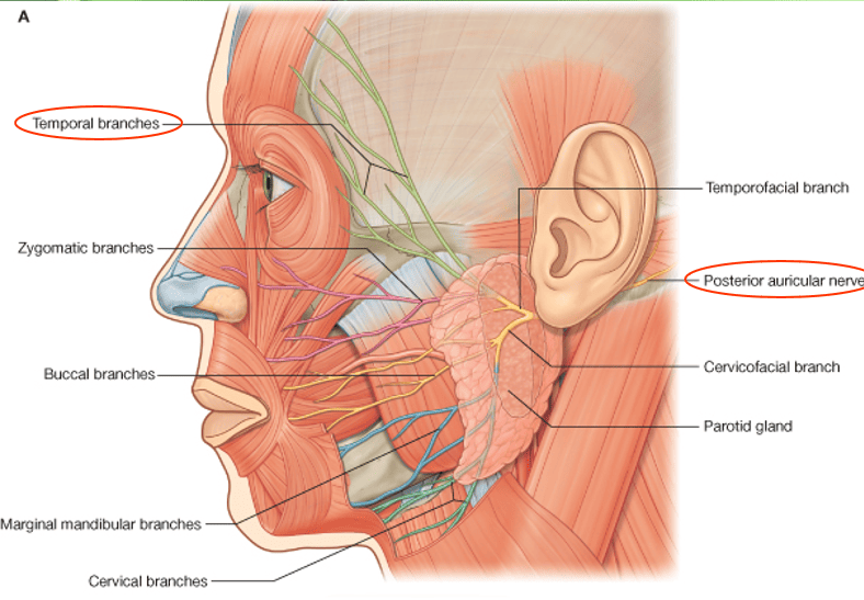
Motor Innervation & Actions
- Motor Nerve Supply: Both bellies are innervated by the Facial Nerve (CN VII):
- Frontal belly: Supplied by the temporal branch of CN VII.
- Occipital belly: Supplied by the posterior auricular branch of CN VII.
- Scalp Sliding Action: Alternate contraction of the occipital and frontal bellies pulls the galea backward and forward, sliding the outer three layers of the scalp (Layers 1, 2, and 3) as a single unit over the loose subaponeurotic tissue.
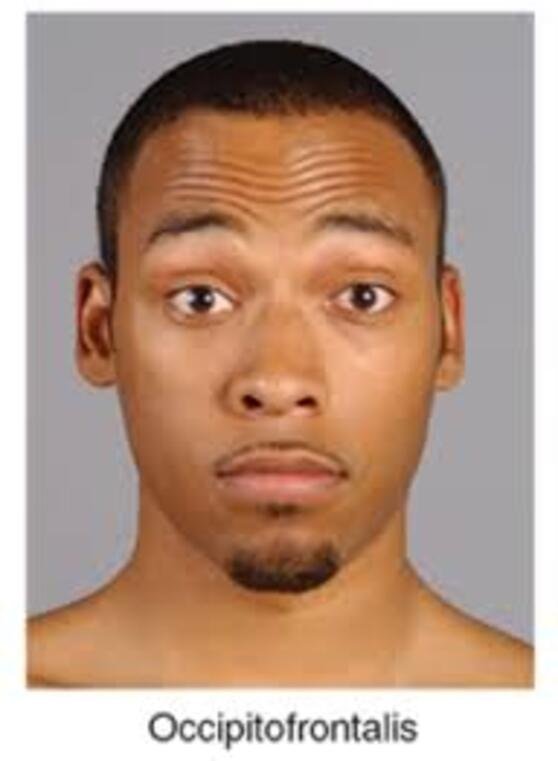
- Forehead Expression: Acting from above, the frontal bellies raise the eyebrows and upper eyelids, producing prominent transverse (horizontal) wrinkles across the forehead in facial expressions of surprise, astonishment, or horror.
Scalp nerves & arteries
The neurovascular supply of the scalp travels within Layer 2 (dense connective tissue). Branches ascend from the margins of the head (orbit, temple, and occiput) toward the vertex, producing extensive collateral anastomoses across the midline and between adjacent vascular territories.
Sensory Innervation: Trigeminal vs. Cervical Spinal Inputs
The sensory innervation of the scalp is divided into two distinct anatomical regions relative to the external ear: anterior to the auricle (innervated by the Trigeminal Nerve, CN V) and posterior to the auricle (innervated by Spinal Cervical Nerves C2 and C3).
Scroll the table sideways on a smaller screen.
| Nerve | Source / Parent Nerve | Course & Cranial Exit | Scalp Sensory Territory |
|---|---|---|---|
| Supratrochlear Nerve | Ophthalmic (CN V1) via frontal nerve | Winds around superior orbital margin medial to supraorbital notch | Forehead close to midline, ascending toward the vertex |
| Supraorbital Nerve | Ophthalmic (CN V1) via frontal nerve | Exits via the supraorbital notch/foramen | Skin of central forehead and scalp as far posteriorly as the vertex |
| Zygomaticotemporal Nerve | Maxillary (CN V2) via zygomatic nerve | Traverses zygomatico-orbital foramen into temporal fossa | Skin over the anterior temporal region |
| Auriculotemporal Nerve | Mandibular (CN V3) posterior division | Ascends from upper border of parotid gland anterior to auricle | Temporal region, scalp superior to auricle, tragus, external acoustic meatus |
| Lesser Occipital Nerve | Cervical plexus (Anterior rami C2) | Ascends along the posterior border of sternocleidomastoid | Lateral part of the occipital region and medial surface of auricle |
| Greater Occipital Nerve | Posterior (dorsal) ramus of C2 | Pierces semispinalis capitis and trapezius, accompanies occipital artery | Extensive skin of posterior scalp as far anteriorly as the vertex |
| Third Occipital Nerve | Posterior (dorsal) ramus of C3 | Pierces trapezius inferior to greater occipital nerve | Skin of lower occiput and suboccipital nuchal region |

Arterial Blood Supply: ICA & ECA Anastomoses
The scalp possesses a rich collateral blood supply derived from branches of both the Internal Carotid Artery (ICA) and the External Carotid Artery (ECA), which anastomose freely across the vertex.

- Branches of the Internal Carotid Artery (via Ophthalmic Artery):
- Supratrochlear Artery: Leaves the orbit through the supratrochlear notch/groove and ascends the medial forehead with the supratrochlear nerve.
- Supraorbital Artery: Emerges through the supraorbital notch/foramen and ascends over the forehead with the supraorbital nerve, supplying tissues up to the vertex.
- Branches of the External Carotid Artery:
- Superficial Temporal Artery: The smaller terminal branch of the external carotid artery. Arises within the substance of the parotid gland, crosses the root of the zygomatic arch immediately anterior to the tragus (where its arterial pulse is easily palpated), and divides into an anterior (frontal) branch and a posterior (parietal) branch.
- Posterior Auricular Artery: Ascends behind the auricle over the mastoid process to supply the scalp superior and posterior to the ear.
- Occipital Artery: Emerges from the posterior triangle of the neck, pierces trapezius, and ascends over the occipital bone with the greater occipital nerve to supply the scalp as far forward as the vertex.
Scalp veins, sinuses & lymph
The venous system of the scalp accompanies the corresponding arteries within Layer 2, forming an anastomotic arcade that drains into both external and internal jugular systems, as well as intracranial dural sinuses.
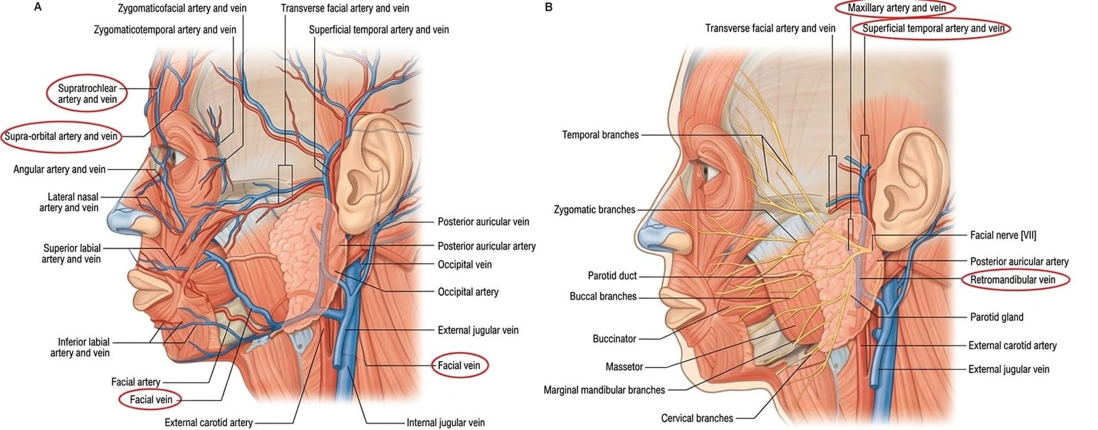
Superficial Venous Drainage Routes
- Anterior Scalp: The supratrochlear and supraorbital veins descend over the forehead and unite near the medial canthus of the eye to form the facial vein (angular vein).
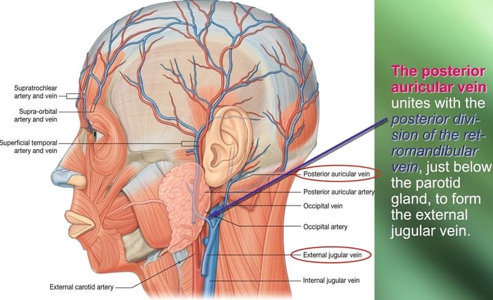
- Lateral Scalp: The superficial temporal vein receives tributaries from the temple and scalp, descends across the zygomatic arch, and enters the parotid gland. Within the gland, it unites with the maxillary vein to form the retromandibular vein.
- The anterior division of the retromandibular vein joins the facial vein to form the common facial vein, which drains into the internal jugular vein (IJV).
- The posterior division of the retromandibular vein unites with the posterior auricular vein below the parotid gland to form the external jugular vein (EJV).
- Posterior Scalp: The occipital vein descends from the back of the head into the suboccipital venous plexus (situated beneath the floor of the upper posterior cervical triangle). From this plexus, venous blood enters the vertebral veins or drains into the internal jugular vein.
Emissary & Diploic Veins: Pathophysiology of Spread
Emissary veins are valveless venous conduits that pass through anatomical apertures in the cranial vault, establishing direct communications between the extracranial veins of the scalp, the diploic veins within the cancellous skull bones, and the intracranial dural venous sinuses:
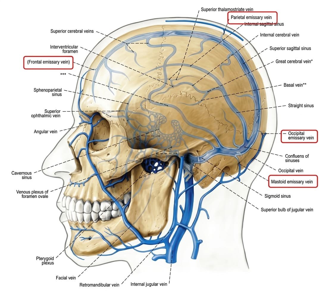
- Parietal Emissary Vein: Traverses the parietal foramen to connect scalp veins with the superior sagittal sinus.
- Mastoid Emissary Vein: Traverses the mastoid foramen to connect posterior auricular/occipital veins with the sigmoid sinus.
- Frontal Emissary Vein: Traverses the foramen cecum (when patent) to connect nasal/forehead veins with the anterior end of the superior sagittal sinus.
- Condylar Emissary Vein: Traverses the condylar canal to connect the suboccipital venous plexus with the sigmoid sinus.
Lymphatic Drainage of the Scalp
The scalp contains no lymph nodes within its substance; lymphatic channels drain radially from the vertex toward a pericervical collar of superficial lymph nodes:

- Anterior Scalp & Forehead: Drains into the submandibular lymph nodes.
- Lateral Scalp (Above Ear): Drains into the superficial parotid (preauricular) lymph nodes.
- Lateral Scalp (Above & Behind Ear): Drains into the mastoid (retroauricular) lymph nodes located over the mastoid process.
- Posterior Scalp (Occiput): Drains into the occipital lymph nodes at the base of the skull.
- Final Terminal Pathway: All of these primary regional pericervical nodes drain secondarily into the deep cervical lymph node chain running along the internal jugular vein (deep to sternocleidomastoid).
Face: features & development
The human face extends superiorly to the normal hairline/superciliary arches, inferiorly to the chin and lower border of the mandible, and laterally to the auricles. It provides the mechanical interface for communication, feeding, vision, and olfaction.
Morphological Characteristics
- Lack of Deep Fascia: Distinct from other anatomical regions, the face possesses no true deep investing fascia (except over the masseter and parotid gland). Facial muscles reside directly within the superficial fascia.
- Cutaneous Mobility: Facial muscles arise from the underlying facial skeleton or fascia and insert directly into the dermis of the skin. Contraction moves the skin, pulling it into mobile folds and creases.
- Orificial Sphincters & Dilators: The facial openings—the orbit, nasal aperture, and oral fissure—are guarded by organized muscle loops that act as functional sphincters (compressors) or dilators.
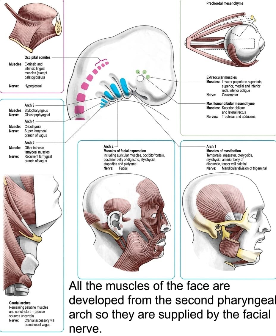
Embryological Derivation
All muscles of facial expression develop from the mesoderm of the second pharyngeal arch (hyoid arch). During embryonic development, this myogenic tissue migrates widely across the skull to form the muscles of the scalp, external ear, eyelids, nose, lips, and neck (platysma). Because they share this embryological origin, every muscle of facial expression is innervated by the cranial nerve of the second arch: the Facial Nerve (CN VII).
Muscles of facial expression
1. Muscles of the Eyelids & Eyebrows
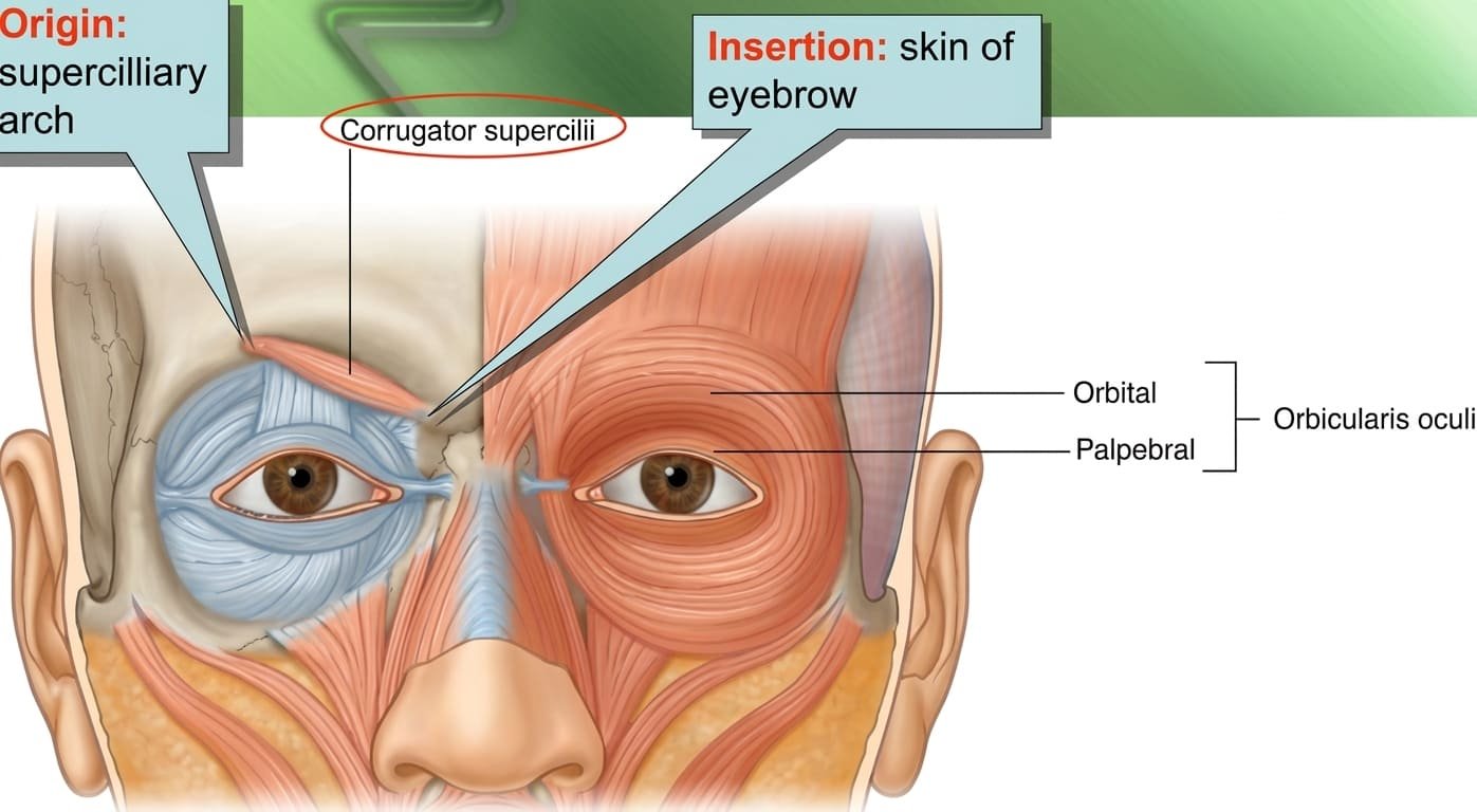
- Orbicularis Oculi: The broad, elliptical sphincter muscle of the palpebral fissure. Composed of two major parts:
- Palpebral Part: Lies within the eyelids. Arises from the medial palpebral ligament, arches across the tarsal plates, and inserts into the lateral palpebral raphe.
Function: Gently closes the eyelids during voluntary blinking and sleep. Its contraction also creates negative suction that dilates the lacrimal sac to facilitate tear drainage. - Orbital Part: A thicker, outer peripheral ring. Arises from the medial palpebral ligament and the adjoining nasal part of the frontal bone and frontal process of the maxilla. Its fibers loop completely around the orbital rim without interruption to re-insert near their origin.
Function: Tightly closes the eyelids, drawing the surrounding skin into folds to shield the globe from bright glare, dust, and trauma.
- Palpebral Part: Lies within the eyelids. Arises from the medial palpebral ligament, arches across the tarsal plates, and inserts into the lateral palpebral raphe.
- Corrugator Supercilii: A small, pyramidal muscle deep to the orbicularis oculi.
- Origin: Medial end of the superciliary arch of the frontal bone.
- Insertion: Skin of the middle part of the eyebrow.
- Action: Pulls the eyebrow medially and downward, producing vertical wrinkles on the forehead above the bridge of the nose (the muscle of frowning, grief, and concentration).
- Eyelid Dilators (Palpebral Openers):
- Levator Palpebrae Superioris: The primary upper eyelid elevator. Originates from the lesser wing of the sphenoid above the optic canal and inserts into the superior tarsal plate and skin of the upper eyelid. Important: Innervated by the Oculomotor Nerve (CN III), not the facial nerve!
- Frontal Belly of Occipitofrontalis: Assists in elevating the upper eyelid and eyebrow during upward gaze or expressions of surprise (CN VII).

2. Muscles of the Nostrils
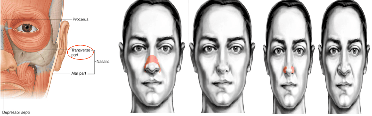
- Compressor Naris (Transverse Part of Nasalis):
- Origin: Maxilla, just lateral to the incisive fossa.
- Insertion: Fibers ascend across the nasal bridge to join an aponeurosis continuous with its fellow muscle.
- Action: Sphincter of the nose; compresses the mobile lateral nasal cartilages, narrowing the aperture.
- Dilator Naris (Alar Part of Nasalis):
- Origin: Maxilla above the lateral incisor tooth.
- Insertion: Greater alar cartilage of the nostril.
- Action: Dilator of the nose; pulls the alar cartilage downward and laterally, widening the anterior nares during deep inspiration or anger.
- Procerus: Arises from the nasal bone and inserts into the skin of the lower forehead between the eyebrows, producing transverse wrinkles over the nasal bridge.
3. Muscles of the Lips, Cheeks & Mouth
The oral orifice is guarded by a complex central sphincter (orbicularis oris) and a radiating array of dilator muscles that adjust lip position, smile, pucker, and speak.

The Oral Sphincter: Orbicularis Oris
- Structural Composition: An intricate muscle encircling the oral aperture. Its deep fibers are derived largely from the buccinator muscle (which decussates at the corner of the mouth), while its superficial fibers are contributed by radiating dilator muscles (levators and depressors of the lips).
- Origins & Insertions: Intrinsic fibers arise near the midline from the alveolar margins of the maxilla and mandible, and from the deep surface of the skin. They insert into the dermis and mucous membrane of the lips.
- Motor Innervation: Buccal and marginal mandibular branches of the facial nerve (CN VII).
- Action: Compresses and closes the lips together; protrudes the lips forward during whistling, blowing, and kissing.
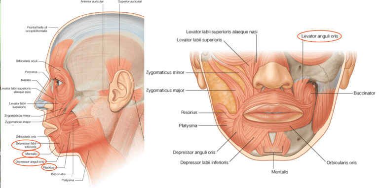
The Dilators of the Lips
Traced in sequence from the side of the nose, around the oral angle, and down to the chin:
- Levator Labii Superioris Alaeque Nasi: Elevates the upper lip and flares the nostril.
- Levator Labii Superioris: Arises from the inferior orbital rim; elevates and everts the upper lip.
- Zygomaticus Minor: Arises from the zygomatic bone anteriorly; elevates the upper lip.
- Zygomaticus Major: Arises from the zygomatic bone; pulls the angle of the mouth upward and laterally (the primary muscle of laughing and smiling).
- Levator Anguli Oris: Lies deep to the zygomatic muscles; elevates the angle of the mouth.
- Risorius: A slender transverse slip arising from parotid fascia; retracts the angle of the mouth laterally (produces a smirk or insincere smile).
- Depressor Anguli Oris: Triangular muscle arising from the mandible; depresses the angle of the mouth (muscle of sadness or weeping).
- Depressor Labii Inferioris: Depresses and draws the lower lip downward and slightly laterally.
- Mentalis: Arises from the incisive fossa of the mandible; elevates and wrinkles the skin of the chin, protruding the lower lip in doubt, pout, or disdain.
The Muscle of the Cheek: Buccinator
The buccinator forms the primary muscular foundation of the cheek and the lateral wall of the oral cavity.
- Origins:
- Outer alveolar borders of the maxilla and mandible opposite the three molar teeth.
- Posteriorly from the pterygomandibular raphe (a tendinous band extending from the pterygoid hamulus to the mylohyoid line of the mandible, separating the buccinator from the superior pharyngeal constrictor).
- Insertion & Decussation: Fibers project forward toward the corner of the mouth. At the modiolus (the dense muscular node at the angle of the mouth):
- Central fibers decussate: lower fibers cross into the upper lip; upper fibers cross into the lower lip.
- Peripheral highest and lowest fibers continue directly into the upper and lower lips without decussating.
- Important Relationship: The buccinator is pierced by the parotid duct (Stensen’s duct) opposite the upper second molar tooth.
- Motor Innervation: Supplied by the buccal branch of the Facial Nerve (CN VII). (Sensory innervation to the overlying skin and underlying cheek mucosa is supplied by the buccal nerve, a branch of the mandibular nerve CN V3).
- Functional Role: Compresses the cheeks against the teeth and gums. During mastication, it prevents food from pooling in the oral vestibule (between teeth and cheek), directing the food bolus between the occlusal surfaces of the teeth. It is also used to expel air forcefully (as in playing a trumpet or whistling).

Sensory innervation (CN V)
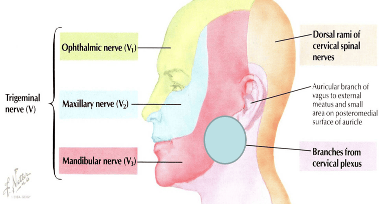
The skin of the face is innervated almost entirely by the three divisions of the Trigeminal Nerve (CN V): the ophthalmic (V1), maxillary (V2), and mandibular (V3) nerves.
Embryological Correlates of the Trigeminal Nerve
- Ophthalmic Nerve (V1): Supplies tissues derived from the embryonic frontonasal prominence.
- Maxillary Nerve (V2): Supplies tissues derived from the maxillary process of the first pharyngeal arch.
- Mandibular Nerve (V3): Supplies tissues derived from the mandibular process of the first pharyngeal arch.
The 11 Cutaneous Branches of the Trigeminal Nerve
The trigeminal nerve gives rise to exactly 11 cutaneous branches on the face and scalp: five from V1, three from V2, and three from V3 (the “5 – 3 – 3” rule).
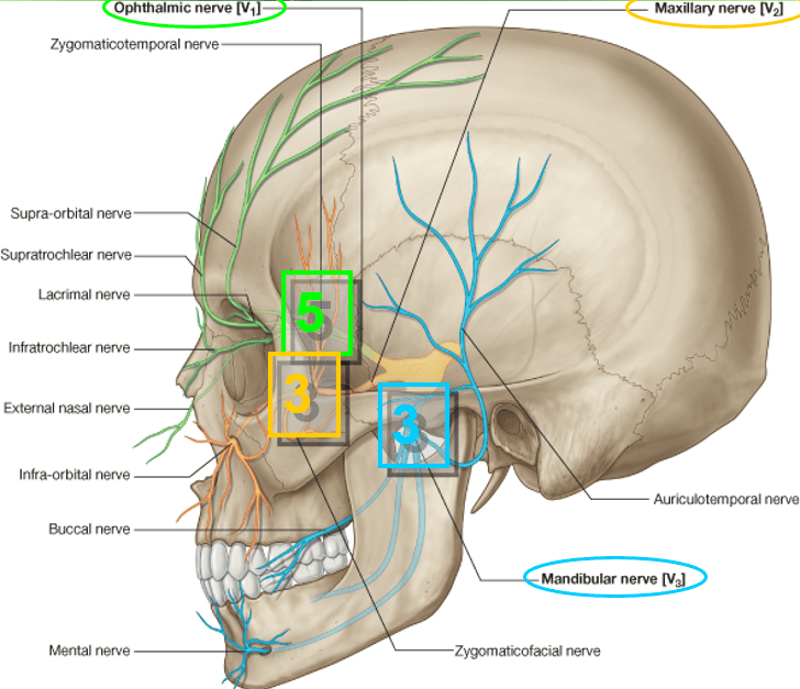
A. Branches of the Ophthalmic Division (V1 – 5 Branches)
- Lacrimal Nerve: Exits the lateral orbit to supply the skin and conjunctiva of the lateral upper eyelid.
- Supraorbital Nerve: Traverses the supraorbital notch/foramen; supplies the skin and conjunctiva of the central upper eyelid and ascends over the forehead to the vertex.
- Supratrochlear Nerve: Emerges medial to the supraorbital notch; supplies the medial upper eyelid and the lower medial forehead.
- Infratrochlear Nerve: Leaves the orbit inferior to the trochlea of the superior oblique; supplies the medial part of the upper eyelid, the caruncle, and the lateral root of the nose.
- External Nasal Nerve: Terminal branch of the anterior ethmoidal nerve; emerges between the nasal bone and upper lateral nasal cartilage to supply the skin on the dorsum of the nose down to the tip.
B. Branches of the Maxillary Division (V2 – 3 Branches)
- Zygomaticotemporal Nerve: Traverses the zygomatic bone to enter the temporal fossa; supplies the skin of the anterior temple.
- Zygomaticofacial Nerve: Emerges through a small foramen on the lateral surface of the zygomatic bone; supplies the skin over the prominence of the cheek.
- Infraorbital Nerve: The direct continuation of the maxillary nerve. Traverses the infraorbital canal and emerges onto the face via the infraorbital foramen. It immediately divides into palpebral, nasal, and superior labial branches that supply the lower eyelid, lateral nose, and upper lip.
C. Branches of the Mandibular Division (V3 – 3 Branches)
- Auriculotemporal Nerve: Ascends from behind the temporomandibular joint, emerging from the upper border of the parotid gland; supplies the tragus, external acoustic meatus, tympanic membrane, and temporal scalp.
- Buccal Nerve (Sensory): Passes forward deep to the ramus of the mandible, emerging from beneath the anterior border of the masseter; supplies the skin over the cheek and the mucous membrane lining the inside of the cheek. (Must not be confused with the motor buccal branch of CN VII!).
- Mental Nerve: Terminal branch of the inferior alveolar nerve. Emerges from the mental foramen of the mandible; supplies the skin of the lower lip and chin, as well as the labial gingiva of the anterior mandibular teeth.
Motor innervation (CN VII)
The Facial Nerve (CN VII) is the sole motor nerve supplying the muscles of facial expression. It leaves the posterior cranial fossa through the internal acoustic meatus, traverses the facial canal within the petrous temporal bone, and exits the base of the skull through the stylomastoid foramen.
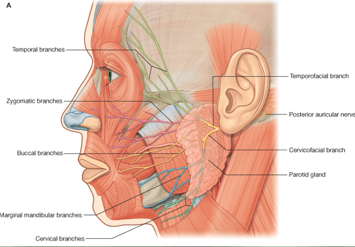
Intraparotid Course & The Five Terminal Branches
After exiting the stylomastoid foramen, the main trunk of the facial nerve enters the posteromedial surface of the parotid salivary gland. Within the parotid substance, it runs superficial to the retromandibular vein and external carotid artery, branching into the parotid plexus (pes anserinus):
1. Temporal Branch
Emerges from the superior border of the parotid gland, crossing the zygomatic arch. Supplies the frontal belly of occipitofrontalis, anterior and superior auricular muscles, the corrugator supercilii, and the upper part of orbicularis oculi.
2. Zygomatic Branch
Crosses the zygomatic bone toward the lateral canthus. Supplies the lower and lateral fibers of orbicularis oculi, facilitating eyelid closure.
3. Buccal Branch
Emerges from the anterior border of the gland below the parotid duct. Runs forward across the cheek to supply the buccator muscle and muscles of the upper lip and nose.
4. Marginal Mandibular Branch
Emerges from the anterior border, often dipping below the inferior margin of the mandible before curving upward across the facial vessels onto the face. Supplies the depressor anguli oris, depressor labii inferioris, and mentalis of the lower lip.
5. Cervical Branch
Emerges from the inferior border of the parotid gland, descending into the neck posterior to the angle of the mandible. Runs beneath the deep cervical fascia to supply the platysma muscle.
Proprioception in Facial Muscles
The facial nerve does not provide sensory innervation to the skin of the face. Proprioceptive afferents from the facial muscles travel retrogradely along communicating anastomotic branches between the terminal twigs of the facial nerve and adjacent sensory branches of the trigeminal nerve, reaching the central nervous system via the trigeminal pathways.
Facial blood supply
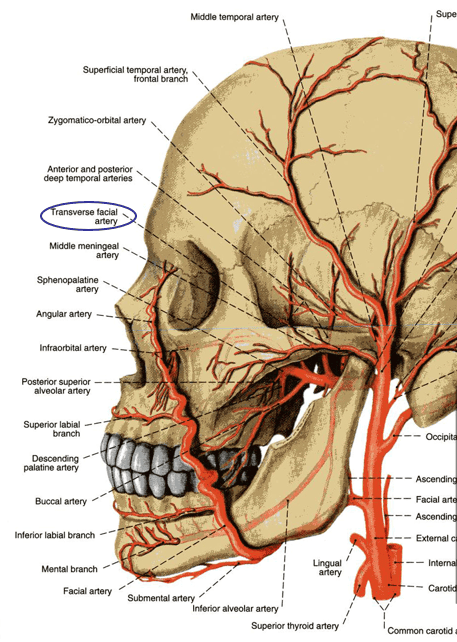
The face receives a rich arterial blood supply primarily through two major vessels: the facial artery and the superficial temporal artery, supplemented by small branches originating from the ophthalmic and maxillary arteries.
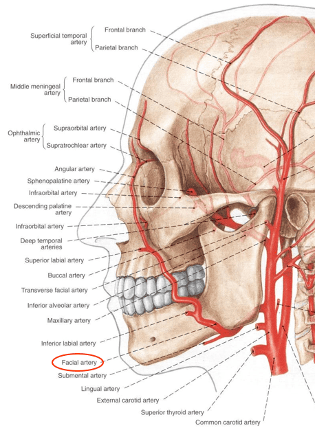
The Facial Artery: Course & Branches
The facial artery arises from the anterior aspect of the external carotid artery within the carotid triangle:
- Cervical Course: It loops upward and deep to the posterior belly of the digastric, arches over the posterior surface of the submandibular salivary gland, and hooks around the inferior border of the mandible at the anterior margin of the masseter muscle. Clinical note: The facial arterial pulse is easily palpable against the mandible at this landmark.
- Facial Course: It ascends tortuously toward the angle of the mouth, covered initially by the platysma. It passes deep to the zygomaticus major, zygomaticus minor, and levator labii superioris, ascending along the lateral margin of the nose to terminate at the medial angle (canthus) of the eye as the angular artery. At the medial canthus, the angular artery forms an anastomosis with the dorsal nasal and supratrochlear branches of the ophthalmic artery, establishing a collateral connection between the external and internal carotid systems.
Primary Branches on the Face
- Submental Artery: Arises beneath the mandible, running forward across the mylohyoid to supply the chin and submandibular region.
- Inferior Labial Artery: Arises near the angle of the mouth; runs medially within the lower lip between the mucous membrane and orbicularis oris, anastomosing across the midline with its contralateral counterpart.
- Superior Labial Artery: Supplies the upper lip and gives off a small septal branch to the anterior nasal septum (contributing to Kiesselbach’s plexus, the primary site of epistaxis).
- Lateral Nasal Artery: Arises alongside the external nose, supplying the skin and cartilages of the ala and dorsum.
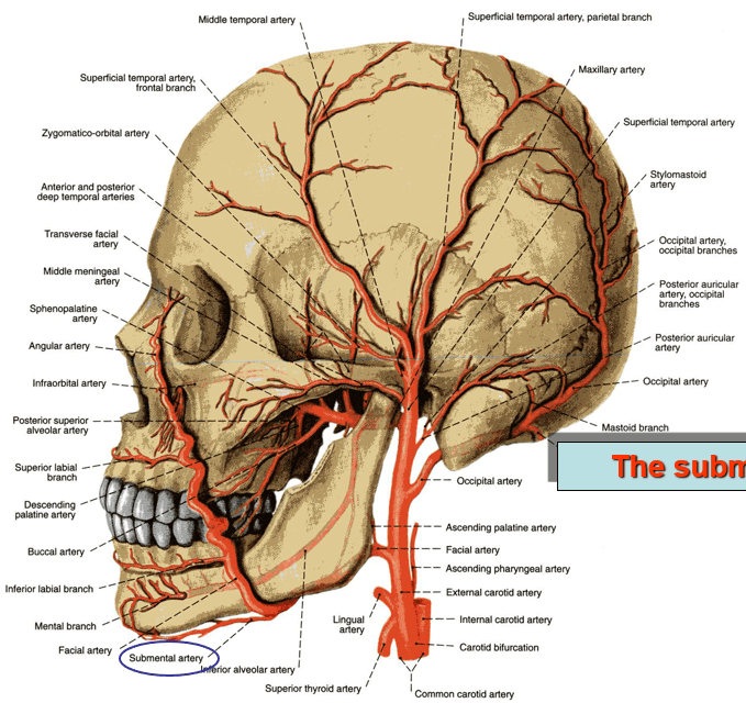
The Superficial Temporal & Supplementary Arteries
- Transverse Facial Artery: Arises from the superficial temporal artery within the parotid gland. Emerges from the anterior border of the parotid and crosses the masseter horizontally between the zygomatic arch and the parotid duct, supplying the parotid gland, parotid duct, masseter, and overlying skin.
- Ophthalmic Artery Branches (ICA): The supraorbital and supratrochlear arteries emerge from the orbit to supply the forehead.
- Maxillary Artery Branches (ECA): The infraorbital artery (emerging via the infraorbital foramen) and the mental artery (emerging via the mental foramen) provide blood to the midface and chin, respectively.
Venous drainage & the danger area
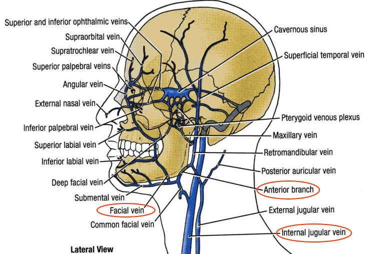
The facial vein is the primary venous conduit of the face. It is characterized by an almost straight course across the face, in contrast to the tortuous path of the facial artery.
Formation, Course & Termination
- Origin: Formed at the medial angle of the eye by the confluence of the supratrochlear and supraorbital veins. Here it is termed the angular vein.
- Course: Descends straight down the face posterior to the facial artery. Passes deep to zygomaticus major and the parotid duct, but runs superficial to the buccinator and submandibular gland.
- Termination: Crosses the inferior border of the mandible, enters the neck, and unites with the anterior division of the retromandibular vein to form the common facial vein, which drains into the internal jugular vein.
The “Danger Area of the Face”
The triangular zone bounded by the bridge of the nose and the corners of the mouth is clinically termed the Danger Area (Danger Triangle) of the face. Its clinical importance stems from two deep venous communications that link the superficial facial vein directly to the intracranial cavernous sinus:
- Superior Route (Via Orbit): The angular vein communicates directly with the superior ophthalmic vein, which passes through the superior orbital fissure to enter the cavernous sinus.
- Deep Route (Via Infratemporal Fossa): The facial vein connects via the deep facial vein (crossing the buccinator) to the pterygoid venous plexus, which communicates with the cavernous sinus through emissary veins traversing the foramen ovale and sphenoidal emissary foramen (of Vesalius).
Lymphatic drainage of the face
The lymphatic vessels of the face follow the course of facial blood vessels, draining into three primary regional nodal basins:

1. Submandibular Lymph Nodes
Receive lymph from the forehead, medial aspects of the eyelids, the external nose, anterior cheek, upper lip, and the lateral parts of the lower lip. A few small buccal (facial) lymph nodes are occasionally scattered along the facial vein over the buccinator to filter this drainage pathway.
2. Superficial Parotid (Preauricular) Lymph Nodes
Receive drainage from the lateral part of the face, including the lateral eyelids, temple, and the anterior surface of the auricle.
3. Submental Lymph Nodes
Receive lymph from the central part of the lower lip, the skin of the chin, the tip of the tongue, and the floor of the mouth. From the submental nodes, lymph drains secondarily into the submandibular and deep cervical chains.
Clinical & surgical pearls
Scroll the table sideways on a smaller screen.
| Clinical Condition / Sign | Anatomical Basis | Pathophysiology & Diagnostic Presentation |
|---|---|---|
| Black Eye (Periorbital Ecchymosis) | Layer 4 (Loose areolar tissue) of scalp tracking anteriorly | Blood or inflammatory exudate accumulating in the subaponeurotic space gravitates anteriorly into the upper eyelids and periorbital tissues because the frontal belly of occipitofrontalis has no bony insertion. |
| Bell’s Palsy | Lower motor neuron lesion of the Facial Nerve (CN VII) | Acute unilateral flaccid paralysis of all muscles of facial expression on the affected side: inability to wrinkle forehead, loss of nasolabial fold, mouth drooping, and lagophthalmos (inability to close eye, risking corneal ulceration). |
| Trigeminal Neuralgia (Tic Douloureux) | Sensory root compression of the Trigeminal Nerve (CN V) | Excruciating, paroxysmal, lancinating pain along the distribution of V2 or V3, triggered by light touch, chewing, or washing the face over discrete trigger zones. |
| Temporal (Giant Cell) Arteritis | Granulomatous vasculitis of the Superficial Temporal Artery | Throbbing temporal headache, tender indurated temporal artery, scalp tenderness when brushing hair, and jaw claudication; risks sudden irreversible blindness from ischemic optic neuropathy if untreated. |
| Corneal Blink Reflex | Afferent: Nasociliary (V1) / Efferent: Temporal and Zygomatic (CN VII) | Touching the cornea elicits bilateral eyelid closure. Tests integrity of the ophthalmic nerve sensory pathway and the facial nerve motor loop to the orbicularis oculi. |
Quick recap
References
The following authoritative reference texts were verified in compiling this study note:
- Snell’s Clinical Anatomy: Splittgerber, R. (2019) Snell’s Clinical Anatomy by Regions. 10th edn. Philadelphia: Wolters Kluwer. [Chapter 11: The Head and Neck – The Scalp and Face, pp. 582–615].
- Moore’s Clinically Oriented Anatomy: Dalley, A.F. and Agur, A.M.R. (2022) Moore’s Clinically Oriented Anatomy. 9th edn. Philadelphia: Wolters Kluwer. [Chapter 8: Head – Cutaneous Nerves, Vessels, and Muscles of Face and Scalp].
- Gray’s Anatomy: Standring, S. (2021) Gray’s Anatomy: The Anatomical Basis of Clinical Practice. 42nd edn. London: Elsevier. [Section 4: Head and Neck; Chapter 27: Face and Scalp].
- Last’s Anatomy: Sinnatamby, C.S. (2011) Last’s Anatomy: Regional and Applied. 12th edn. Edinburgh: Churchill Livingstone / Elsevier. [Chapter 6: Head and Neck and Spine – Face and Scalp].