
The temporomandibular joint: functional anatomy, biomechanics & muscles of mastication
On this page
Overview & joint classification
The temporomandibular joint (TMJ) is a paired, highly specialized articulation connecting the mandible to the cranium. Classified structurally as a modified hinge type of synovial joint (a ginglymoarthrodial joint), it permits both hinge-like rotational movement in the sagittal plane and complex sliding or translational gliding movements.
Key Anatomical Features
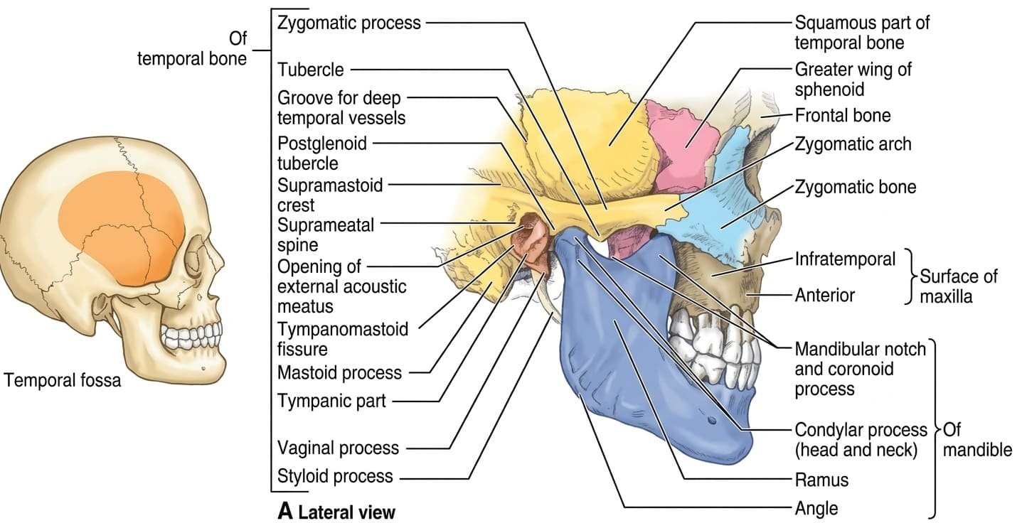
- Articular Components: Formed by the elliptical head (condylar process) of the mandible articulating superiorly with the mandibular fossa (glenoid fossa) and the convex articular tubercle (articular eminence) of the squamous temporal bone.
- Fibrocartilage Covering: Unlike typical adult synovial joints that are capped by hyaline cartilage, all articular surfaces of the TMJ (as well as its intra-articular disc) are lined by dense fibrocartilage containing type I collagen. This structural adaptation reflects the intramembranous developmental origin of the mandible and temporal bones and grants exceptional resistance to heavy compressive and shearing masticatory loads.
- Bilateral Interdependence: The left and right TMJs form a linked bicondylar unit; neither joint can move completely independently of the other during physiological jaw movements.
Osteological foundations
Mastery of TMJ biomechanics requires a thorough understanding of the bony features of the mandible and temporal bone that define the limits of joint movement.

1. The Mandible
- Head (Condyle) of the Mandible: An elliptical transverse cylinder (measuring approximately 20 mm mediolaterally by 10 mm anteroposteriorly). The long axes of the two condyles are oriented obliquely, projecting posteromedially toward the anterior margin of the foramen magnum.
- Neck & Pterygoid Fovea: The constricted bone supporting the condyle. Its anterior surface features a distinct depression—the pterygoid fovea—which serves as the primary insertion site for the inferior head of the lateral pterygoid muscle.
- Coronoid Process: The thin, triangular anterior projection of the mandibular ramus that receives the insertion of the powerful temporalis muscle.
- Masseteric & Pterygoid Tuberosities: Rough bony ridges located at the mandibular angle:
- Masseteric tuberosity: On the lateral surface, providing firm insertion for the masseter.
- Pterygoid tuberosity: On the internal medial surface, receiving the insertion of the medial pterygoid.
2. The Temporal Bone
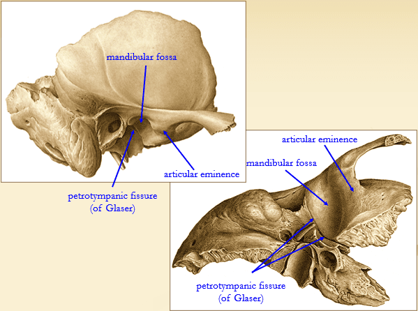
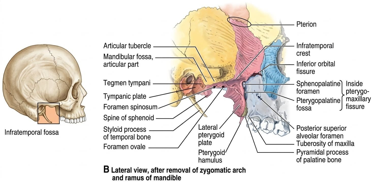
- Mandibular Fossa (Glenoid Fossa): A deep, concave depression on the inferior surface of the squamous temporal bone. Only its anterior portion participates in articulation with the mandibular condyle.
- Articular Tubercle (Articular Eminence): A transverse bony ridge located immediately anterior to the mandibular fossa. It forms the anterior boundary of the joint and provides a smooth surface over which the condyle and disc glide during mouth opening.
- Petrotympanic Fissure (Fissure of Glaser): A narrow crevice that separates the anterior articular part of the mandibular fossa (squamous part) from the non-articular posterior part (tympanic plate). It transmits the chorda tympani nerve (from CN VII) and the anterior tympanic artery into the middle ear cavity.
- Postglenoid Tubercle: A conical bony crest immediately anterior to the external acoustic meatus that forms the posterior wall of the mandibular fossa, functioning as a physical barrier against posterior condylar displacement.
The articular disc & compartments
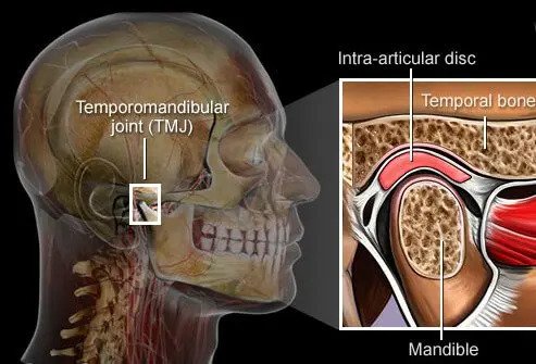
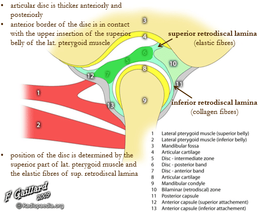
The joint cavity is divided completely into two functional synovial compartments by an oval, fibrous intra-articular partition: the articular disc (meniscus).
1. The Two Joint Compartments
- Discotemporal (Superior) Compartment: The space between the temporal bone (mandibular fossa and articular tubercle) and the superior surface of the articular disc. Functions as a plane joint where translational gliding movements occur (the condyle and disc move together forward and downward onto the articular tubercle).
- Discomandibular (Inferior) Compartment: The space between the inferior surface of the articular disc and the condylar head of the mandible. Functions as a hinge joint where rotational movements take place around a transverse bicondylar axis during early mouth opening.
2. Morphology & Microarchitecture of the Disc
The articular disc is a dense, biconcave fibrocartilaginous plate with distinct regions from anterior to posterior:
- Anterior Band: A moderately thickened region (approximately 2 mm) whose anterior border blends with the joint capsule and receives the insertion of the superior head of the lateral pterygoid muscle.
- Intermediate Zone: The central, thinnest segment (approximately 1 mm) of the disc. Highly compressed between the convex condylar head and the convex articular tubercle. It is completely avascular and aneural, adapted to bear high compressive biting loads without pain or tissue ischemia.
- Posterior Band: The thickest portion of the disc (approximately 3 to 4 mm), situated over the condyle when the jaw is in its neutral closed position.
- Bilaminar (Retrodiscal) Zone: Located behind the posterior band, composed of two divergent connective tissue leaves separated by loose, vascularized, and richly innervated tissue:
- Superior Retrodiscal Lamina: Rich in elastic fibers; attaches the posterior band of the disc to the squamotympanic fissure and postglenoid tubercle. Its passive elastic recoil pulls the disc posteriorly during mouth closure.
- Inferior Retrodiscal Lamina: Inelastic, rich in collagen fibers; anchors the lower border of the posterior band directly to the posterior neck of the mandibular condyle, preventing anterior over-translation of the disc relative to the bone.
Capsule & ligamentous apparatus
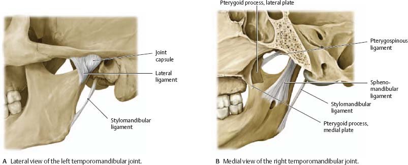
The stability of the TMJ relies on an articular fibrous capsule supported by one intrinsic primary ligament and two accessory extrinsic ligaments.
1. The Fibrous Capsule
The TMJ is enveloped by a loose, thin fibrous capsule. It attaches superiorly to the temporal bone around the perimeter of the mandibular fossa and articular tubercle, and inferiorly around the circumference of the neck of the mandible. The capsule is lined internally by synovial membrane that lines both non-articular compartments.
2. Intrinsic Ligament
- Lateral (Temporomandibular) Ligament: A strong, fan-shaped band that forms an intrinsic thickening of the lateral joint capsule. Extends from the lateral surface of the zygomatic process of the temporal bone and articular tubercle downward and backward to attach to the lateral and posterior aspect of the condylar neck.
Function: It is the strongest and functionally most critical ligament of the joint. It reinforces the capsule laterally and, in concert with the postglenoid tubercle, prevents excessive posterior displacement of the condyle, which protects the retrodiscal tissue, middle ear cavity, and external acoustic meatus from impact injury.
3. Extrinsic Accessory Ligaments
- Sphenomandibular Ligament: A flat, thin fibrous band running from the spine of the sphenoid bone downward and laterally to the lingula of the mandibular foramen.
Embryology & Biomechanics: Represents the persistent fibrous remnant of the perichondrium of Meckel’s cartilage (first pharyngeal arch). Because its insertion at the lingula lies close to the neutral rotational axis of the mandible, it remains under relatively constant tension throughout opening and closing, acting as a supportive fulcrum or swing without actively limiting normal excursions. - Stylomandibular Ligament: A specialized thickening of the deep investing layer of deep cervical fascia covering the parotid gland. Runs from the tip of the styloid process to the posterior border and angle of the mandible.
Function: Does not contribute significantly to TMJ structural strength, but limits excessive protrusion of the mandible.
Scroll the table sideways on a smaller screen.
| Ligament | Classification | Proximal & Distal Attachments | Primary Mechanical Role |
|---|---|---|---|
| Lateral (Temporomandibular) Ligament | Intrinsic capsular thickening | Articular tubercle & zygomatic arch → Postero-lateral neck of condyle | Primary joint stabilizer; limits posterior displacement; halts rotation at 15° to initiate translation |
| Sphenomandibular Ligament | Extrinsic accessory (Meckel’s remnant) | Spine of sphenoid bone → Lingula of mandibular foramen | Acts as a supportive suspensory fulcrum; protects inferior alveolar neurovascular bundle |
| Stylomandibular Ligament | Extrinsic accessory (Fascial thickening) | Styloid process → Inferior posterior border of mandibular angle | Weak secondary support; limits excessive anterior protrusion of the mandible |
Neurovascular supply
The TMJ is supplied by arterial branches of the external carotid system and sensory fibers of the mandibular division of the trigeminal nerve (CN V3).
1. Arterial Vascular Supply
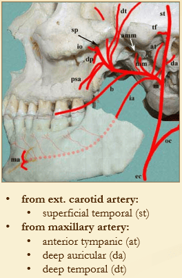
The fibrous capsule and retrodiscal tissue are supplied by an anastomotic vascular network arising from:
- Superficial Temporal Artery: A terminal branch of the external carotid artery, providing direct articular twigs to the posterior and lateral capsule.
- Maxillary Artery: The larger terminal branch of the external carotid artery, contributing via:
- Anterior Tympanic Artery: Traverses the petrotympanic fissure to supply the retrodiscal pad and middle ear.
- Deep Auricular Artery: Ascends to supply the external acoustic meatus and the posterior capsule.
- Deep Temporal & Masseteric Arteries: Supply the anterior capsule and adjacent pterygoid and masseter muscle attachments.

2. Nerve Supply
Innervation follows Hilton’s law (the nerves supplying the muscles that cross and move a joint also supply sensory innervation to that joint and the overlying skin):
- Auriculotemporal Nerve (CN V3): Forms the primary sensory innervation of the joint. Loops around the neck of the condyle, providing articular branches to the posterior, medial, and lateral aspects of the capsule and the vascular retrodiscal pad.
- Masseteric Nerve (CN V3): Sends sensory branches to the anteromedial aspect of the joint capsule as it passes through the mandibular notch.
- Deep Temporal Nerves (CN V3): Supply sensory twigs to the anterior joint capsule.
- Facial Nerve (CN VII) Contribution: Small accessory articular twigs from the temporal and zygomatic branches of the facial nerve contribute to sensory or proprioceptive innervation of the superficial lateral capsule in approximately 50% of individuals.
Muscles of mastication
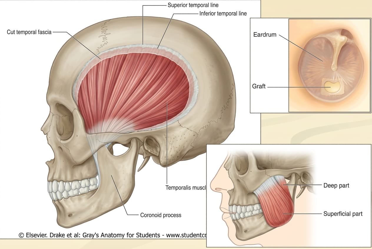
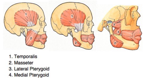
The muscles of mastication comprise four paired muscles that develop embryologically from the mesoderm of the first pharyngeal (mandibular) arch. Consequently, all four muscles are innervated by motor branches of the mandibular nerve (CN V3), the third division of the trigeminal nerve.
Scroll the table sideways on a smaller screen.
| Muscle | Origin | Insertion | Principal Actions | Innervation (CN V3) |
|---|---|---|---|---|
| Masseter |
• Superficial part: Anterior two-thirds of lower border of zygomatic arch. • Deep part: Posterior third of lower border and deep surface of zygomatic arch. |
Masseteric tuberosity on lateral surface of ramus and angle of mandible. | Powerful elevation (jaw closing); superficial fibers assist in mandibular protrusion. | Masseteric nerve (exits infratemporal fossa via mandibular notch). |
| Temporalis | Floor of temporal fossa (inferior to inferior temporal line) and deep layer of temporal fascia. | Apex and medial surface of coronoid process, extending down anterior border of mandibular ramus. | Powerful elevation (vertical anterior fibers); retrusion / retraction of protruded chin (horizontal posterior fibers); unilateral lateral movement. | Deep temporal nerves (anterior and posterior branches). |
| Medial Pterygoid |
• Deep head: Medial surface of lateral pterygoid plate and pterygoid fossa. • Superficial head: Tuberosity of maxilla and pyramidal process of palatine bone. |
Pterygoid tuberosity on medial (internal) surface of angle and ramus of mandible. | Elevates mandible (working with masseter as a mechanical sling); assists in protrusion; unilateral contraction drives contralateral grinding. | Medial pterygoid nerve (direct branch from main trunk of V3). |
| Lateral Pterygoid |
• Superior head: Infratemporal crest and infratemporal surface of greater wing of sphenoid. • Inferior head: Lateral surface of lateral pterygoid plate. |
• Superior head: Joint capsule and anterior margin of articular disc. • Inferior head: Pterygoid fovea on anterior surface of condylar neck. |
• Bilateral: Initiates depression (mouth opening) by drawing condyle and disc forward; strong protrusion. • Unilateral: Swings chin to opposite side (lateral excursion for grinding). |
Lateral pterygoid nerve (from anterior division of V3). |
Biomechanics of mastication
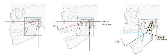
The TMJ executes three primary types of movement during functional mastication and speech: depression-elevation (open-close), protrusion-retrusion (translation along a sagittal line), and lateral grinding (lateral excursion around variable vertical axes).
1. The Dual-Phase Open-Close Movement
Mouth opening (depression of the mandible) occurs in two distinct, sequential phases:
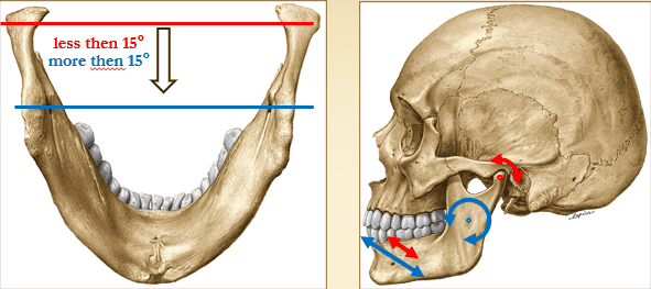
- First Phase (≤15° Opening / Pure Rotation): During modest opening (up to approximately 15 degrees of rotation, producing about 20 mm of interincisal clearance as used during normal speech), movement is restricted to pure rotation within the inferior discomandibular joint. The condylar head rotates like a hinge against the concave inferior surface of the articular disc around a fixed horizontal bicondylar axis.
- Transition at 15°: At approximately 15 degrees of depression, the strong lateral temporomandibular ligament becomes taut, mechanically arresting further pure rotational movement.
- Second Phase (>15° Opening / Gliding Translation): To achieve wide mouth opening (up to 40–50 mm), the movement shifts to gliding translation within the superior discotemporal joint. The condyle and articular disc slide together downward and forward along the slope of the articular eminence. The effective functional axis of rotation shifts downward and forward, aligning along a transverse axis connecting the two mandibular foramina. This positioning prevents over-stretching of the entering inferior alveolar neurovascular bundle.

2. Translation: Protrusion & Retrusion
- Protrusion: Both condyles and discs slide forward and downward along the articular tubercles in the discotemporal joint, driven by bilateral contraction of the lateral pterygoids (assisted by superficial masseter and medial pterygoid fibers). The mouth must open slightly during protrusion so the lower incisors can clear the overlap of the upper teeth.
- Retrusion (Retraction): The condyles slide backward into the mandibular fossa via the contraction of the posterior horizontal fibers of the temporalis and deep masseter fibers. Retrusion from the neutral resting position is anatomically limited because the condyle is arrested by the postglenoid tubercle and the tympanic plate.
3. Lateral Grinding (Masticatory Excursion)
Grinding relies on rotation of the mandible around vertical axes perpendicular to the intercondylar line:
- The Resting (Working) Condyle: Remains within the mandibular fossa and rotates around a vertical axis passing through its center.
- The Swinging (Balancing) Condyle: Translates forward, downward, and medially onto the articular tubercle.
- Muscle Coordination: Contraction of the lateral and medial pterygoids on one side swings the chin toward the opposite (contralateral) side. Mastication alternates this unilateral contraction in synchrony with ipsilateral temporalis and masseter stabilization.
TMJ disorders & inflammatory disease
The TMJ is vulnerable to mechanical derangements, degenerative changes, and local or hematogenous inflammatory insults.
1. Primary Bone & Joint Inflammatory Conditions
- Infectious & Traumatic Etiologies: Primary bone and inflammatory conditions of the TMJ can arise from:
- Hematogenous osteomyelitis of the condylar process of the mandible.
- Birth trauma to the TMJ (e.g., difficult forceps delivery causing condylar crushing or hemarthrosis).
- Intra- and extra-articular fractures of the condylar neck.
- Acute traumatic arthritis following blunt facial impact.
- Odontogenic osteomyelitis spreading directly from lower molar apical abscesses to the ramus and condyle.
- Otogenic Spread: Acute purulent otitis media or mastoiditis tracking through the petrotympanic fissure, thin roof of the mandibular fossa, or vascular retrodiscal tissue into the joint.
2. Temporomandibular Disorder (TMD) & Osteoarthritis
- Temporomandibular Joint Dysfunction (TMD): A common disorder characterized by abnormal joint mobility (either hypermobility or restricted opening), clicking or crepitus during movement, lateral deviation of the mandible upon opening, and myofascial pain radiating across the trigeminal distribution. Often triggered by malocclusion, bruxism (nocturnal tooth grinding), stress-induced masseter spasm, or internal disc displacement.
Internal Derangement: In anterior disc displacement with reduction, the disc slips anterior to the condyle when closed; as the mouth opens, the condyle clicks back onto the intermediate zone (“reciprocal click”). In displacement without reduction (“closed lock”), the disc stays stuck anteriorly, blocking translation and limiting opening to ≤20 mm. - Osteoarthritis of the TMJ: Develops secondary to chronic biomechanical overload, untreated internal derangements, trauma, or systemic arthritides (rheumatoid arthritis, gout). The fibrocartilage wears away, the articular disc thins and tears, and subchondral bony sclerosis and osteophytes form, leading to chronic joint pain, crepitus, and limited jaw movement.
3. TMJ Ankylosis & Trismus
- Joint Ankylosis (Contracture): Fibrous tissue proliferation or complete bony fusion uniting the condyle to the temporal bone. Most commonly follows childhood intracapsular condylar fractures or untreated neonatal purulent septic arthritis. Leads to severe, permanent inability to open the mouth and, in growing children, retrognathia (“bird-face deformity”) due to damage to the condylar secondary growth cartilage.
- Trismus (“Lockjaw”): Painful, acute spasm of the muscles of mastication (principally the masseter) causing restricted mouth opening. Clinical causes include acute pericoronitis around an impacted mandibular third molar, peritonsillar abscess (quinsy), tetanus (via tetanospasmin blocking glycine release), or irritation following inferior alveolar nerve blocks.
TMJ dislocation & reduction
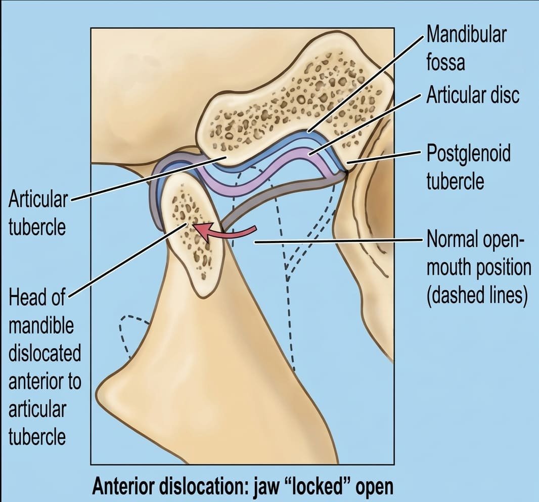
1. Mechanism of Anterior Dislocation
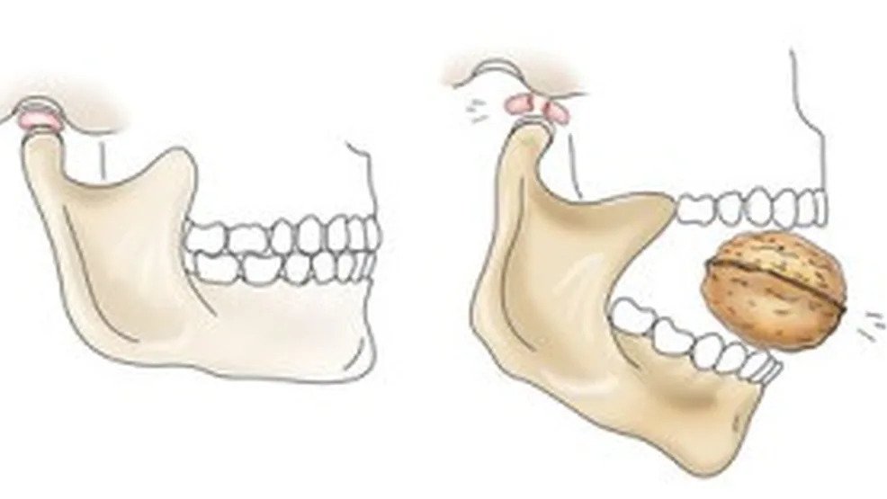
Anterior dislocation is the most common form of TMJ displacement. It often occurs during yawning, laughing widely, or taking an exceptionally large bite, or as a complication of dental extractions or endotracheal intubation:
- Pathophysiology: The condylar head translates forward past the apex of the articular tubercle into the infratemporal fossa.
- The “Locked Open” Jaw: Once the condyle slips anterior to the tubercle, the masseter and temporalis muscles reflexively enter severe spasm. This pulls the ramus superiorly, wedging the condyle in front of the articular eminence and preventing it from sliding back into the mandibular fossa.
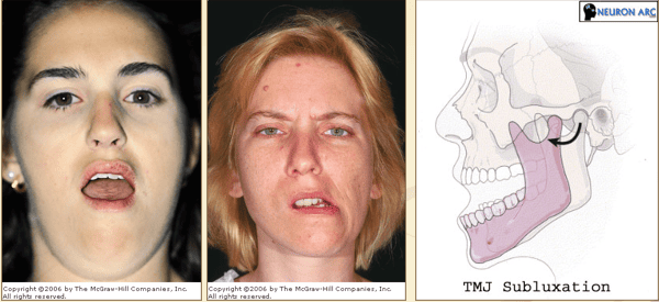
2. Clinical Presentation: Bilateral vs. Unilateral
- Bilateral Dislocation: The patient presents with their mouth fixed wide open, an inability to approximate the incisors or speak clearly, drooling of saliva, and hollow depressions visible and palpable over the preauricular areas where the condyles normally sit.
- Unilateral Dislocation (More Common): Only one condyle slips anterior to the tubercle. Because the contralateral intact joint acts as a hinge while the dislocated condyle is fixed anteriorly, the chin deviates toward the unaffected (contralateral) side, accompanied by an open bite on the affected side.
Quick recap
References
The following anatomical and surgical reference texts were verified in compiling this study note:
- Gray’s Anatomy: Standring, S. (2021) Gray’s Anatomy: The Anatomical Basis of Clinical Practice. 42nd edn. London: Elsevier. [Chapter 31: Infratemporal and Pterygopalatine Fossae and Temporomandibular Joint].
- Clinically Oriented Anatomy: Dalley, A.F. and Agur, A.M.R. (2023) Moore’s Clinically Oriented Anatomy. 9th edn. Philadelphia: Wolters Kluwer. [Chapter 8: Head – Temporomandibular Joint].
- Last’s Anatomy: Sinnatamby, C.S. (2011) Last’s Anatomy: Regional and Applied. 12th edn. Edinburgh: Churchill Livingstone / Elsevier. [Chapter 6: Head and Neck and Spine – Temporomandibular Joint].
- Snell’s Clinical Anatomy by Regions: Splittgerber, R. (2019) Snell’s Clinical Anatomy by Regions. 10th edn. Philadelphia: Wolters Kluwer. [Chapter 11: The Head – The Infratemporal Fossa].