
Mycobacteria, Spirochetes and Introduction to Mycology: Laboratory Diagnosis of the Atypicals
On this page
Overview
Mycobacteria: the acid-fast family
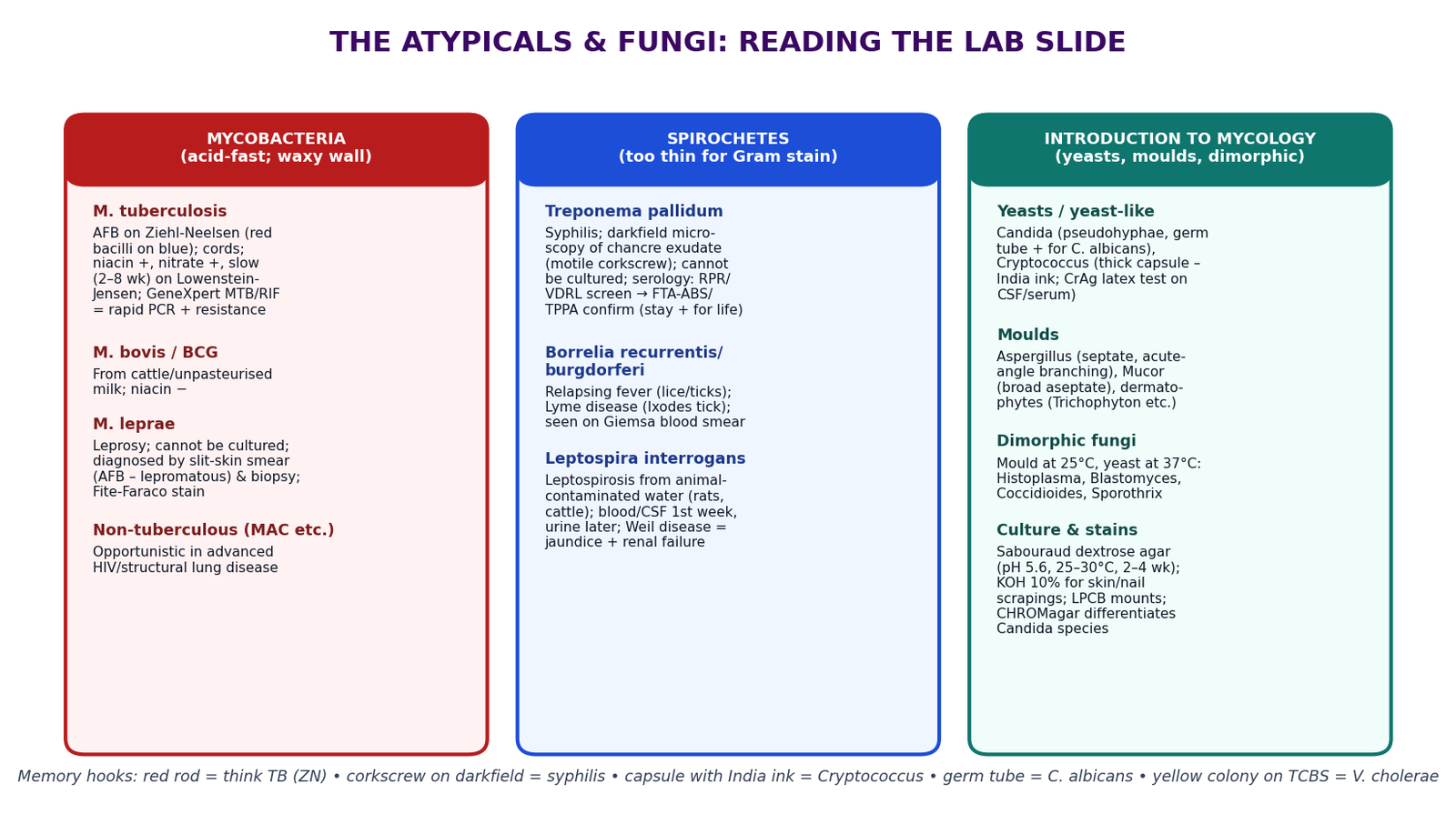
Mycobacteria are slender, slightly curved, non-motile, non-sporing rods whose cell wall is extraordinarily rich in mycolic acids — long-chain fatty acids that make the wall waxy. Consequences: they stain poorly with Gram stain; once stained (with hot carbol fuchsin) they resist decolourisation by acid-alcohol — hence "acid-fast"; and they grow slowly (dividing every 12–24 h vs ~20 min for E. coli).
- M. tuberculosis complex (M. tuberculosis, M. bovis from cattle/unpasteurised milk, M. africanum) — tuberculosis.
- M. leprae — leprosy; cannot be cultured in vitro (grows only in cooler tissues and in armadillos/mouse footpads).
- Non-tuberculous mycobacteria (NTM) — M. avium complex (advanced HIV), M. kansasii and others; environmental organisms causing opportunistic, often harder-to-treat disease.
Virulence anchors: cord factor (trehalose dimycolate — serpentine "cords" of bacilli on microscopy and a granuloma-sustaining lipid), the mycobacterial capsule, and intracellular survival inside macrophages (blocks phagosome–lysosome fusion) — which is why TB is a cell-mediated immunity battle and why the purified protein derivative (PPD/Mantoux) test reflects T-cell memory.
Laboratory diagnosis of tuberculosis
Scroll the table sideways on a smaller screen.
| Test | Principle | Reading / interpretation | Limitations |
|---|---|---|---|
| Ziehl-Neelsen (ZN) smear | Hot carbol fuchsin drives stain into mycolic acids; acid-alcohol cannot remove it; methylene blue counterstain | Red/bright pink slender rods against a blue background = acid-fast bacilli (AFB); report graded (scanty, 1+, 2+, 3+) by fields examined | Needs ~10⁴–10⁵ bacilli/mL (smear-negative TB exists — children, HIV); cannot distinguish TB from NTM; says nothing about resistance |
| Fluorescence (auramine) microscopy | Auramine binds mycolic acids; bacilli fluoresce against dark background | Same grading, faster scanning, ~10% more sensitive than ZN | Requires fluorescence microscope |
| GeneXpert MTB/RIF | Automated real-time PCR detecting M. tuberculosis DNA and rifampicin-resistance mutations (rpoB) | Result within ~2 hours: "MTB detected / not detected" + RIF resistance; recommended first test for suspected pulmonary TB, including HIV and children | Does not replace culture for full drug-susceptibility; costly cartridges |
| Culture | Specimens decontaminated (N-acetyl cysteine–NaOH) then inoculated on Löwenstein–Jensen (egg-based) or liquid (MGIT) media | Definitive diagnosis; enables full drug-susceptibility testing (DST); LJ colonies in 2–8 weeks (rough, buff, tough); cord formation typical of M. tuberculosis | Slow; needs biosafety level 2/3 containment |
| Biochemical identification (where cultures done) | Niacin accumulation (M. tuberculosis niacin positive; M. bovis negative), nitrate reduction (MTB +), pigment and growth rate | Separates species within the complex and from NTM | Largely superseded by molecular line-probe assays |
| Tuberculin skin test (Mantoux/PPD) & IGRA | Cell-mediated (type IV) hypersensitivity to mycobacterial antigens | Induration ≥10 mm (≥5 mm in HIV/immunosuppressed) read at 48–72 h = infection (latent or active), not activity; IGRA (blood) not distorted by BCG | Cannot distinguish latent from active TB; BCG/vanilla NTM can falsely boost TST |
Leprosy (M. leprae)
A chronic granulomatous infection of skin, peripheral nerves and mucosa, curable with multidrug therapy (MDT: dapsone + rifampicin ± clofazimine per national/WHO leprosy programme). Two poles, with the patient's cell-mediated immunity deciding where they sit: tuberculoid (paucibacillary) — strong immunity, few well-defined hypopigmented anaesthetic patches, granulomas, skin smear negative; lepromatous (multibacillary) — weak immunity, widespread symmetric lesions, nodular facies (leonine), slit-skin smears positive for acid-fast M. leprae (best taken from the earlobe/lesion edge), nerve thickening and damage. Diagnosis is clinical (anaesthetic patch + peripheral nerve involvement) supported by smear/biopsy; the organism cannot be cultured — a favourite exam fact. Complications: claw hand, foot drop, trophic ulcers, eye damage (lagophthalmos) — early MDT prevents disability.
Spirochetes: Treponema, Borrelia, Leptospira
Slender, helically coiled gram-negative bacteria with endoflagella (axial filaments) in the periplasmic space — the engine of their corkscrew motility. They are too thin for routine Gram staining; the laboratory answers with darkfield microscopy, silver stains and serology.
Scroll the table sideways on a smaller screen.
| Organism | Disease & stages | Transmission | Laboratory diagnosis |
|---|---|---|---|
| Treponema pallidum subsp. pallidum | Syphilis — primary: painless chancre (highly infectious) with regional lymphadenopathy; secondary (weeks–months): disseminated rash incl. palms/soles, condylomata lata, fever; latent: serology only; tertiary (years): gummata, cardiovascular (aortitis), neurosyphilis (tabes dorsalis, general paresis); congenital: miscarriage, stigmata | Sexual contact; transplacental (congenital); the non-venereal treponematoses (yaws T. p. pertenue, bejel, pinta) spread by skin contact | Cannot be cultured. Darkfield microscopy of chancre/condyloma exudate: motile, tightly coiled spirochetes with bending/flexing (best in primary/secondary moist lesions; oral lesions unreliable due to commensal spirochetes); serology: non-treponemal RPR/VDRL screen (titre tracks activity, falls with treatment, can be falsely +ve in pregnancy/autoimmune disease) → treponemal TPPA/FTA-ABS/EIA confirm (stay positive for life) |
| Borrelia (B. recurrentis; Lyme group B. burgdorferi) | Relapsing fever: recurrent febrile episodes separated by afebrile intervals — each relapse expresses a new antigenic variant (antigenic variation outruns antibody); Lyme disease (where tick-borne borreliosis occurs): erythema migrans → arthritis, neuro and cardiac features | Louse-borne (B. recurrentis, crowding/poverty) and tick-borne relapsing fever; Ixodes tick for Lyme | Giemsa/Wright-stained blood smears during febrile peaks — visible spirochetes between RBCs; serology (ELISA) for Lyme |
| Leptospira interrogans | Leptospirosis: biphasic — febrile septicaemic phase (headache, myalgia esp. calves, conjunctival suffusion) then immune phase; severe form = Weil disease (jaundice + renal failure + haemorrhage) | Zoonosis: water/soil contaminated with urine of rats, cattle, dogs; enters through skin breaks or mucosa — farmers, fishermen, sewer workers, flooded areas | Specimen by week: blood/CSF 1st week, urine later; darkfield of blood (skilled); serology — microscopic agglutination test (MAT) gold standard; culture on Fletcher/EMJH medium (weeks, reference labs) |
Introduction to mycology: yeasts, moulds, dimorphic fungi
Fungi are eukaryotes: a chitin + β-glucan cell wall (echinocandin target) and an ergosterol membrane (azoles block its synthesis; polyenes bind it — this is why antifungals don't touch human cells). Four morphological groups:
Scroll the table sideways on a smaller screen.
| Group | Form | Examples & diseases |
|---|---|---|
| Yeasts / yeast-like | Unicellular, budding | Candida albicans (thrush, vaginitis, invasive candidiasis in ICU) — forms pseudohyphae and a positive germ tube; Cryptococcus neoformans — thick polysaccharide capsule, cryptococcal meningitis in advanced HIV (India ink halo; CrAg test) |
| Moulds (filamentous) | Hyphae → mycelium; reproduce by spores | Aspergillus (septate hyphae with acute-angle (~45°) branching — invasive pulmonary aspergillosis in neutropenia), Mucorales (broad, ribbon-like, sparsely septate — mucormycosis in diabetic ketoacidosis), dermatophytes Trichophyton/Microsporum/Epidermophyton (tinea capitis/corporis/pedis/unguium) |
| Dimorphic | Mould in the environment/at 25°C, yeast (or spherule) in tissue/at 37°C | Histoplasma capsulatum, Blastomyces, Coccidioides (spherules), Sporothrix schenckii (rose-thorn sporotrichosis — lymphocutaneous nodules); mostly regional/systemic, relevant in advanced HIV |
| Pneumocystis jirovecii (special case) | Fungus that cannot be cultured | Pneumocystis pneumonia (PCP) in advanced HIV — diagnosed by induced sputum/BAL stain; co-trimoxazole treatment and prophylaxis |
Laboratory diagnosis of fungal infection
- Direct microscopy: 10–20% KOH mount of skin/nail/hair scrapings digests keratin, leaving refractile hyphae/spores visible — the office test for tinea; India ink (or nigrosin) negative stain of CSF showing the encapsulated yeast → Cryptococcus; calcofluor white binds chitin and fluoresces (most sensitive rapid stain); Gram stain shows yeasts gram-positive; tissue stains: PAS and Gomori methenamine silver (GMS) blacken fungi in biopsy sections.
- Culture: Sabouraud dextrose agar (pH ~5.6, high glucose) selects fungi over bacteria; add antibiotics (chloramphenicol/gentamicin) and cycloheximide (suppresses saprophytic moulds) as needed; incubate 25–30°C for 1–4 weeks; identify by colony colour/texture and lactophenol cotton blue (LPCB) mounts of hyphae and conidia; CHROMagar differentiates Candida species by colony colour; germ tube test identifies C. albicans within 2–3 hours of serum incubation.
- Antigen/serology: cryptococcal antigen (CrAg) latex agglutination or lateral flow on CSF/serum — the standard first-line test for cryptococcal meningitis; galactomannan for Aspergillus; β-D-glucan as a pan-fungal screen (not species-specific).
- Molecular: PCR and MALDI-TOF mass spectrometry (rapid species ID from colonies); 18S/ITS sequencing in reference labs.
Clinical scenarios
Scenario 1 — The smear, the Xpert and the X-ray
A 45-year-old with 3 weeks of cough, night sweats and weight loss has a chest X-ray showing apical cavitation. Sputum ZN shows 2+ acid-fast bacilli — red slender rods. You do not wait for culture: the Xpert confirms MTB detected, rifampicin resistance not detected, so he starts the standard first-line regimen and you register him in the TB programme. You counsel on adherence (directly observed support where available), infection control at home, and HIV testing (routine in all TB patients). You explain the difference between tests: the smear says "how infectious, roughly", the Xpert says "TB DNA and rifampicin resistance", and culture/DST will confirm everything and catch resistance the Xpert missed.
Scenario 2 — A painless ulcer and a two-step serology
A 26-year-old truck driver has a painless indurated genital ulcer of 3 weeks with rubbery inguinal nodes. Darkfield microscopy is unavailable, so you take blood for RPR (reactive, titre 1:32) and TPPA (reactive) — confirming syphilis, and the high RPR titre supports active secondary-stage disease. You treat with benzathine penicillin G per the STI protocol, warn about the Jarisch–Herxheimer reaction (transient fever and flare as spirochetes die — reassure, do not stop treatment), trace and treat partners, and document that the TPPA will stay positive for life — future "positive syphilis tests" will not prove reinfection on their own; rising RPR titre would.
Scenario 3 — Headache and fever in advanced HIV
A 34-year-old with newly diagnosed HIV (CD4 28) has 2 weeks of headache, fever and confusion. You suspect cryptococcal meningitis and send CSF for cryptococcal antigen (rapid, sensitive) and India ink (encapsulated yeasts with a halo). The CrAg comes back positive within the hour. You start induction therapy per the national guideline (amphotericin-based combination), manage raised intracranial pressure with therapeutic lumbar punctures, and defer ART for several weeks to avoid IRIS. You note the partnership with the laboratory: the antigen test made the diagnosis before any fungus grew — in mycology, serology often beats culture for speed.
Key points
References
- Medical Microbiology (Baron, ed.), NCBI Bookshelf — Mycobacteria, Spirochetes and Introduction to Mycology chapters (Library reference.)
- WHO consolidated guidelines on tuberculosis: diagnosis (smear, Xpert MTB/RIF) (Library reference.)
- Stop TB Partnership — Laboratory Diagnosis of Tuberculosis by Sputum Microscopy handbook (Library reference.)
- Jawetz, Melnick & Adelberg's Medical Microbiology (Library reference.)
- WHO Guidelines for Diagnosing, Preventing and Treating Cryptococcal Disease among People Living with HIV (Library reference.)
Further reading
For deeper study, compare this lesson with current guidance and standard references from recognised medical and scientific organisations.
- WHO Global Tuberculosis Programme
- NCBI Bookshelf — Introduction to Mycology
- CDC DPDx — parasite and fungal identification library
- WHO Cryptococcal Disease Guidelines
- Clinical Medicine Year 1, Semester 2 curriculum
For education only. TB diagnosis and treatment follow the national TB programme; leprosy MDT and cryptococcal protocols follow national/WHO guidance.