
Autacoid pharmacology: histamine, serotonin, bradykinin, ergot alkaloids, eicosanoids & NSAIDs
On this page 19 sections
Overview of autacoids
Autacoids (derived from the Greek autos [self] and acos [remedy, relief, or drug]) are a heterogeneous family of biologically active endogenous substances synthesized, stored, and released locally in response to tissue injury, chemical irritation, or immunological stimuli. Functioning as “local hormones” or autocrine/paracrine signaling mediators, their physiological and pathological actions are predominantly restricted to the microenvironment immediately adjacent to their site of release.
Autacoids vs. Endocrine Hormones
- Site of Synthesis: True endocrine hormones (e.g., insulin, cortisol, thyroxine) are produced by specialized, anatomically discrete glandular organs. Autacoids are synthesized by diffuse, non-glandular cell populations across virtually all vascularized tissues (e.g., mast cells, vascular endothelium, platelets, mucosal enterocytes).
- Mechanism of Delivery: Endocrine hormones are released into the systemic bloodstream to act upon distant target organs. Autacoids act locally near their site of synthesis (paracrine and autocrine modulation).
- Biological Half-Life: Autacoids possess extremely short biological half-lives (seconds to minutes) and are rapidly inactivated by local tissue enzymes or during first-pass pulmonary clearance, preventing uncontrolled systemic spillover under normal conditions.
Physiological & Pathological Roles
- Peripheral Tissues (Predominant Effects): Acute inflammation, type I hypersensitivity and anaphylaxis, smooth muscle tone regulation, vascular permeability, hemostasis/thrombosis, and gastric acid secretion.
- Central Nervous System (CNS): Neurotransmission, neuroendocrine regulation, sleep-wake cycles, circadian rhythmicity, core body temperature setting, appetite suppression, thirst modulation, blood pressure autoregulation, and central nociceptive perception.
| Biochemical Category | Precursor Substrate | Representative Autacoid Molecules | Primary Clinical Significance |
|---|---|---|---|
| Amine-Derived | Natural Amino Acids (L-Histidine, L-Tryptophan) | Histamine, Serotonin (5-HT) | Allergic anaphylaxis, gastric acid secretion, neurotransmission, migraine, carcinoid syndrome. |
| Peptide-Derived | Circulating Plasma & Tissue Kininogens / Angiotensinogen | Bradykinin, Kallidin, Angiotensin II | Vasodilation, vascular permeability, pain generation, ACE inhibitor cough, renal autoregulation. |
| Lipid-Derived (Eicosanoids) | Membrane Phospholipids (Arachidonic Acid, C20:4) | Prostaglandins (PGE2, PGF2α), Prostacyclin (PGI2), Thromboxane A2 (TXA2), Leukotrienes (LTB4, LTC4, LTD4, LTE4), Platelet-Activating Factor (PAF) | Inflammation, platelet aggregation, fever, dysmenorrhea, bronchospasm, gastric cytoprotection, ductus arteriosus patency. |
Histamine: synthesis, storage & release
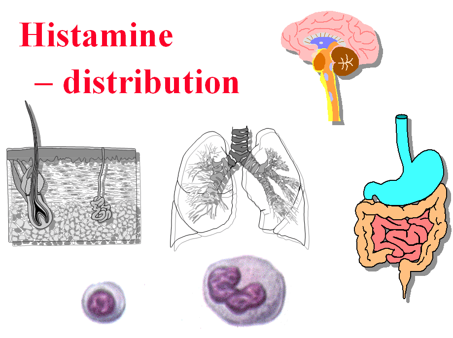
Histamine (2-[4-imidazolyl]ethylamine) is an endogenous basic amine occurring ubiquitously across animal and plant tissues. In humans, high concentrations reside within epithelial boundary tissues directly interfacing with the external environment: the skin epidermis and dermis, bronchial and pulmonary mucosa, and the gastrointestinal tract mucosa.
1. Biosynthesis of Histamine
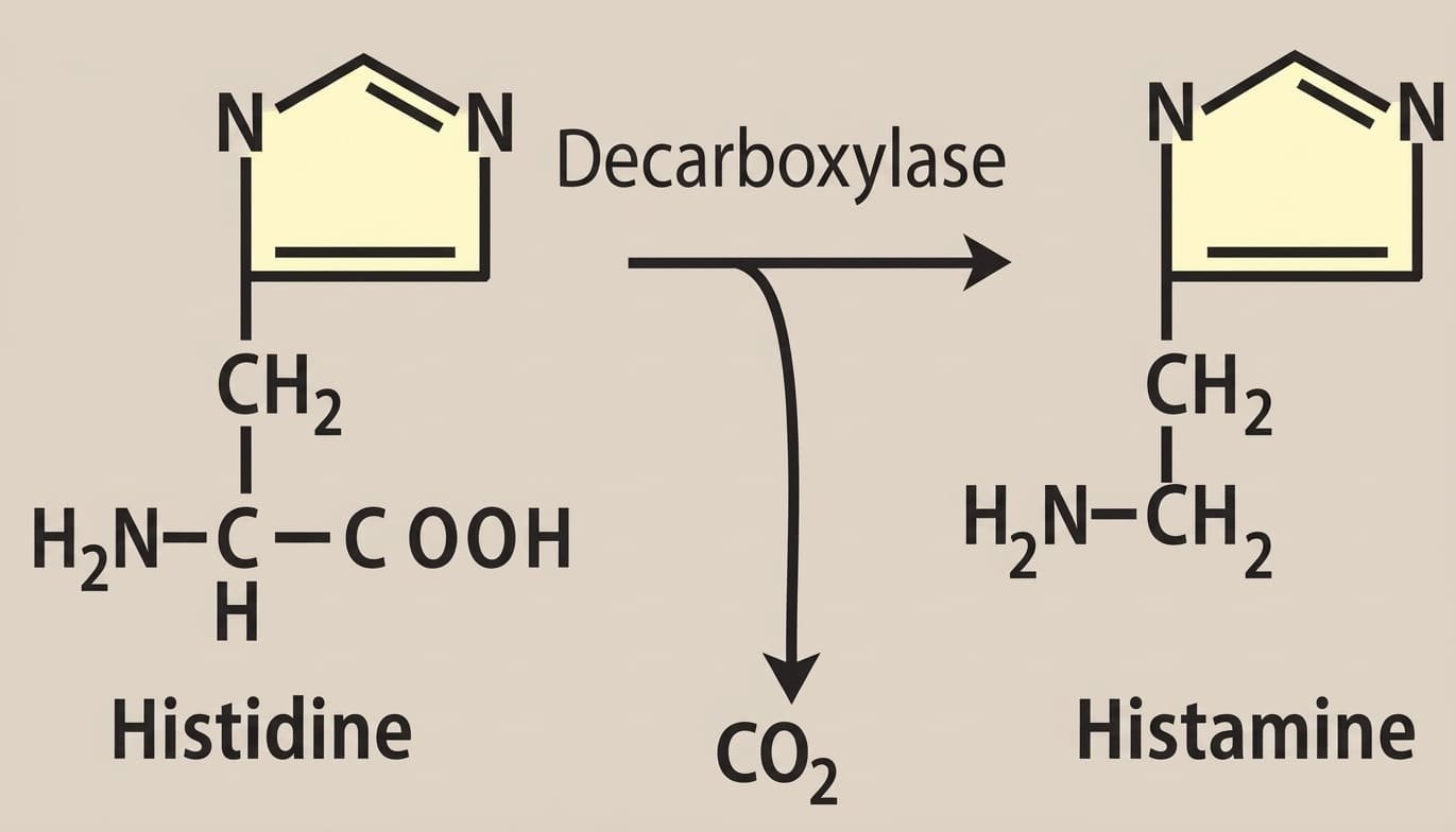
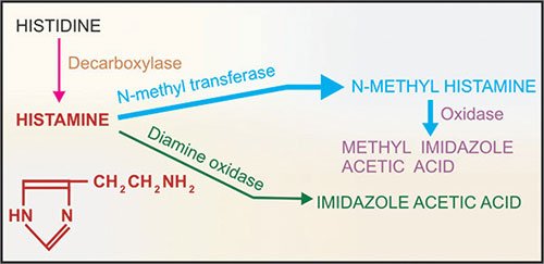
Histamine is synthesized from the essential dietary amino acid L-histidine in a single enzymatic step. The reaction involves decarboxylation catalyzed by the cytosolic enzyme histidine decarboxylase (HDC), which requires pyridoxal 5′-phosphate (Vitamin B6) as an obligate cofactor.
2. Cellular Pools & Storage
- Mast Cell and Basophil Pool (Slow Turnover Pool): Histamine is sequestered inside dense, electron-rich secretory granules of tissue mast cells and circulating basophils. It is ionically bound in an electrostatically inactive complex with acidic, polyanionic proteoglycans (heparin sulfate in mast cells; chondroitin sulfate in basophils) and neutral proteases. Mast cells concentrate densely at sites prone to mechanical trauma or microbial entry: the nose, mouth, feet, internal mucosal surfaces, and along blood vessel bifurcations.
- Non-Mast Cell Pool (Fast Turnover Pool): Histamine is synthesized continuously and released immediately without long-term granular storage:
- Enterochromaffin-Like (ECL) Cells: Located in the gastric fundic oxyntic mucosa; release histamine upon gastrin and vagal muscarinic (M1/M3) stimulation to activate adjacent acid-secreting parietal cells via H2 receptors.
- Central Histaminergic Neurons: Cell bodies localized strictly in the tuberomammillary nucleus of the posterior hypothalamus. They project diffusely across the neuroaxis, regulating wakefulness, circadian arousal, appetite suppression, neuroendocrine release, and body temperature.
3. Mechanisms of Histamine Release
A. Immunological Release (Antigen-Mediated / Type I Hypersensitivity)
Requires prior sensitization of the adaptive immune system:
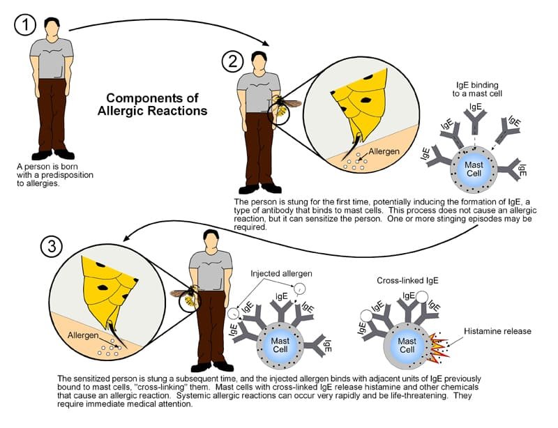
- Sensitization Phase: Exposure to an initial allergen stimulates helper T-cells (TH2) and B-lymphocytes to produce antigen-specific Immunoglobulin E (IgE) antibodies. These IgE molecules attach with high affinity to FcεRI receptors on the surface of mast cells and basophils.
- Degranulation Phase: Upon re-exposure, the bivalent or multivalent allergen binds and cross-links adjacent membrane-bound IgE-FcεRI complexes. This activates membrane phospholipase C and tyrosine kinases, provoking rapid intracellular Ca2+ mobilization. Secretory granules fuse with the plasma membrane, explosively exocytosing histamine, heparin, proteases, and newly formed leukotrienes into the interstitial space.
- Negative Feedback Autoregulation: Histamine inhibits its own release in skin mast cells and blood basophils by binding to cell-surface H2 receptors (raising cAMP via Gs), limiting allergic intensity. Critical note: This negative feedback loop does NOT occur in pulmonary lung mast cells.
B. Chemical & Mechanical Release (Non-Immunological / Non-IgE Mediated)
Occurs without prior immunological sensitization, foreign antibodies, or energy expenditure:
- Direct Chemical Displacement: Highly basic, positively charged organic amines, alkaloids, and polycationic macromolecules displace histamine from its ionic complex with heparin inside the granule. Examples include morphine, codeine, d-tubocurarine, radiocontrast media, vancomycin (“Red Man Syndrome”), polymyxin B, and carbohydrate plasma expanders (dextran). Extracellular sodium ions (Na+) rapidly exchange with and liberate the amine.
- Mechanical & Physical Injury: Direct physical disruption of mast cell membranes provoked by thermal burns, frostbite, skin scratching (dermographism), ionizing radiation, and bacterial pore-forming endotoxins.
- Venoms and Stings: Basic polypeptides present in bee, wasp, and snake venoms activate mast cell degranulation directly via MAS-related G-protein coupled receptor X2 (MRGPRX2).
- Neoplastic Proliferation: Marked systemic mast cell accumulation and uncontrolled histamine release occur in systemic mastocytosis, urticaria pigmentosa, gastric carcinoids, and basophilic myelogenous leukemia.

4. Metabolism & Inactivation of Histamine
Circulating and local histamine is rapidly metabolized by two distinct enzymatic pathways:
- 1. Histamine N-Methyltransferase (HNMT) Pathway (Primary Pathway, ~70%): Prevalent in parenchymal tissues (especially brain, liver, and kidneys), but completely absent in circulating blood. HNMT transfers a methyl group from S-adenosylmethionine (SAM) to the imidazole nitrogen, yielding 1-methylhistamine (tele-methylhistamine). Monoamine oxidase B (MAO-B) and aldehyde dehydrogenase subsequently oxidize it into 1-methylimidazole-4-acetic acid, which is excreted in the urine.
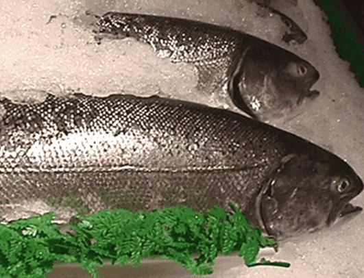
- 2. Diamine Oxidase (DAO / Histaminase) Pathway (Secondary Pathway, ~30%): Present in intestinal mucosa, liver, kidney, and circulating plasma. Catalyzes oxidative deamination of the primary aliphatic amine group, forming imidazole-4-acetic acid and its riboside conjugate. DAO plays an essential defense role in catabolizing high concentrations of histamine ingested in fermented or spoiled foods (preventing dietary histamine toxicity).
Histamine receptors & pharmacodynamics
Histamine acts through four distinct, non-selective G-protein coupled receptor (GPCR) subtypes designated H1, H2, H3, and H4. Structurally, H1 receptors share close phylogenetic homology with muscarinic acetylcholine receptors, whereas H2 receptors share sequence alignment with serotonergic 5-HT1 receptors.
1. H1 Receptor (Gq/11 Coupled → ↑ IP3, DAG, ↑ Intracellular Ca2+)
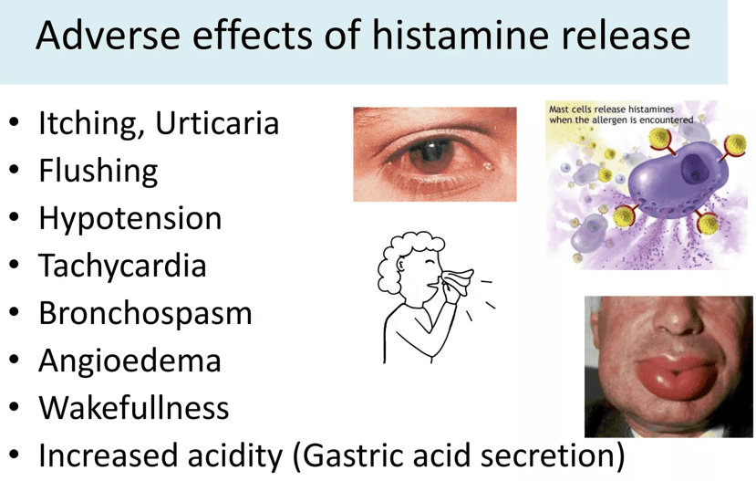
- Endothelial Contraction & Edema: Stimulates microvascular endothelial cells in postcapillary venules to contract and separate, forming intercellular gaps that expose the basement membrane. This produces marked plasma protein exudation, fluid transudation, and local tissue edema.
- Smooth Muscle Contraction: Directly contracts non-vascular visceral smooth muscle, causing powerful bronchoconstriction (severe in asthmatics), intestinal cramping, and uterine contraction.
- Endothelial Vasodilation: Stimulates endothelial nitric oxide synthase (eNOS) to generate nitric oxide (NO), which diffuses into vascular smooth muscle to elevate cGMP, producing rapid, transient arteriolar vasodilation, cutaneous flushing, and throbbing headache.
- Sensory Nerve Terminals: Depolarizes unmyelinated C-fiber nociceptive and pruriceptive sensory nerve endings, provoking intense pruritus (itching) and localized burning pain.
- The Classic Triple Response of Lewis: Intradermal injection of histamine produces three sequential vascular phenomena:
- Localized Red Spot (Flush): Develops within seconds; direct capillary/venular vasodilation mediated by endothelial H1 and smooth muscle H2 receptors.
- Surrounding Bright Red Flare: Appears slowly, extending irregularly beyond the red spot; mediated by an axon reflex where sensory impulses propagate antidromically into adjacent collateral arterioles, releasing vasodilating calcitonin gene-related peptide (CGRP) and substance P.
- Central Wheal (Edema): Appears within 1 to 2 minutes; circumscribed swelling caused by H1-mediated postcapillary venular endothelial gap formation and plasma extravasation.
2. H2 Receptor (Gs Coupled → ↑ Adenylyl Cyclase, ↑ cAMP, ↑ PKA)
- Gastric Acid Secretion: Concentrated on gastric mucosal parietal cells. Binding of histamine triggers adenylyl cyclase → cAMP elevation → PKA activation, driving vesicle fusion and activating the apical H+/K+-ATPase proton pump to extrude hydrogen ions into the gastric lumen (the primary driver of nocturnal and basal acid secretion).
- Vascular Smooth Muscle: Located directly on vascular smooth muscle cells; mediates sustained, slow-onset vasodilation via cAMP-dependent protein kinase phosphorylation.
- Cardiac Stimulation: Increases pacemaker automaticity in the sinoatrial node (positive chronotropy) and enhances myocardial contractility (positive inotropy).
3. H3 Receptor (Gi/o Coupled → ↓ cAMP, ↓ Ca2+ Influx, ↑ K+ Current)
- Autoreceptors: Located on presynaptic histaminergic nerve terminals in the CNS and myenteric plexus; provides autoinhibitory negative feedback, suppressing further histamine synthesis and exocytosis.
- Heteroreceptors: Located presynaptically on non-histaminergic neurons; inhibits the release of diverse neurotransmitters including acetylcholine, norepinephrine, dopamine, serotonin, and GABA.
- Therapeutic Horizon: Promising cognitive target for narcolepsy, ADHD, Alzheimer’s disease, and schizophrenia.
4. H4 Receptor (Gi/o Coupled → ↓ cAMP, ↑ Intracellular Ca2+)
- Immunological Chemotaxis: Expressed selectively on hematopoietic and immunocompetent lineages: eosinophils, neutrophils, basophils, mast cells, and CD4+ T-lymphocytes.
- Inflammatory Signaling: Mediates directional leukocyte chemotaxis, upregulation of adhesion molecules, and cytokine secretion; actively investigated for chronic inflammatory conditions (severe asthma, rheumatoid arthritis, atopic dermatitis).
| Receptor | G-Protein Coupling & Effector | Primary Anatomical Distribution | Partially Selective Agonists | Selective Antagonists / Inverse Agonists |
|---|---|---|---|---|
| H1 | Gq/11 (↑ IP3, ↑ DAG, ↑ Ca2+) | Smooth muscle, vascular endothelium, sensory nerves, CNS | Histaprodifen, 2-(m-chlorophenyl)histamine | Mepyramine, Triprolidine, Diphenhydramine, Cetirizine |
| H2 | Gs (↑ cAMP, ↑ PKA) | Gastric parietal cells, cardiac myocytes, vascular smooth muscle, mast cells | Amthamine, Dimaprit, Betazole | Cimetidine, Ranitidine, Famotidine, Nizatidine |
| H3 | Gi/o (↓ cAMP, ↓ Ca2+) | Presynaptic neurons in CNS, myenteric plexus, sensory endings | (R)-α-methylhistamine, Immepip, Imetit, Proxyfan | Pitolisant (approved for narcolepsy), Thioperamide, Ciproxifan, Clobenpropit |
| H4 | Gi/o (↓ cAMP, ↑ Ca2+) | Eosinophils, neutrophils, CD4+ T-cells, bone marrow, spleen | Clobenpropit (partial), Imetit, VUF-8430 | Thioperamide, JNJ-7777120, Toreforant |
Histamine antagonists & blockers
The pathophysiological actions of histamine can be pharmacologically attenuated via three distinct clinical mechanisms: physiological antagonism, mast cell release inhibition, and receptor antagonism (inverse agonism).
1. Physiological Antagonists
Adrenaline (Epinephrine): Operates as the ultimate life-saving physiological antagonist. It does not interact with histamine receptors; instead, it binds to β2-adrenoceptors on bronchial smooth muscle (reversing bronchospasm), α1-adrenoceptors on vascular smooth muscle (reversing vasodilation and mucosal edema, elevating blood pressure), and β2 receptors on mast cells (suppressing further mediator release). It is the mandatory, immediate first-line drug of choice for systemic anaphylaxis (0.3 to 0.5 mg IM in adults).
2. Release Inhibitors (Mast Cell Stabilizers)
Cromolyn Sodium & Nedocromil: Inhibit chloride channels in the mast cell membrane, blocking the antigen-stimulated transmembrane influx of Ca2+ required for exocytic degranulation. They prevent the release of histamine, leukotrienes, and secondary autacoids, while suppressing leukocyte chemotaxis. Critical Caveat: They possess zero direct bronchodilator or antihistaminic action; they are exclusively prophylactic agents and are ineffective if administered during an acute allergic or asthmatic attack.
3. H1-Receptor Antagonists (Antihistamines)
All modern H1 blockers act molecularly as inverse agonists that bind and stabilize the inactive conformation of the H1 receptor, downregulating constitutive basal signaling. They are categorized into three generations:
First-Generation H1 Antagonists
- Key Characteristics: Highly lipophilic, neutral molecules at physiological pH; rapidly cross the blood-brain barrier into the CNS. Possess significant off-target antagonist activity at muscarinic acetylcholine receptors, α1-adrenoceptors, and serotonin (5-HT2) receptors, alongside cardiac/neuronal sodium channel blocking (local anesthetic) properties.
- Chemical Classes & Prototypes:
- Ethanolamines: Diphenhydramine, Dimenhydrinate, Clemastine, Carbinoxamine. Marked sedation and powerful anticholinergic/anti-motion sickness actions.
- Ethylenediamines: Pyrilamine, Mepyramine. Moderate sedation, common gastrointestinal irritation.
- Alkylamines: Chlorpheniramine, Pheniramine, Brompheniramine. Minimal sedation, potent peripheral H1 blockade.
- Piperazines: Cyclizine, Meclizine, Hydroxyzine, Chlorcyclizine. Long duration of action; cyclizine and meclizine are first-line for motion sickness and vestibular vertigo.
- Phenothiazines: Promethazine. Very strong sedation, marked antiemetic, potent local anesthetic action.
- Piperidines: Cyproheptadine. Potent H1 blocker with strong 5-HT2 antagonism; used for cold-induced urticaria, carcinoid syndrome, and appetite stimulation.
Second-Generation H1 Antagonists
- Key Characteristics: Lipophobic molecules containing zwitterionic or carboxylated polar groups; substrates for the P-glycoprotein efflux transporter at the blood-brain barrier. They do not penetrate the CNS at therapeutic doses, causing negligible daytime sedation. Highly selective for H1 receptors; lack anticholinergic, anti-alpha, or local anesthetic side effects.
- Representative Drugs: Loratadine, Cetirizine, and Fexofenadine. (Note: Loratadine is metabolized via CYP3A4 and CYP2D6; cetirizine is cleared mostly unchanged by kidneys).
Third-Generation H1 Antagonists
- Key Characteristics: Active single enantiomers or purified active metabolites of second-generation drugs, engineered to achieve higher receptor affinity, rapid onset, prolonged duration, and freedom from hepatic CYP-mediated cardiotoxicity.
- Representative Drugs: Desloratadine (active metabolite of loratadine), Levocetirizine (active R-enantiomer of cetirizine), and Fexofenadine (active carboxylic acid metabolite of terfenadine; completely lacks the cardiotoxic HERG channel-blocking property that caused fatal torsades de pointes with terfenadine and astemizole).
| Drug Entity | Generation & Class | Sedative Potential | Anti-Motion Sickness Activity | Anticholinergic (Atropine-like) Activity |
|---|---|---|---|---|
| Diphenhydramine | 1st Gen (Ethanolamine) | Marked (High) | Moderate to Marked | Marked (Dry mouth, urinary retention) |
| Promethazine | 1st Gen (Phenothiazine) | Marked (Very High) | Marked | Marked (Pronounced dry mouth) |
| Chlorpheniramine | 1st Gen (Alkylamine) | Low to Moderate | Low | Moderate |
| Meclizine / Cyclizine | 1st Gen (Piperazine) | Moderate | Marked (Preferred for vertigo) | Moderate |
| Loratadine | 2nd Gen (Piperidine) | None / Negligible | None | None |
| Cetirizine | 2nd Gen (Piperazine) | Low (Mild in ~10% patients) | None | None |
| Fexofenadine | 2nd/3rd Gen (Piperidine) | None | None | None |
4. H2-Receptor Antagonists
Competitive, reversible antagonists at parietal cell H2 receptors: Cimetidine, Ranitidine, Famotidine, and Nizatidine.
- Relative Potencies: Ranitidine is 5 times more potent than cimetidine; Famotidine is 5 times more potent than ranitidine (25-fold more potent than cimetidine).
- Therapeutic Indications: Duodenal ulcers, benign gastric ulcers, gastroesophageal reflux disease (GERD), and prevention of stress-related mucosal bleeding.
- Cimetidine Adverse Effects & Toxicity:
- Antiandrogenic Effects: Binds androgen receptors and inhibits dihydrotestosterone, while stimulating prolactin secretion; chronic therapy produces gynecomastia, galactorrhea, and reduced sperm count/impotence in men.
- CYP450 Enzyme Inhibition: Potent inhibitor of CYP1A2, CYP2C9, CYP2D6, and CYP3A4, dangerously elevating serum levels of co-administered narrow-therapeutic-index drugs (warfarin, theophylline, phenytoin, lidocaine). Ranitidine exhibits negligible CYP inhibition; famotidine exhibits zero CYP inhibition.
Serotonin: synthesis, distribution & storage
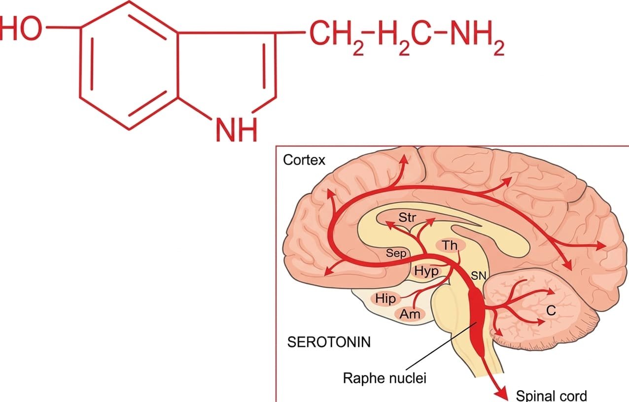
Serotonin (5-Hydroxytryptamine, 5-HT) is an endogenous indoleethylamine autacoid and neurotransmitter synthesized widely across animal tissues, plants, fruits, and venoms.
1. Anatomical Distribution in Humans
- Gastrointestinal Enterochromaffin (EC) Cells (~90% of Total Body Pool): Synthesized and stored within mucosal enterochromaffin cells of the stomach and small intestine; regulates peristaltic motor reflexes and secretory activity.
- Circulating Blood Platelets (~8% of Total Body Pool): Platelets do not synthesize serotonin (they lack tryptophan hydroxylase). They take up free 5-HT from intestinal mesenteric circulation via high-affinity Serotonin Transporters (SERT) and store it in dense core granules; released during platelet activation to promote local vasoconstriction and hemostatic plug formation.
- Central Nervous System Neurons (~1–2% of Total Body Pool): Synthesized and stored in the raphe nuclei of the midbrain, pons, and medulla; modulates mood, sleep, appetite, vomiting, nociception, and thermoregulation.
2. Biosynthesis & Degradation
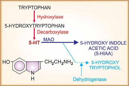
Serotonin is synthesized from essential dietary L-tryptophan via two sequential enzymatic steps:
- Hydroxylation (Rate-Limiting Step): Tryptophan is hydroxylated at the 5-position by the rate-limiting enzyme tryptophan hydroxylase (TPH) to yield 5-hydroxytryptophan (5-HTP). TPH is confined to 5-HT-producing cells and is absent in platelets.
- Decarboxylation: 5-HTP is rapidly converted to 5-hydroxytryptamine (5-HT) by ubiquitous aromatic L-amino acid decarboxylase (dopa decarboxylase).
Following release, 5-HT is taken back up via SERT and degraded primarily by monoamine oxidase A (MAO-A) and aldehyde dehydrogenase into 5-hydroxyindoleacetic acid (5-HIAA), which is excreted in urine. A minor alternative pathway forms 5-hydroxytryptophol via alcohol dehydrogenase.
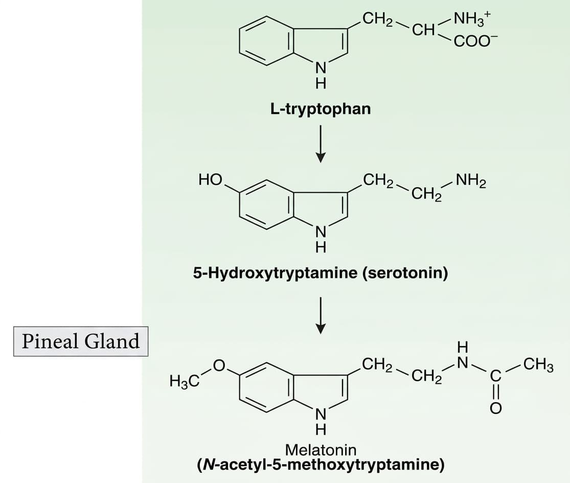
3. Storage & Vesicular Transport
Cytosolic 5-HT is transported into dense intracellular storage vesicles by the Vesicular Monoamine Transporter (VMAT-2). The plant alkaloid reserpine irreversibly inhibits VMAT-2, depleting vesicular serotonin stores across both platelets and central serotonergic terminals (mimicking its depletion of norepinephrine in adrenergic terminals).
Serotonin receptor families
Serotonin acts upon seven distinct receptor families (5-HT1 through 5-HT7) comprising at least 14 molecular subtypes. All are G-protein coupled receptors, with one critical exception: the 5-HT3 receptor is a ligand-gated ion channel.
| Receptor Subtype | Receptor Class & Mechanism | Primary Tissue Distribution | Selective Agonists | Selective Antagonists |
|---|---|---|---|---|
| 5-HT1A | GPCR (Gi/o → ↓ cAMP, opens K+ channels) | Raphe nuclei (autoreceptor), hippocampus, cortex | Buspirone (partial), 8-OH-DPAT | WAY-100635 |
| 5-HT1B | GPCR (Gi/o → ↓ cAMP) | Cranial blood vessels (vasoconstriction), basal ganglia | Sumatriptan, Zolmitriptan | GR-127935 |
| 5-HT1D | GPCR (Gi/o → ↓ cAMP) | Trigeminal presynaptic nerve endings (inhibits CGRP release) | Sumatriptan, Naratriptan, Eletriptan | BRL-15572 |
| 5-HT2A | GPCR (Gq/11 → ↑ IP3, DAG, ↑ Ca2+) | Vascular smooth muscle, platelets (aggregation), cerebral cortex | LSD (hallucinogen), α-methyl-5-HT | Ketanserin, Cyproheptadine, Clozapine, Risperidone |
| 5-HT2B | GPCR (Gq/11 → ↑ IP3, DAG) | Stomach fundus, cardiac valves, vascular endothelium | BW-723C86 | RS-127445 (valvular fibrosis culprit) |
| 5-HT2C | GPCR (Gq/11 → ↑ IP3, DAG) | Choroid plexus, substantia nigra, hypothalamus (appetite) | Lorcaserin (anti-obesity) | Mesulergine, Mirtazapine |
| 5-HT3 | Ligand-Gated Cation Channel (Na+/K+ flux) | Area postrema (CTZ), vagal visceral afferents, myenteric plexus | 2-Methyl-5-HT | Ondansetron, Granisetron, Dolasetron, Palonosetron |
| 5-HT4 | GPCR (Gs → ↑ cAMP, PKA) | Enteric myenteric neurons, visceral smooth muscle, heart | Tegaserod, Cisapride, Prucalopride | GR-113808 |
| 5-HT6 & 5-HT7 | GPCR (Gs → ↑ cAMP) | Limbic system, cortex, hypothalamus, GI tract | LP-44 | Clozapine (high affinity for 5-HT7) |
Carcinoid syndrome & migraine
1. Carcinoid Syndrome
Carcinoid syndrome is a clinical disorder associated with neuroendocrine tumors arising from enterochromaffin (Kulchitsky) cells of the gastrointestinal tract (most commonly the terminal ileum or appendix). Because primitive gut hormones drain directly into the portal circulation and are metabolized by hepatic monoamine oxidases, clinical carcinoid syndrome typically emerges only after the tumor has metastasized to the liver (allowing mediators direct access to hepatic veins and systemic circulation) or originates in primary extra-portal sites (e.g., bronchial carcinoids).
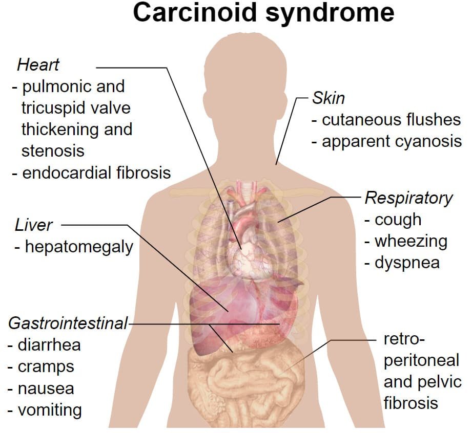
- Secreted Mediators: 5-Hydroxytryptamine is the predominant mediator, accompanied by tachykinins (Substance P, Neurokinin A), histamine, prostaglandins, kallikrein, and motilin.
- Clinical Manifestations: Paroxysmal cutaneous flushing of the face and upper chest; explosive, watery diarrhea and abdominal cramping; asthmatic wheezing and dyspnea; hypotension; and right-sided cardiac valvular lesions (fibrotic thickening of the tricuspid and pulmonic valves leading to right heart failure). Left heart valves are protected because serotonin is catabolized by pulmonary endothelial MAO-A.
- Medical Management:
- Octreotide: A synthetic somatostatin analog that binds somatostatin (sstr2) receptors, suppressing tumor secretion of 5-HT and peptides (the definitive drug of choice).
- Cyproheptadine: A combined 5-HT2 and H1 receptor antagonist that relieves intestinal hypermotility and diarrhea.
2. Pathophysiology of Migraine Headache
Migraine is a neurovascular disorder characterized by severe, throbbing, unilateral headache attacks accompanied by photophobia, phonophobia, nausea, and vomiting. Approximately one-third of patients experience an antecedent aura phase (scintillating scotomas, visual field loss, paresthesias) driven by a self-propagating wave of cortical neuronal and glial depolarization termed cortical spreading depression (CSD).
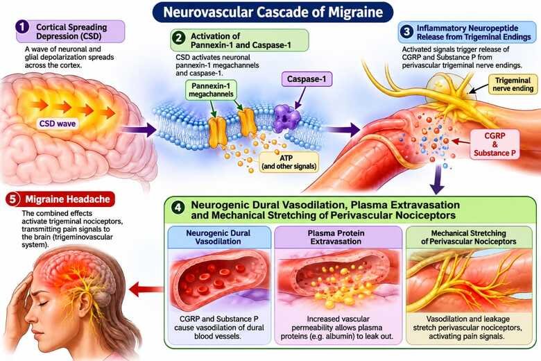
- The Trigeminovascular Cascade: CSD triggers opening of neuronal pannexin-1 megachannels, activating caspase-1 and liberating pro-inflammatory cytokines that stimulate nuclear factor kappa-B (NF-κB). This inflammatory cascade excites perivascular sensory nerve fibers of the ophthalmic division of the trigeminal nerve (CN V1) encircling pial and dural vessels.
- Neurogenic Inflammation & Neuropeptides: Activated trigeminal afferents release potent vasoactive neuropeptides: Calcitonin Gene-Related Peptide (CGRP), Substance P, and Neurokinin A. CGRP induces marked dural arteriolar vasodilation, while Substance P promotes microvascular leakage and mast cell degranulation. The resulting mechanical distension of dural vessels stimulates perivascular nociceptors, transmitting throbbing pain signals into the trigeminal nucleus caudalis and sensory cortex.
- Role of 5-HT in Migraine: Plasma 5-HT falls precipitously during the onset of a migraine attack. A baseline deficit in central serotonergic pain-inhibition systems allows uninhibited trigeminal nociceptive firing.
Serotonergic drugs & agonists
1. 5-HT1D/1B Receptor Agonists (The Triptans)
The triptans (Sumatriptan [prototype], Naratriptan, Rizatriptan, Zolmitriptan, Eletriptan) are the first-line pharmacotherapy for acute, moderate-to-severe migraine attacks and cluster headaches.
- Dual Mechanism of Action:
- Presynaptic 5-HT1D Agonism: Binds Gi-coupled 5-HT1D autoreceptors on perivascular trigeminal sensory nerve terminals, blocking the exocytosis of CGRP, Substance P, and pro-inflammatory neuropeptides, halting neurogenic inflammation.
- Postsynaptic 5-HT1B Agonism: Directly stimulates 5-HT1B receptors on vascular smooth muscle of distended intracranial and meningeal blood vessels, inducing selective vasoconstriction to prevent mechanical stretching of nociceptors.
- Adverse Effects: Paresthesias, warmth, dizziness, and “triptan chest tightness” (transient retrosternal tightness or throat constriction).
- Cardiovascular Contraindications: Strictly contraindicated in patients with documented ischemic coronary artery disease, angina pectoris, prior myocardial infarction, uncontrolled hypertension, peripheral vascular disease, or basilar/hemiplegic migraine, as they can precipitate coronary vasospasm and acute myocardial ischemia.
2. 5-HT1A Partial Agonists
Buspirone: A partial agonist at somatodendritic 5-HT1A autoreceptors in the dorsal raphe and postsynaptic receptors in the limbic cortex. Used as a non-benzodiazepine anxiolytic for generalized anxiety disorder (GAD). Does not interact with GABA receptors; lacks sedative, anticonvulsant, and muscle-relaxant actions; and carries zero potential for tolerance, dependence, or physical abuse.
3. 5-HT2 Receptor Antagonists
- Cyproheptadine: Blocks 5-HT2A and histamine H1 receptors, with significant anticholinergic actions. Indicated for controlling diarrhea and flushing in carcinoid syndrome, cold-induced urticaria, and as an appetite stimulant. It is also the specific drug of choice for managing mild-to-moderate Serotonin Syndrome.
- Ketanserin: Potent competitive 5-HT2A antagonist with concurrent α1-adrenoceptor blocking actions; inhibits platelet aggregation and lowers blood pressure via α1 vasodilation.
- Methysergide & Pizotifen: Prophylactic migraine agents. Methysergide was withdrawn due to its dangerous propensity to cause retroperitoneal, pleural, and cardiac valvular fibrotic reactions leading to obstructive uropathy and renal failure.
- Clozapine & Atypical Antipsychotics: Antagonize 5-HT2A/2C and dopamine D2 receptors; treats refractory schizophrenia with reduced extrapyramidal motor side effects.
4. 5-HT3 Receptor Antagonists
Ondansetron, Granisetron, Dolasetron, Tropisetron, Palonosetron: Competitive antagonists at ligand-gated 5-HT3 receptors located in the chemoreceptor trigger zone (area postrema) and peripheral vagal afferents in the upper gastrointestinal tract. They are first-line antiemetics for preventing chemotherapy-induced nausea and vomiting (CINV), post-radiation emesis, and postoperative nausea and vomiting (PONV).
5. 5-HT4 Receptor Agonists (Prokinetics)
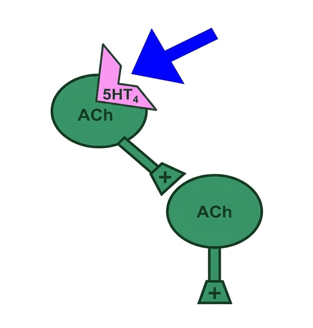
Tegaserod & Cisapride: Stimulate 5-HT4 receptors on prejunctional cholinergic motor neurons of the myenteric plexus, promoting acetylcholine release to enhance coordinated gastrointestinal peristalsis. Indicated historically for irritable bowel syndrome with constipation (IBS-C). Cisapride was restricted due to inhibition of cardiac HERG K+ channels, causing QT prolongation and fatal torsades de pointes.
6. Selective Serotonin Reuptake Inhibitors (SSRIs)
Fluoxetine, Sertraline, Paroxetine, Fluvoxamine, Citalopram, Escitalopram: Selectively inhibit the serotonin transporter (SERT), preventing 5-HT reuptake and elevating synaptic serotonin concentrations throughout the CNS. First-line pharmacotherapy for major depressive disorder, generalized anxiety, panic disorder, obsessive-compulsive disorder (OCD), and bulimia nervosa.
The kallikrein-kinin system
Bradykinin is a potent vasoactive nonapeptide (Arg-Pro-Pro-Gly-Phe-Ser-Pro-Phe-Arg) generated within circulating plasma and inflamed tissues. It serves as a cardinal chemical mediator of acute inflammation, pain sensation, and hemodynamic autoregulation.
1. Synthesis & Degradation
- Enzymatic Cleavage: Bradykinin is derived from high-molecular-weight kininogen (HMWK) in plasma and low-molecular-weight kininogen (LMWK) in tissue lymph. Activated coagulation Factor XIIa (Hageman factor) converts plasma prekallikrein into kallikrein, a serine protease that cleaves kininogens into bradykinin (in plasma) and kallidin (lysyl-bradykinin, in tissues).
- Rapid Inactivation: Bradykinin has an elimination half-life of <30 seconds. Over 90% is degraded on first pass through the pulmonary vascular bed by kininase II, an enzyme biochemically identical to Angiotensin-Converting Enzyme (ACE). Neutral endopeptidase (neprilysin) also catabolizes kinins.
2. Receptor Subtypes & Pharmacodynamics
- B1 Receptor: Minimally expressed under normal physiological conditions; rapidly upregulated (induced) by pro-inflammatory cytokines (IL-1β, TNF-α) and tissue injury. Mediates chronic inflammatory pain and sustained hyperalgesia.
- B2 Receptor: A constitutively active GPCR coupled to Gq/11 (activating PLC → IP3, DAG → intracellular Ca2+ and protein kinase C). Mediates virtually all acute physiological and inflammatory actions of bradykinin:
- Vasodilation & Hypotension: Stimulates endothelial B2 receptors to release nitric oxide (NO) and prostacyclin (PGI2), causing potent arteriolar dilation. In the kidneys, it promotes renal medullary vasodilation and natriuresis by inhibiting tubular sodium reabsorption.
- Increased Microvascular Permeability: Induces contraction of postcapillary venular endothelial cells, causing massive plasma extravasation, blister formation, and angioedema.
- Nociception (Algesia): Directly excites nociceptive primary sensory afferents and provokes the release of substance P and CGRP, generating severe, sharp pain amplified synergistically by prostaglandins.
- Bronchoconstriction: Powerful spasmogen of airway smooth muscle; inhaled bradykinin induces severe bronchospasm in asthmatic patients.
3. Clinical Therapeutics & Pathology
- Aprotinin (Trasylol): A natural broad-spectrum serine protease inhibitor that blocks plasma and tissue kallikreins, plasmin, and trypsin. Used clinically to reduce perioperative blood loss and transfusion requirements during coronary artery bypass graft (CABG) surgery.
- Icatibant: A synthetic decapeptide that acts as a competitive, selective B2 receptor antagonist. Indicated for the subcutaneous treatment of acute, life-threatening attacks of Hereditary Angioedema (HAE) caused by C1-esterase inhibitor deficiency.
- ACE-Inhibitor-Induced Cough and Angioedema: ACE inhibitors (captopril, enalapril) block kininase II, preventing bradykinin degradation. Bradykinin and substance P accumulate in the upper respiratory tract, triggering a persistent, intractable dry cough in up to 15% of patients, and potentially fatal angioedema of the lips, tongue, and larynx.
Ergot alkaloids
Ergot alkaloids are complex, nitrogenous polycyclic compounds produced by Claviceps purpurea, a parasitic fungus that infests rye and other cereal grains. Historically, ingestion of contaminated grain caused epidemics of “St. Anthony’s Fire” (ergotism), characterized by intense burning pain, hallucinations, severe peripheral vasoconstriction, and ischemic gangrene of the extremities.
1. Chemical Classification
- Amine Alkaloids: Simple lysergic acid amide derivatives: Ergonovine (ergometrine), Methylergonovine, Methysergide, and the psychedelic hallucinogen Lysergic acid diethylamide (LSD).
- Peptide (Amino Acid) Alkaloids: Complex cyclic tripeptide moieties attached to lysergic acid: Ergotamine, α-Ergocryptine, and Bromocriptine.
2. Receptor Pharmacology & Mechanism
Ergot alkaloids are notorious for non-selective, promiscuous receptor interactions, acting as agonists, partial agonists, or antagonists across three major biogenic amine GPCR families:
- α-Adrenoceptors: Agonist and partial agonist activity drives potent, long-lasting peripheral and cerebral arterial vasoconstriction; antagonist activity at higher concentrations.
- Serotonin (5-HT) Receptors: High affinity for 5-HT1A, 5-HT1D, and 5-HT2 receptors; mediates acute antimigraine efficacy and smooth muscle spasm.
- Dopamine Receptors: Selective agonists or partial agonists at central dopamine D2 receptors (particularly bromocriptine and cabergoline).
3. Organ System Effects & Clinical Applications
A. Obstetric Applications (Postpartum Hemorrhage)
Ergonovine & Methylergonovine: Cause rapid, powerful, sustained tetanic contractions of uterine myometrium by activating α1 and 5-HT receptors. The myometrial sensitivity increases dramatically as pregnancy progresses due to estrogen-driven upregulation of α1 receptors; the uterus at term is exquisitely sensitive.
Clinical Use: Administered intramuscularly or orally to control postpartum hemorrhage (PPH) and maintain uterine tone following the complete delivery of the placenta. Contraindication: NEVER use for induction of labor, as tetanic contractions cause fetal asphyxia and uterine rupture.
B. Acute Migraine Management
Ergotamine Tartrate & Dihydroergotamine (DHE): Cause sustained vasoconstriction of dilated intracranial pulsation beds and presynaptically inhibit trigeminal neuropeptide exocytosis via 5-HT1D agonism. Highly specific for migraine; lack non-specific analgesic activity for other pain conditions. Ergotamine is administered sublingually, orally with caffeine, or rectally; DHE is administered parenterally (IV, IM, SC) or intranasally.
C. Neuroendocrine Control (Hyperprolactinemia)
Bromocriptine & Cabergoline: Potent dopamine D2 receptor agonists acting on anterior pituitary lactotroph cells to suppress prolactin gene transcription and exocytosis. First-line medical therapy for prolactin-secreting pituitary adenomas (prolactinomas), amenorrhea-galactorrhea syndrome, and suppression of physiological postpartum lactation.
4. Adverse Effects & Contraindications
- Vasospasm & Gangrene: Ergotamine induces prolonged, cumulative arterial spasm. Overdose or co-administration with strong CYP3A4 inhibitors (macrolides, protease inhibitors) causes severe ischemic pain, absent pulses, and dry gangrene of fingers and toes.
- Gastrointestinal Disturbance: Nausea, vomiting, and abdominal pain are common due to direct activation of the medullary chemoreceptor trigger zone (CTZ).
- Contraindications: Absolutely contraindicated in pregnancy (potent abortifacient action), uncontrolled hypertension, peripheral vascular disease, coronary artery disease, and severe hepatic/renal impairment.
The eicosanoid cascade
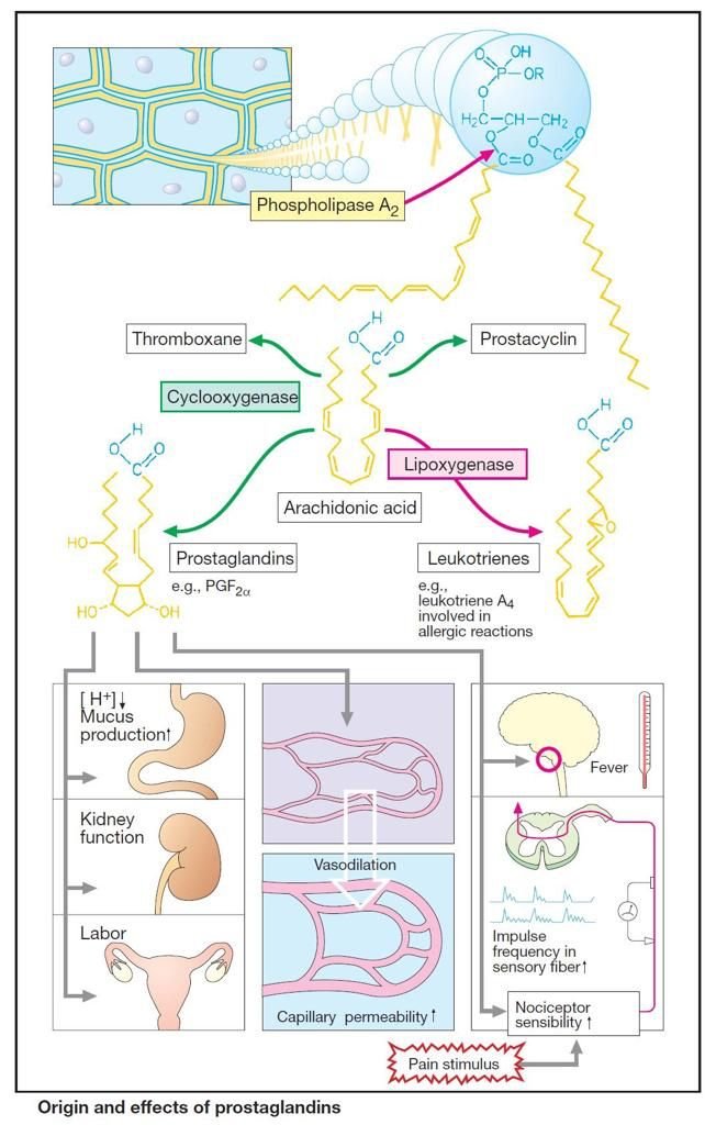
The eicosanoids (Greek eikosi, twenty) are oxygenated, polyunsaturated 20-carbon fatty acid autacoids synthesized de novo from cell membrane phospholipids in response to mechanical trauma, cytokines, or chemical stimuli. They are not stored preformed inside cellular granules.
1. Liberation of Arachidonic Acid
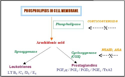
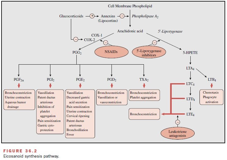
The universal obligate precursor is arachidonic acid (5,8,11,14-eicosatetraenoic acid), an essential ω-6 fatty acid esterified at the sn-2 position of membrane phospholipids. Cellular activation triggers the calcium-dependent enzyme phospholipase A2 (PLA2) to cleave and release free arachidonic acid into the cytoplasm. Glucocorticoids inhibit this step by inducing the synthesis of inhibitory proteins termed annexins (lipocortins).
2. The Cyclooxygenase (COX) Pathway
Free arachidonic acid is converted by the bifunctional enzyme cyclooxygenase (endoperoxide synthase) via a two-step reaction: (1) cyclooxygenase adds molecular oxygen to yield unstable PGG2, and (2) peroxidase reduces PGG2 into unstable PGH2. Tissue-specific synthases convert PGH2 into active prostanoids:
- Prostacyclin (PGI2): Synthesized primarily by vascular endothelial cells via prostacyclin synthase. Acts on IP receptors (Gs → ↑ cAMP) to induce potent vasodilation, inhibit platelet aggregation, maintain patency of the ductus arteriosus, and promote gastric mucosal cytoprotection.
- Thromboxane A2 (TXA2): Synthesized exclusively in platelets via thromboxane synthase. Acts on TP receptors (Gq → ↑ IP3/Ca2+) to induce platelet aggregation, intense vasoconstriction, and bronchoconstriction. PGI2 and TXA2 form a critical physiological antagonistic balance regulating vascular hemostasis.
- Prostaglandin E2 (PGE2): Ubiquitously synthesized; acts on four distinct G-protein coupled receptors:
- EP1 (Gq): Smooth muscle contraction, hyperalgesia.
- EP2 & EP4 (Gs → ↑ cAMP): Vasodilation, bronchodilation, maintenance of renal blood flow, cervical ripening.
- EP3 (Gi → ↓ cAMP): Inhibits gastric acid secretion, stimulates protective mucin and bicarbonate secretion, contracts uterine smooth muscle, and elevates anterior hypothalamic thermoregulatory set-point (inducing fever in response to IL-1 and pyrogens).
- Prostaglandin F2α (PGF2α): Synthesized in the myometrium, lungs, and eye; acts on FP receptors (Gq) to contract the pregnant uterus, contract bronchioles, and increase uveoscleral outflow of aqueous humor.
- Prostaglandin D2 (PGD2): Produced by mast cells and brain; acts on DP receptors to induce bronchoconstriction, vasodilation, and non-REM sleep.
3. The Lipoxygenase (5-LOX) Pathway
In inflammatory leukocytes (neutrophils, eosinophils, basophils, macrophages), arachidonic acid is oxygenated by 5-lipoxygenase (5-LOX) in the presence of 5-lipoxygenase activating protein (FLAP) to form 5-HPETE, which is converted to unstable leukotriene A4 (LTA4):
- Leukotriene B4 (LTB4): Produced via LTA4 hydrolase; acts on BLT1 receptors as an extraordinarily potent chemoattractant for neutrophils, stimulating leukocyte adhesion to endothelium, trans-endothelial migration, degranulation, and lysosomal enzyme release.
- Cysteinyl Leukotrienes (LTC4, LTD4, LTE4): Formed by conjugating glutathione to LTA4 (peptidoleukotrienes). Formerly designated “Slow-Reacting Substance of Anaphylaxis” (SRS-A). Act on CysLT1 receptors as potent bronchoconstrictors (1000-fold more potent than histamine), promoting airway mucosal edema, plasma exudation, and hypersecretion in bronchial asthma.
Clinical eicosanoid agonists
1. Reproductive & Obstetric Pharmacology
- Dinoprostone (Synthetic PGE2): Administered topically as an endocervical gel or vaginal insert for cervical ripening and labor induction at term; promotes collagen degradation and cervical softening. Also used as an abortifacient in second-trimester termination.
- Misoprostol (Synthetic PGE1 Methyl Ester Analog): Orally and vaginally active. Combined with the progesterone receptor antagonist mifepristone for medical termination of early pregnancy (≤70 days gestation). Widely used off-label for induction of labor and prevention/treatment of postpartum hemorrhage (PPH).
- Carboprost Tromethamine (15-Methyl-PGF2α): Administered intramuscularly for controlling severe, refractory postpartum hemorrhage due to uterine atony unresponsive to oxytocin and ergometrine; also used as a second-trimester abortifacient.
- Gemeprost: A PGE1 analog administered as a vaginal pessary for cervical dilation prior to vacuum aspiration and early pregnancy termination.
2. Gastrointestinal Cytoprotection
- Misoprostol: Specifically indicated for the prevention and treatment of NSAID-induced and steroid-induced gastric and duodenal ulcers. Stimulates EP3 receptors on parietal cells to inhibit acid secretion while promoting mucus and bicarbonate secretion. Adverse Effects: Dose-dependent abdominal cramping and diarrhea; strictly contraindicated in pregnancy due to abortifacient risk.
3. Cardiovascular & Pediatric Applications
- Alprostadil (PGE1):
- Patent Ductus Arteriosus (PDA) Maintenance: Administered as a continuous intravenous infusion in neonates born with ductus-dependent congenital heart defects (e.g., transposition of great vessels, pulmonary atresia, coarctation of aorta) to maintain patency of the ductus arteriosus until corrective cardiac surgery can be performed.
- Erectile Dysfunction: Injected intracavernosally or administered as a transurethral pellet; induces direct relaxation of trabecular smooth muscle and cavernous arterioles, facilitating penile erection.
- Epoprostenol & Treprostinil (PGI2 Analogs): Potent pulmonary vasodilators and inhibitors of platelet aggregation administered via continuous IV/subcutaneous infusion or inhalation for primary pulmonary arterial hypertension (PAH), lowering pulmonary arterial pressure and vascular resistance.
4. Ophthalmic Applications (Glaucoma)
- Latanoprost, Bimatoprost, Travoprost, Unoprostone: Stable PGF2α isopropyl ester prodrugs administered as once-daily evening eye drops. They stimulate ocular FP receptors, relaxing the ciliary muscle to dramatically increase uveoscleral outflow of aqueous humor, lowering intraocular pressure (IOP) in open-angle glaucoma and ocular hypertension.
- Characteristic Adverse Effects: Irreversible brown pigmentation of the iris (due to increased melanin synthesis within melanocytes), hypertrichosis (increased length, thickness, and darkness of eyelashes), conjunctival hyperemia, and ocular irritation.
5. Leukotriene Pathway Modifiers (Asthma)
- Zileuton: Orally active selective inhibitor of 5-lipoxygenase (5-LOX); prevents synthesis of all leukotrienes (LTB4 and cysteinyl leukotrienes). Used for chronic asthma prophylaxis. Caution: Can induce hepatotoxicity; liver function tests must be monitored.
- Montelukast & Zafirlukast: Competitive antagonists at the CysLT1 receptor; block LTC4-, LTD4-, and LTE4-mediated bronchoconstriction, airway edema, and mucus plugging. Highly effective oral prophylactic controllers for chronic asthma and aspirin-exacerbated respiratory disease.
| Drug Name | Eicosanoid Class | Receptor Target | Primary Clinical Application |
|---|---|---|---|
| Alprostadil | PGE1 analog | EP2, EP4 | Maintains patent ductus arteriosus in neonates; intracavernosal therapy for erectile dysfunction. |
| Misoprostol | PGE1 derivative | EP3, EP1 | Prevention of NSAID gastropathy; medical termination of pregnancy (with mifepristone); postpartum hemorrhage. |
| Dinoprostone | PGE2 preparation | EP1, EP2, EP3 | Cervical ripening; labor induction; second-trimester therapeutic abortion. |
| Carboprost Tromethamine | 15-methyl-PGF2α | FP | Refractory postpartum hemorrhage due to uterine atony; second-trimester abortion. |
| Epoprostenol / Treprostinil | PGI2 (Prostacyclin) | IP | Severe pulmonary arterial hypertension (PAH); inhibition of platelet aggregation during hemodialysis. |
| Latanoprost / Bimatoprost | PGF2α analogs | FP | First-line topical reduction of intraocular pressure in open-angle glaucoma via uveoscleral outflow. |
| Montelukast / Zafirlukast | CysLT1 Antagonist | CysLT1 | Prophylactic oral maintenance therapy for bronchial asthma; allergic rhinitis; aspirin-sensitive asthma. |
| Zileuton | 5-LOX Inhibitor | 5-Lipoxygenase | Chronic prophylactic treatment of asthma (blocks both LTB4 and cysteinyl leukotriene synthesis). |
Inflammation & COX enzymes
Inflammation is a protective homeostatic vascular and cellular response to tissue injury caused by physical trauma, noxious chemicals, or invading microbiological pathogens. Its physiological objective is to isolate and destroy the inciting insult, clear necrotic debris, and initiate tissue repair. If over-vigorous or dysregulated, acute inflammation can transition into debilitating chronic inflammatory and autoimmune pathology.
1. Cardinal Signs of Acute Inflammation
- Redness (Rubor): Local microvascular arteriolar vasodilation increasing regional blood flow (hyperemia); mediated by histamine, prostaglandins (PGI2, PGE2), and nitric oxide.
- Heat (Calor): Increased local temperature due to sustained vascular hyperemia and elevated perfusion.
- Pain (Dolor): Hyperalgesia resulting from direct nociceptor stimulation by bradykinin and 5-HT, markedly sensitized by PGE2 and PGI2.
- Swelling (Tumor): Increased microvascular permeability causing postcapillary venular endothelial gap formation, allowing plasma protein leakage into the extravascular interstitial space (edema).
- Loss of Function (Functio Laesa): Functional limitation produced by local mechanical swelling and reflex pain inhibition.
2. The Cyclooxygenase Isozymes
Cyclooxygenase exists in three distinct structural and functional isoforms:
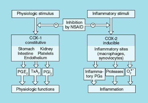
COX-1 (Constitutive / “Housekeeping” Isozyme)
- Expression: Expressed constitutively across virtually all healthy tissues under basal physiological conditions.
- Physiological Functions:
- Gastric Mucosal Cytoprotection: Synthesizes basal PGE2 and PGI2, which inhibit parietal cell acid secretion, stimulate protective mucus and bicarbonate release, and sustain mucosal blood flow.
- Hemostasis: Platelet COX-1 synthesizes Thromboxane A2, driving platelet aggregation and primary hemostasis.
- Renal Hemodynamics: Maintains medullary blood flow and glomerular filtration rate during physiological stress.
COX-2 (Inducible / Inflammatory Isozyme)
- Expression: Expressed at very low basal levels in most tissues; rapidly and dramatically induced in inflammatory cells (macrophages, synoviocytes, endothelial cells, fibroblasts) by pro-inflammatory cytokines (IL-1β, TNF-α), bacterial lipopolysaccharide (LPS), and growth factors.
- Constitutive Sites: Expressed constitutively in the brain (cortex, hippocampus), kidneys (macula densa, medullary interstitial cells), vascular endothelium, and female reproductive tract.
- Pathological Role: The principal enzymatic driver of inflammatory prostanoid synthesis, generating PGE2 that causes peripheral erythema, edema, inflammatory hyperalgesia, and hypothalamic fever. In vascular endothelium, constitutive COX-2 is the primary source of anti-thrombotic Prostacyclin (PGI2).
COX-3 (Central Splice Variant)
- Expression & Nature: An alternative splicing variant of the COX-1 gene expressed predominantly in the central nervous system (cerebral cortex) and cardiac tissue.
- Pharmacology: Highly sensitive to inhibition by paracetamol (acetaminophen), phenacetin, and metamizole, explaining central analgesia and antipyresis in the absence of significant peripheral anti-inflammatory action.
Classification & PK of NSAIDs
Non-Steroidal Anti-Inflammatory Drugs (NSAIDs) are among the most widely prescribed therapeutic agents worldwide. They share three primary clinical actions: analgesia (pain relief), antipyresis (fever reduction), and anti-inflammatory activity. They act fundamentally by inhibiting cyclooxygenase enzymes, preventing eicosanoid biosynthesis.
1. Chemical Classification of NSAIDs
- Salicylates: Acetylsalicylic acid (Aspirin), Sodium salicylate, Diflunisal, Sulfasalazine.
- Propionic Acid Derivatives: Ibuprofen, Naproxen, Ketoprofen, Flurbiprofen, Fenoprofen, Oxaprozin.
- Acetic Acid Derivatives:
- Indole Acetic Acids: Indomethacin, Sulindac.
- Phenylacetic Acids: Diclofenac.
- Pyrrolo-Pyrrole Derivatives: Ketorolac.
- Oxicam Derivatives: Piroxicam, Tenoxicam, Meloxicam.
- Fenamates (Anthranilic Acids): Mefenamic acid, Meclofenamate.
- Pyrazolone Derivatives: Phenylbutazone, Oxyphenbutazone, Sulfinpyrazone, Metamizole.
- Sulfonanilide Derivatives: Nimesulide.
- Selective COX-2 Inhibitors (Coxibs): Celecoxib, Etoricoxib, Parecoxib.
- Para-Aminophenol Derivatives: Paracetamol (Acetaminophen).
2. General Pharmacokinetics of NSAIDs
- Absorption: Administered orally, parenterally, rectally, and topically. Most are weak organic acids (pKa 3.0 to 5.0). Completely absorbed from the stomach and upper small intestine; oral bioavailability is high (>80%) as they undergo minimal first-pass hepatic metabolism. Food may delay absorption rate, but does not decrease total bioavailability.
- Plasma Protein Binding: Extensively bound to plasma albumin (≥95% to 99%). Highly prone to competitive displacement interactions, displacing co-administered drugs like warfarin, sulfonylureas (glibenclamide), and methotrexate, potentially triggering toxicity.
- Tissue Distribution & Synovial Penetration: Distribute broadly into extracellular fluids and inflame joints. All NSAIDs penetrate readily into synovial fluid; drugs with short elimination half-lives (e.g., ibuprofen, diclofenac) persist in synovial compartments long after plasma concentrations decline, maintaining prolonged clinical joint efficacy.
- Metabolism & Excretion: Extensively metabolized in the liver via phase I cytochrome P450 oxidation (predominantly CYP2C9 and CYP3A4) followed by phase II glucuronide conjugation. Cleared predominantly by renal excretion. Many agents (e.g., indomethacin, sulindac, piroxicam) undergo significant enterohepatic recirculation, which correlates with severe lower gastrointestinal mucosal damage and ulceration.
| Drug | Elimination Half-Life (Hours) | Urinary Excretion of Unchanged Drug (%) | Standard Anti-Inflammatory Dose |
|---|---|---|---|
| Aspirin | 0.25 (Salicylate: 2 to 19h) | <2% | 1200 – 1500 mg TID |
| Ibuprofen | 2.0 | <1% | 600 – 800 mg QID (Max: 2400–3200 mg/day) |
| Diclofenac | 1.1 | <1% | 50 – 75 mg TID / BID |
| Indomethacin | 4.0 – 5.0 | 16% | 50 – 75 mg TID |
| Naproxen | 14.0 | <1% | 375 – 500 mg BID |
| Ketorolac | 4.0 – 10.0 | 58% | 10 mg QID (Max 5 days duration) |
| Meloxicam | 20.0 | Low | 7.5 – 15 mg QD |
| Celecoxib | 11.0 | 27% | 100 – 200 mg BID (or 200 mg QD) |
| Piroxicam | 57.0 | 4% – 10% | 20 mg QD |
| Oxaprozin | 58.0 | 1% – 4% | 1200 – 1800 mg QD |
Aspirin & non-selective NSAIDs
1. Aspirin: The Irreversible Prototype
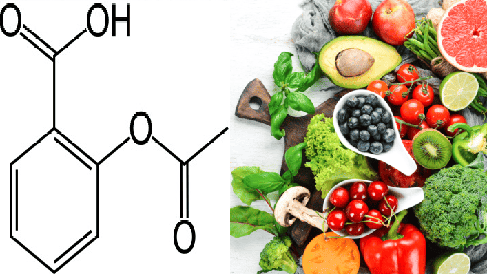
Aspirin (Acetylsalicylic Acid, ASA) is the historic prototype to which all NSAIDs are compared. Its molecular mechanism is unique among NSAIDs:
- Covalent Irreversible Inactivation: Aspirin covalently acetylates a specific serine residue (Serine-529 in COX-1; Serine-516 in COX-2) located near the catalytic active site. This sterically obstructs arachidonic acid from entering the cyclooxygenase channel, permanently abolishing enzymatic activity. All other traditional NSAIDs (ibuprofen, naproxen, indomethacin) are reversible competitive inhibitors.
- The Platelet Thromboxane Paradox: Blood platelets possess COX-1 to synthesize pro-aggregatory TXA2. Because mature platelets lack nuclei, they cannot transcribe mRNA or synthesize new protein; inhibition of platelet COX-1 is permanent and lasts for the entire lifespan of the platelet (7 to 10 days). Conversely, vascular endothelial cells have nuclei and regenerate new COX-1/COX-2 within hours, restoring anti-aggregatory prostacyclin (PGI2) synthesis. Therefore, low-dose aspirin (75 to 100 mg/day) produces selective, cardioprotective antiplatelet inhibition without suppressing endothelial PGI2.
- Dose-Dependent Spectrum of Action:
- Low Doses (75 to 150 mg/day): Selective irreversible antiplatelet aggregation; secondary prevention of myocardial infarction, ischemic stroke, and transient ischemic attacks (TIAs).
- Moderate Doses (300 to 2400 mg/day): Analgesic and antipyretic actions.
- High Anti-Inflammatory Doses (3.2 to 4.5 g/day): Effective anti-inflammatory activity for acute rheumatic fever and severe rheumatoid arthritis (frequently causes salicylism).
- Uric Acid Biphasic Actions: Low-to-moderate doses (<2 to 3 g/day) compete with uric acid for active renal tubular secretion, inducing hyperuricemia (worsening gout); high doses (>5 g/day) inhibit renal tubular reabsorption of uric acid, producing uricosuria. Rule: Aspirin is avoided in gout.
2. Adverse Effects of Aspirin & Traditional NSAIDs
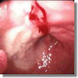
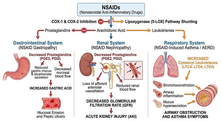
- Gastric Mucosal Ulceration & Ion Trapping: Prostaglandin deficiency removes mucosal cytoprotection. In addition, aspirin is a weak acid (pKa 3.5) that remains non-ionized and lipophilic within acidic gastric juice (pH 1.5 to 2.0). It diffuses across the gastric epithelial cell membrane into neutral cytoplasm (pH 7.1), where it immediately ionizes into charged salicylate and H+ ions. These charged ions cannot diffuse out, becoming “ion trapped” inside the mucosal cell, causing cell edema, necrosis, subepithelial hemorrhage, and peptic ulceration.
- Aspirin-Exacerbated Respiratory Disease (AERD / Samter’s Triad): A non-IgE pseudoallergic reaction occurring in up to 10–20% of asthmatics with nasal polyps and chronic rhinosinusitis. Blocking COX shunts arachidonic acid entirely down the 5-lipoxygenase pathway, generating a massive surge in cysteinyl leukotrienes (LTC4, LTD4) that triggers severe, life-threatening bronchospasm.
- Reye’s Syndrome: Administration of aspirin to children and adolescents during febrile viral illnesses (influenza A/B, varicella/chickenpox) is strongly linked to Reye’s syndrome: acute mitochondrial injury causing microvesicular hepatic steatosis, fulminant hepatic failure, hyperammonemia, and lethal cerebral edema. Aspirin is strictly contraindicated in pediatric patients with viral fever; paracetamol or ibuprofen must be used instead.
- Renal Toxicity: In high-risk states (congestive heart failure, cirrhosis, hypovolemia, pre-existing chronic kidney disease), renal perfusion relies on vasodilating prostaglandins (PGE2, PGI2) maintaining afferent arteriolar dilation. NSAIDs block these prostaglandins, precipitating acute renal vasoconstriction, acute kidney injury (AKI), sodium and water retention (blunting antihypertensives and diuretics), hyperkalemia, and chronic renal papillary necrosis.
3. Acute Salicylate Poisoning (Salicylism)
- Toxic Doses: Serious toxicity occurs above 150 mg/kg; lethal doses range from 15 to 30 grams in adults (lower in children).
- Clinical Progression: Early toxicity (salicylism) presents with tinnitus, high-frequency hearing loss, vertigo, nausea, vomiting, and diaphoresis. Salicylates directly stimulate the medullary respiratory center, causing hyperventilation (hyperpnea) and initial respiratory alkalosis. Subsequently, toxic concentrations uncouple mitochondrial oxidative phosphorylation, causing anaerobic glycolysis, accumulation of lactic acid and pyruvic acid, and hyperthermia. Combined with circulating salicylate ketoacids, this produces a severe, life-threatening mixed respiratory alkalosis and high anion-gap metabolic acidosis, central respiratory depression, coma, and cardiorespiratory arrest.
- Emergency Management:
- Gastric decontamination: Gastric lavage and repeated doses of activated charcoal.
- Aggressive intravenous fluid resuscitation to correct dehydration and hypokalemia.
- Urinary Alkalinization: Intravenous infusion of sodium bicarbonate (NaHCO3) to titrate urine pH to 7.5–8.5. Alkalinization ionizes salicylic acid into polar salicylate inside renal tubular lumens, preventing reabsorption (“ion trapping”) and increasing renal excretion up to twenty-fold.
- Hemodialysis for severe poisoning (salicylate level >100 mg/dL, refractory acidosis, or renal failure).
- Intramuscular Vitamin K to correct hypoprothrombinemia.
4. Individual Non-Selective NSAIDs
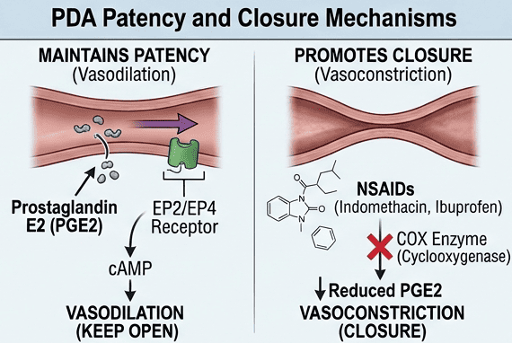
- Ibuprofen: Phenylpropionic acid derivative with the best overall gastrointestinal safety profile among non-selective NSAIDs. Doses of 2400 mg/day equal 4 g of aspirin in anti-inflammatory efficacy; lower doses (<2400 mg/day) are analgesic/antipyretic. Available as topical creams, oral liquid gels, and IV infusions. Used intravenously to close patent ductus arteriosus (PDA) in premature infants with efficacy equal to indomethacin and less renal impairment. Drug Interaction: Reversibly blocks the platelet COX-1 binding site, antagonizing the irreversible antiplatelet action of aspirin if taken prior to aspirin.
- Indomethacin: Indole acetic acid derivative; exceptionally potent non-selective COX inhibitor that also directly inhibits neutrophil motility. First-line therapy for acute gouty arthritis, ankylosing spondylitis, and medical closure of patent ductus arteriosus. Characterized by high toxicity: severe frontal headaches (in 25–50% of patients), dizziness, confusion, depression, acute psychosis with hallucinations, pancreatitis, and renal papillary necrosis. Probenecid inhibits its renal and biliary clearance.
- Diclofenac: Phenylacetic acid derivative with high potency. Concentrates extensively in synovial fluid. Causes significant gastrointestinal ulceration; often co-formulated with misoprostol (Arthrotec) or omeprazole. Topical 3% gel is effective for actinic (solar) keratoses; 0.1% ophthalmic drops prevent postoperative ocular inflammation. Carries higher cardiovascular risk than naproxen.
- Ketorolac: Potent analgesic with weak anti-inflammatory action; administered parenterally (IM/IV) or orally for severe acute postoperative pain. Because of extreme nephrotoxicity and gastrointestinal ulceration risk, therapy is strictly limited to a maximum duration of 5 days.
- Naproxen: Propionic acid derivative with a long half-life (14 hours, permitting BID dosing). Exhibits the lowest cardiovascular risk profile among traditional NSAIDs; preferred in patients with high baseline cardiovascular disease.
- Piroxicam: Oxicam derivative with an exceptionally long half-life (57 hours, once-daily dosing); associated with a very high incidence of gastrointestinal ulceration and cutaneous reactions.
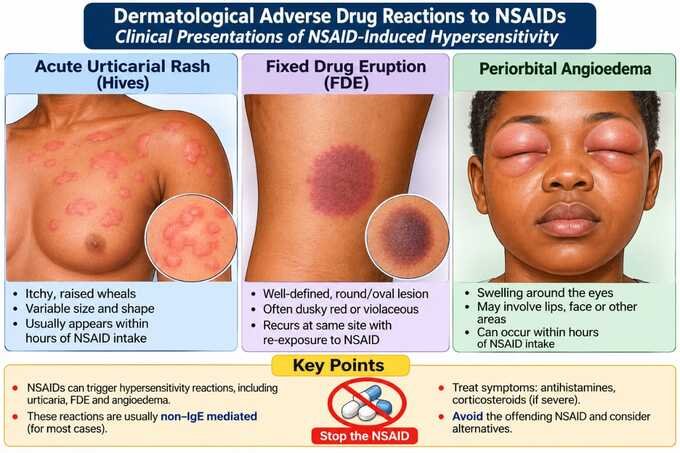
Selective COX-2 inhibitors (coxibs)
Selective COX-2 inhibitors (coxibs) were engineered to inhibit the inducible COX-2 enzyme at sites of inflammation while sparing the cytoprotective COX-1 enzyme in the gastrointestinal mucosa and platelets. The clinical prototype is Celecoxib; other agents include Etoricoxib and the injectable prodrug Parecoxib.
1. Clinical Benefits & Gastrointestinal Safety
- Analgesic & Anti-Inflammatory Efficacy: Display therapeutic potency identical to non-selective NSAIDs in osteoarthritis, rheumatoid arthritis, and acute pain.
- Reduced Gastropathy: By preserving gastric mucosal COX-1, coxibs reduce the incidence of endoscopically confirmed gastroduodenal ulcers, gastrointestinal perforations, and bleeding by approximately 50% compared to traditional NSAIDs.
- No Antiplatelet Action: At therapeutic doses, coxibs do not inhibit platelet COX-1 or reduce TXA2 production, exerting no effect on bleeding time or primary hemostasis.
2. The Cardiovascular Thrombotic Paradox
Pathophysiological Mechanism of Cardiovascular Risk
Under normal conditions, vascular homeostasis is governed by a dynamic balance between platelet COX-1-derived thromboxane A2 (which promotes vasoconstriction and platelet aggregation) and endothelial COX-2-derived prostacyclin (PGI2) (which inhibits aggregation and causes vasodilation):
- Selective COX-2 inhibitors suppress endothelial PGI2 synthesis without inhibiting platelet TXA2 synthesis.
- This creates an unopposed prothrombotic state favoring platelet hyper-reactivity, systemic thrombosis, and vasoconstriction.
- Large randomized clinical trials confirmed a significantly increased incidence of myocardial infarction, ischemic stroke, and cardiovascular death, resulting in the worldwide market withdrawal of rofecoxib (Vioxx) and valdecoxib (Bextra).
- Clinical Rule: Coxibs do not offer cardioprotection; patients requiring cardiovascular prophylaxis must take low-dose aspirin concomitantly.
Renal Toxicity of Coxibs
Because COX-2 is constitutively expressed in the renal cortex and medulla, selective COX-2 inhibitors produce identical renal adverse effects to traditional non-selective NSAIDs: sodium and water retention, peripheral edema, destabilization of arterial hypertension, hyperkalemia, and decreased glomerular filtration. They are contraindicated in severe renal impairment.
3. Celecoxib Properties & Preferential COX-2 Inhibitors
- Celecoxib Pharmacokinetics: Readily absorbed (peak concentration in 3 hours); highly protein-bound; elimination half-life is ~11 hours (dosed once or twice daily). Extensively metabolized by hepatic CYP2C9; interacts with warfarin, requiring INR monitoring.
- Sulfonamide Cross-Allergy: Celecoxib contains a sulfonamide moiety in its chemical structure; can cause skin rashes and is contraindicated in patients with documented severe sulfonamide hypersensitivity.
- Preferential COX-2 Inhibitors:
- Meloxicam: An oxicam derivative that preferentially inhibits COX-2 over COX-1, particularly at the low anti-arthritic dose of 7.5 mg/day. Demonstrates significantly lower gastrointestinal complications than piroxicam, diclofenac, or naproxen, and does not prolong in vivo bleeding time. Selectivity is lost at higher doses (15 mg/day).
- Nimesulide: A sulfonanilide preferential COX-2 inhibitor; associated with severe, idiosyncratic hepatotoxicity (restricted or banned in multiple jurisdictions).
- Nabumetone: A non-acidic prodrug converted in the liver to the active 6-MNA metabolite; preferentially inhibits COX-2, producing very low rates of gastric ulceration.
Paracetamol & acute toxicity
Paracetamol (Acetaminophen, APAP) is the active metabolite of phenacetin and the world’s most widely utilized over-the-counter analgesic and antipyretic. Unlike NSAIDs, paracetamol possesses negligible peripheral anti-inflammatory activity and does not impair platelet aggregation, damage the gastric mucosa, or disrupt renal blood flow.
1. Mechanism of Action
Paracetamol acts primarily within the central nervous system. It inhibits central cyclooxygenase activity (acting on the putative COX-3 / peroxidase catalytic site) to decrease hypothalamic PGE2 synthesis, resetting the elevated thermoregulatory set-point to produce antipyresis and raising the central pain threshold. It is inactive at peripheral inflammatory sites because inflamed tissues contain high concentrations of lipid peroxides that override paracetamol’s inhibitory action on COX enzymes.
2. Clinical Advantages over Aspirin
- Does not damage the gastric mucosal barrier (drug of choice in peptic ulcer disease and gastritis).
- Does not inhibit platelet COX-1 or increase bleeding time (safe in hemophilia, thrombocytopenia, and preoperatively).
- Does not affect renal uric acid tubular excretion (safe in gout patients taking probenecid).
- Does not trigger bronchospasm in patients with aspirin-exacerbated respiratory disease (AERD).
- Does NOT cause Reye’s syndrome; the first-line antipyretic and analgesic for infants, children, and adolescents with viral infections.
3. Acute Paracetamol Poisoning & Hepatotoxicity
Metabolism & Pathogenesis of Hepatic Necrosis
- Normal Therapeutic Metabolism: Over 90% to 95% of therapeutic paracetamol is metabolized in the liver via non-toxic Phase II glucuronidation (55%) and sulfation (30%), forming water-soluble conjugates excreted in urine. A small fraction (~5%) is oxidized by microsomal cytochrome P450 enzymes (predominantly CYP2E1, alongside CYP1A2 and CYP3A4) into a highly toxic, electrophilic, reactive arylating intermediate: N-acetyl-p-benzoquinone imine (NAPQI / NABQI). Under normal conditions, NAPQI is rapidly conjugated with endogenous hepatic glutathione into non-toxic mercapturic acid and cysteine derivatives excreted renally.
- Overdose Mechanics: Toxic ingestion (>150 mg/kg in children or >10 grams in adults; lethal dose ~250 mg/kg):
- Phase II glucuronide and sulfate conjugation pathways become completely saturated.
- Excess paracetamol is shunted into the CYP2E1 pathway, producing massive quantities of NAPQI.
- Hepatic glutathione stores are rapidly depleted (below 30% of normal reserves).
- Unconjugated NAPQI binds covalently to nucleophilic sulfhydryl macromolecules and mitochondrial proteins within hepatocytes, inducing oxidative stress, mitochondrial permeability transition, and irreversible centrilobular hepatic necrosis and acute renal tubular necrosis.
- The Chronic Alcohol Warning: In chronic alcoholics, chronic ethanol consumption induces hepatic CYP2E1 expression while poor nutrition depletes basal glutathione reserves. In this population, hepatotoxicity can occur at therapeutic or sub-toxic doses (5 to 6 grams/day).
Clinical Stages & Antidotal Management
- Clinical Course:
- Stage 1 (0–24 Hours): Asymptomatic or mild nausea, vomiting, diaphoresis, malaise, and pallor.
- Stage 2 (24–72 Hours): Initial symptoms subside; right upper quadrant abdominal tenderness, hepatomegaly, rising serum transaminases (AST/ALT can exceed 10,000 IU/L), bilirubin, and prothrombin time (PT/INR).
- Stage 3 (72–96 Hours): Peak hepatotoxicity; jaundice, coagulopathy, hepatic encephalopathy, acute renal failure, hypoglycemia, and multiorgan failure.
- Stage 4 (4–14 Days): Resolution or death from fulminant hepatic necrosis.
- Specific Antidote – N-Acetylcysteine (NAC):
- Mechanism: Acts as a direct precursor for glutathione synthesis and directly conjugates with NAPQI as a glutathione substitute, neutralizing the reactive metabolite.
- Administration: Administered intravenously (150 mg/kg IV loading dose over 60 minutes, followed by 50 mg/kg over 4 hours and 100 mg/kg over 16 hours) or orally. Most effective when initiated within the first 8 to 10 hours post-ingestion; treatment should still be administered up to 24+ hours or in established acute liver failure.
- Activated Charcoal: Administered if the patient presents within 1 to 2 hours of acute oral ingestion.
Quick recap
References
The following authoritative biomedical reference works and national guidelines were verified in the compilation of this study note:
- Goodman & Gilman’s: Brunton, L.L., Hilal-Dandan, R. and Knollmann, B.C. (2018) Goodman & Gilman’s The Pharmacological Basis of Therapeutics. 13th edn. New York: McGraw-Hill Education. [Chapter 32: Histamine, Bradykinin, and Their Antagonists; Chapter 33: 5-Hydroxytryptamine (Serotonin) and Dopamine; Chapter 37: Prostaglandins and Eicosanoids; Chapter 38: Analgesic-Antipyretic and Anti-inflammatory Agents].
- Katzung’s Pharmacology: Katzung, B.G. and Vanderah, T.W. (2021) Basic & Clinical Pharmacology. 15th edn. New York: McGraw-Hill Education. [Chapter 16: Histamine, Serotonin, and the Ergot Alkaloids; Chapter 18: The Eicosanoids; Chapter 36: Nonsteroidal Anti-Inflammatory Drugs].
- Rang & Dale’s Pharmacology: Ritter, J.M., Flower, R., Henderson, G., Loke, Y.K., MacEwan, D. and Rang, H.P. (2020) Rang & Dale’s Pharmacology. 9th edn. Edinburgh: Elsevier. [Chapter 18: Chemical Mediators and the Cellular Mechanisms of Inflammation and Allergy; Chapter 27: Anti-inflammatory and Immunosuppressive Drugs].
- Lippincott Illustrated Reviews: Whalen, K. (2019) Lippincott Illustrated Reviews: Pharmacology. 7th edn. Philadelphia: Wolters Kluwer. [Chapter 42: Autacoids and Autacoid Antagonists; Chapter 44: Anti-inflammatory, Antipyretic, and Analgesic Agents].
- Uganda Clinical Guidelines: Ministry of Health Uganda (2023) Uganda Clinical Guidelines: National Guidelines for the Management of Common Conditions. Kampala: Ministry of Health. [Section 1.8: Management of Acute Poisoning and Anaphylaxis; Section 16: Postpartum Hemorrhage Management Protocols].